DIGESTIVE ORGANS: GASTROINTESTINAL TRACT (ANATOMICAL MICROSCOPY)
19.1
Vermiform appendix
Specimen:
Specimen Details:
Organ: Appendix
Origin: Human
Staining: Hematoxylin - Eosin (H&E)
Method and Specimen Description:
This is a normal histological section of the human vermiform appendix, stained with the general H&E method, which highlights nuclei in blue-purple (hematoxylin) and cytoplasmic and connective tissue elements in varying shades of pink (eosin).
Objective of the Examination:
This is a normal histological section of the human vermiform appendix, stained with the general H&E method, which highlights nuclei in blue-purple (hematoxylin) and cytoplasmic and connective tissue elements in varying shades of pink (eosin).
Special Features of the Specimen:
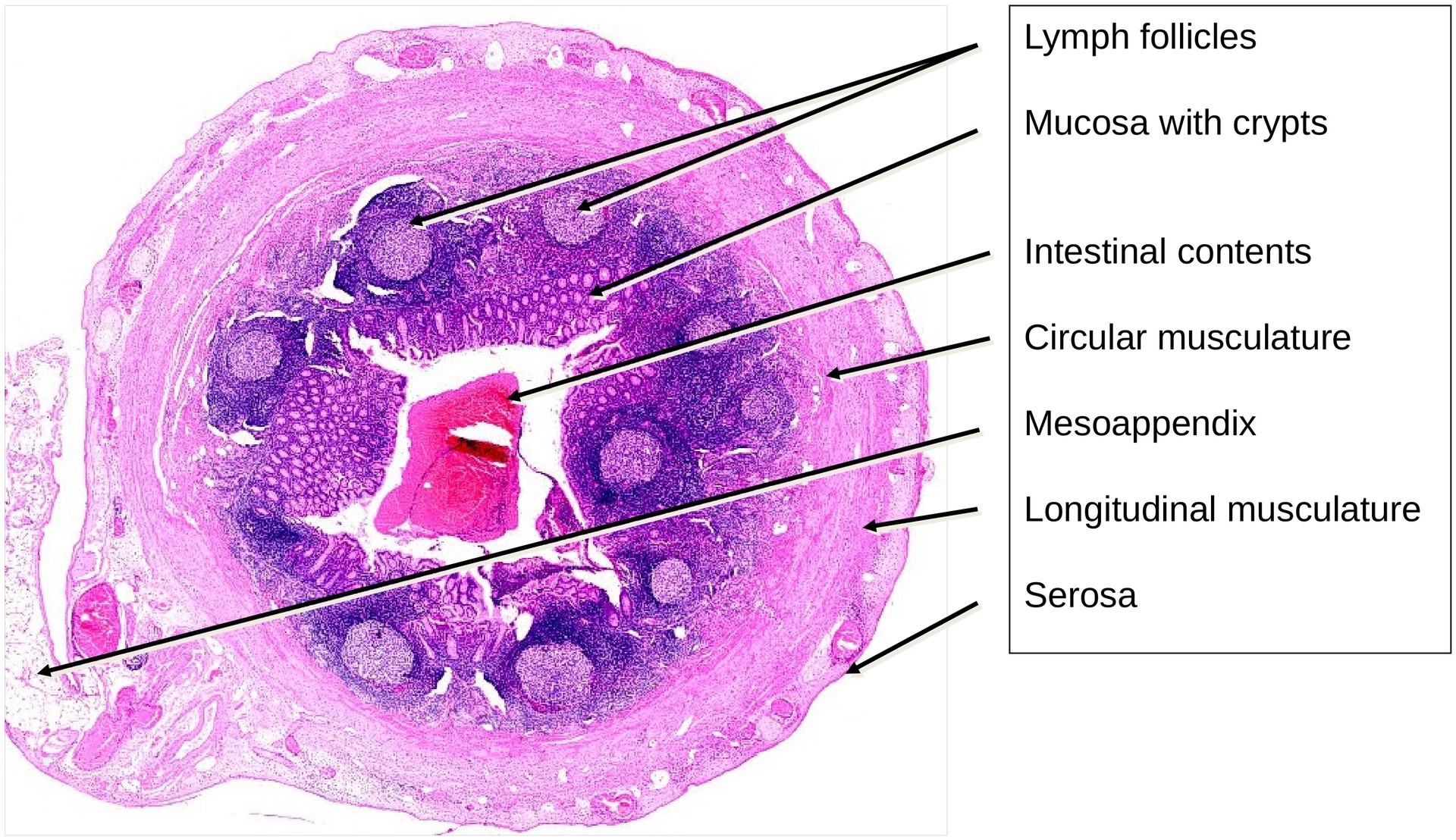
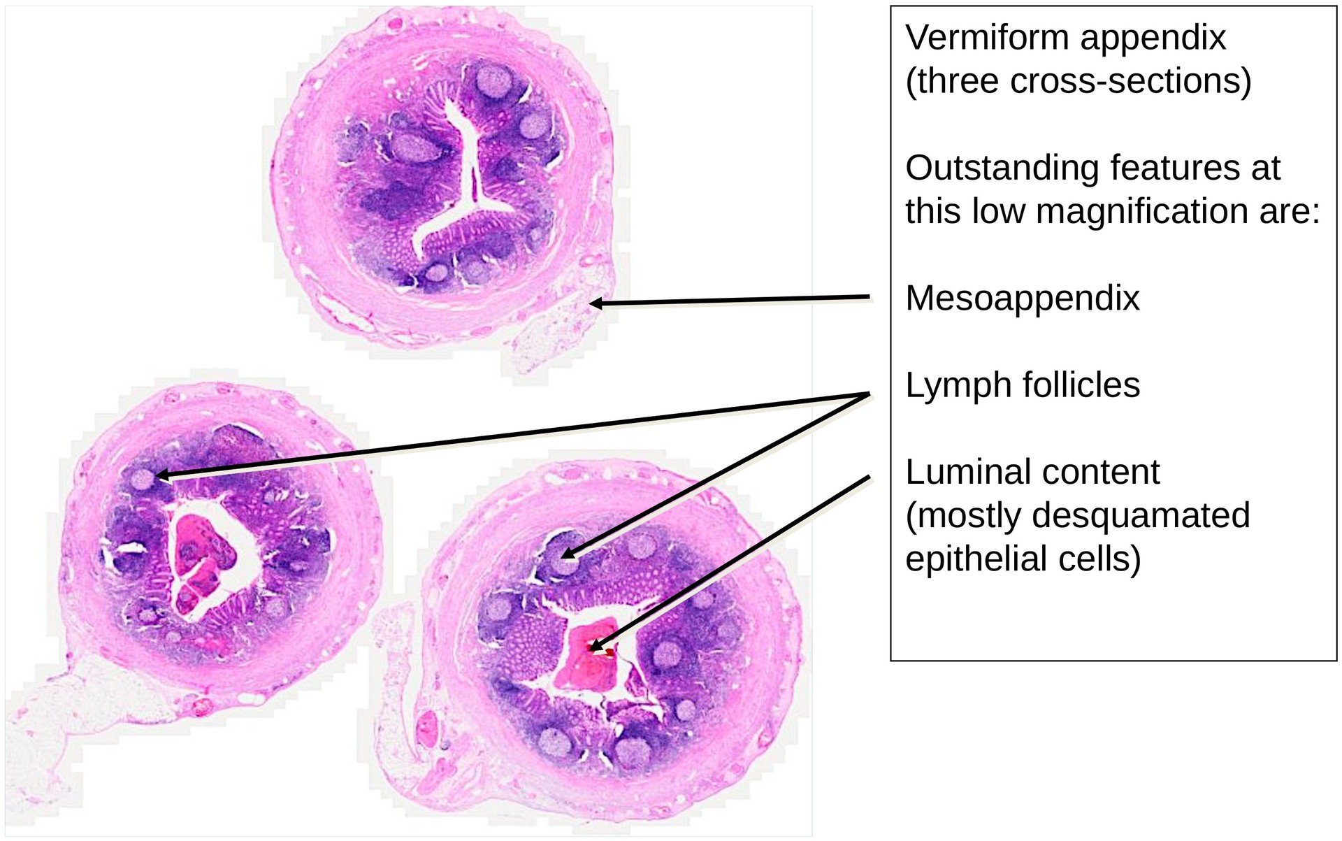
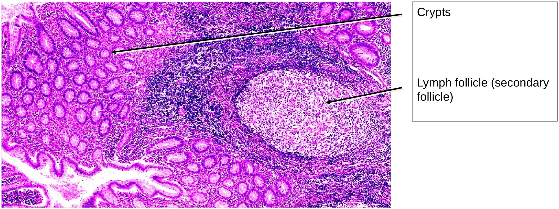
General: The vermiform appendix shares many structural similarities with the large intestine, including the presence of crypts (crypts of Lieberkühn) and the absence of villi. However, in contrast to the colon, the outer longitudinal muscle layer forms a continuous sheath rather than being limited to taeniae coli.
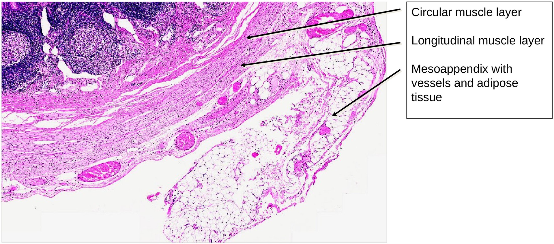
The appendix lies within the peritoneal cavity and is therefore completely surrounded by serosa, lined by mesothelial cells, and attached to the caecum via the mesoappendix.
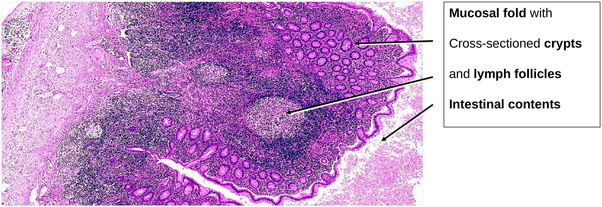
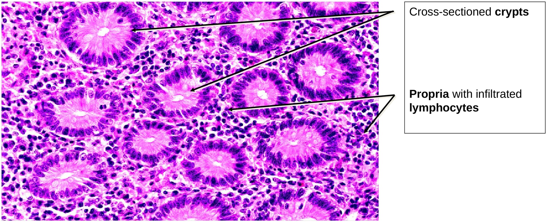
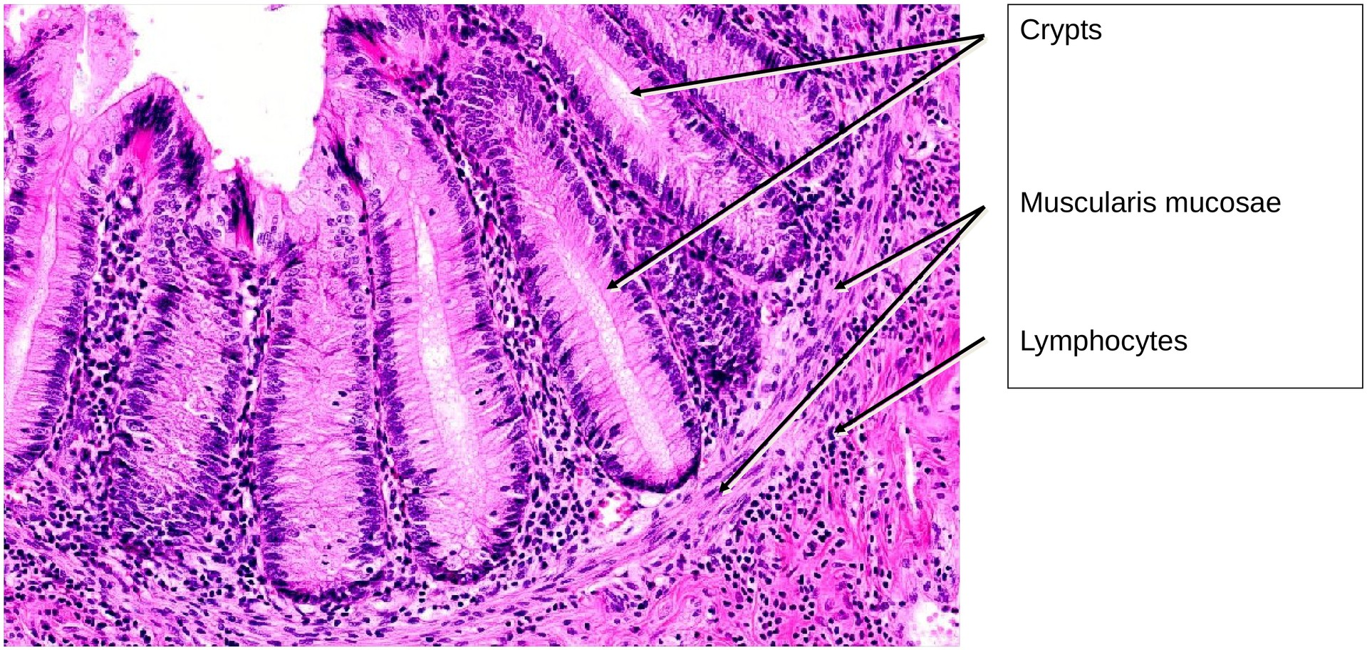
The mucosa is lined by a simple columnar epithelium with numerous goblet cells, opening into intestinal crypts. A key feature is the abundance of lymphoid follicles located in the lamina propria and submucosa, sometimes extending through the muscularis mucosae. These follicles often elevate the overlying epithelium towards the lumen, forming dome epithelium regions where crypts are absent and antigen sampling occurs.
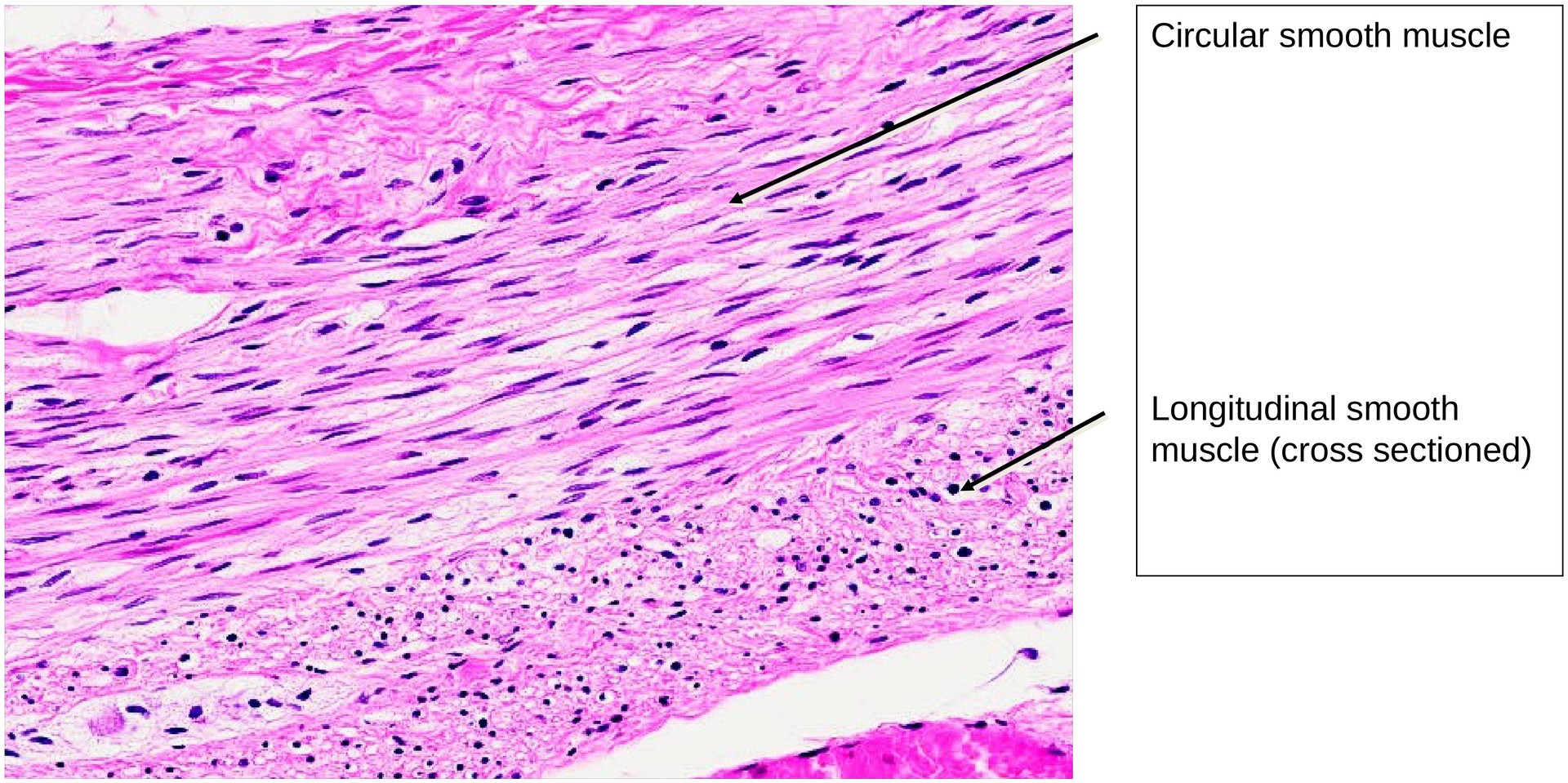
The muscularis externa consists of an inner circular and outer longitudinal layer of smooth muscle, both relatively thin compared with the colon. The submucosa may contain vessels, nerves, and connective tissue, and the serosa forms a delicate peritoneal covering.
Nerve plexuses (myenteric and submucosal) are poorly developed in this organ. Occasionally, intestinal contents or desquamated epithelial and immune cells may be seen within the lumen. In areas of prominent lymphoid tissue, the muscularis mucosae may be discontinuous or absent.
Tasks:
- Identify the overall organ type under low magnification.
- What features confirm that this is a large intestine–type structure?
- Locate the mesoappendix and identify structures within it (vessels, connective tissue, nerves).
- Observe regions where the muscularis mucosae is clearly visible, and others where it is obscured by lymphoid tissue.
- Examine the serosa, noting its mesothelial covering and underlying connective tissue.
- Microscope individual lymphoid follicles and determine whether they are primary (uniformly dark) or secondary (with pale germinal centers).
- Assess the muscularis externa: identify the inner circular and outer longitudinal layers, noting the continuous sheath of the latter.
- Evaluate the epithelial covering above lymphoid follicles:
- What structural features are absent in the dome epithelium compared to normal mucosal regions?
License
University of Basel
Downloads