FEMALE REPRODUCTIVE ORGANS (ANATOMICAL MICROSCOPY)
10.13
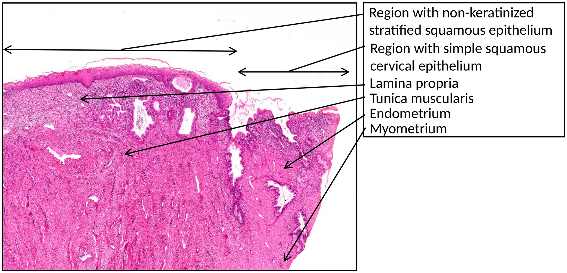
Vaginal portion of the cervix
Specimen:
SPECIMEN DETAILS:
Organ: Cervix (vaginal portion)
Origin: Human
Staining: Haematoxylin Eosin (H&E)
METHOD AND SPECIMEN DESCRIPTION:
Normal histological section of the vaginal portion of the cervix, illustrating the epithelial transition from the endocervix to the vagina.
OBJECTIVE OF THE EXAMINATION:
To examine the junction between the cervical epithelium (endocervical mucosa) and the vaginal epithelium, including associated glandular structures.
SPECIAL FEATURES OF THE SPECIMEN:
General:
The cervix uteri forms the lower part of the uterus and is divided into two regions:
-
a supravaginal portion (cranial), and
-
a vaginal portion (portio vaginalis cervicis), which projects into the vagina.
The cervical canal runs through the cervix and opens into the vagina at the external cervical os.
-
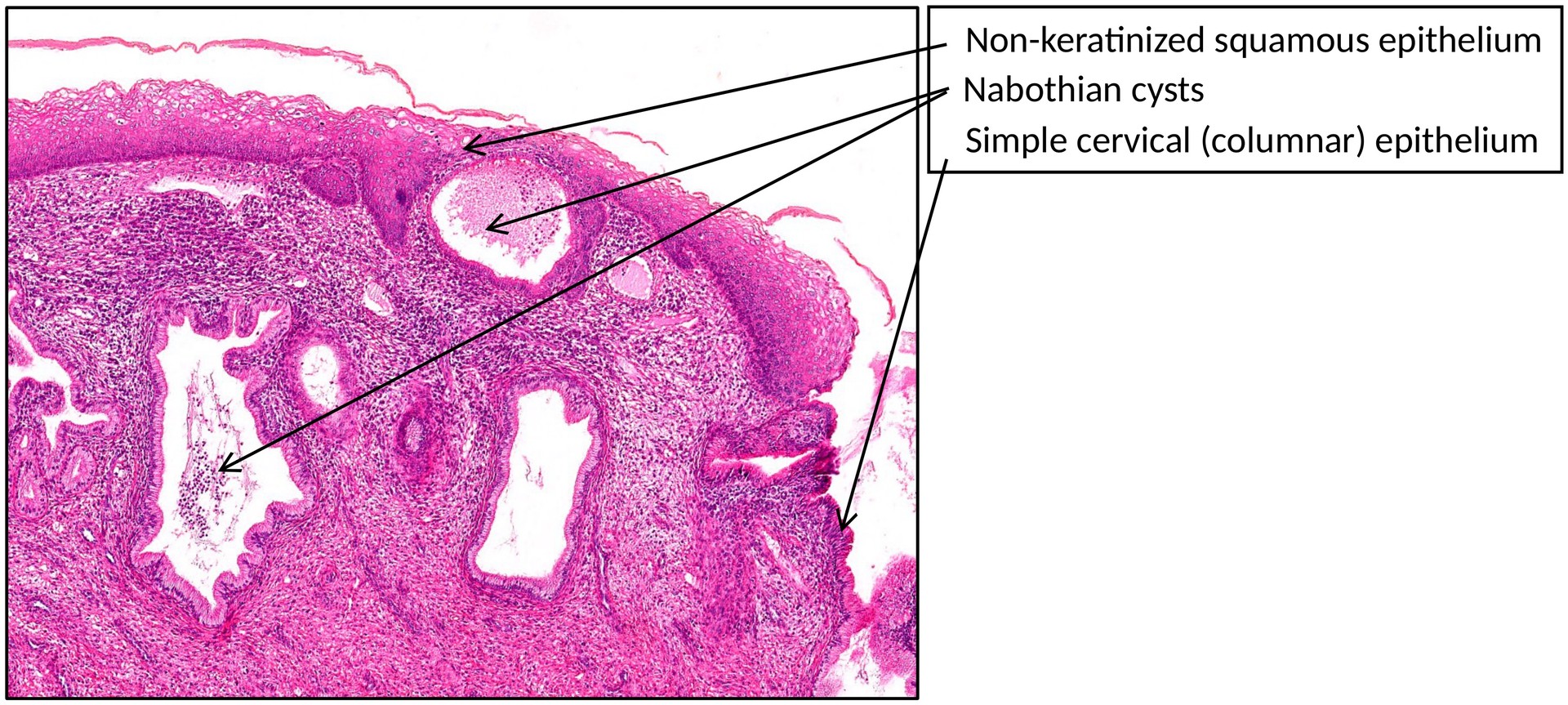
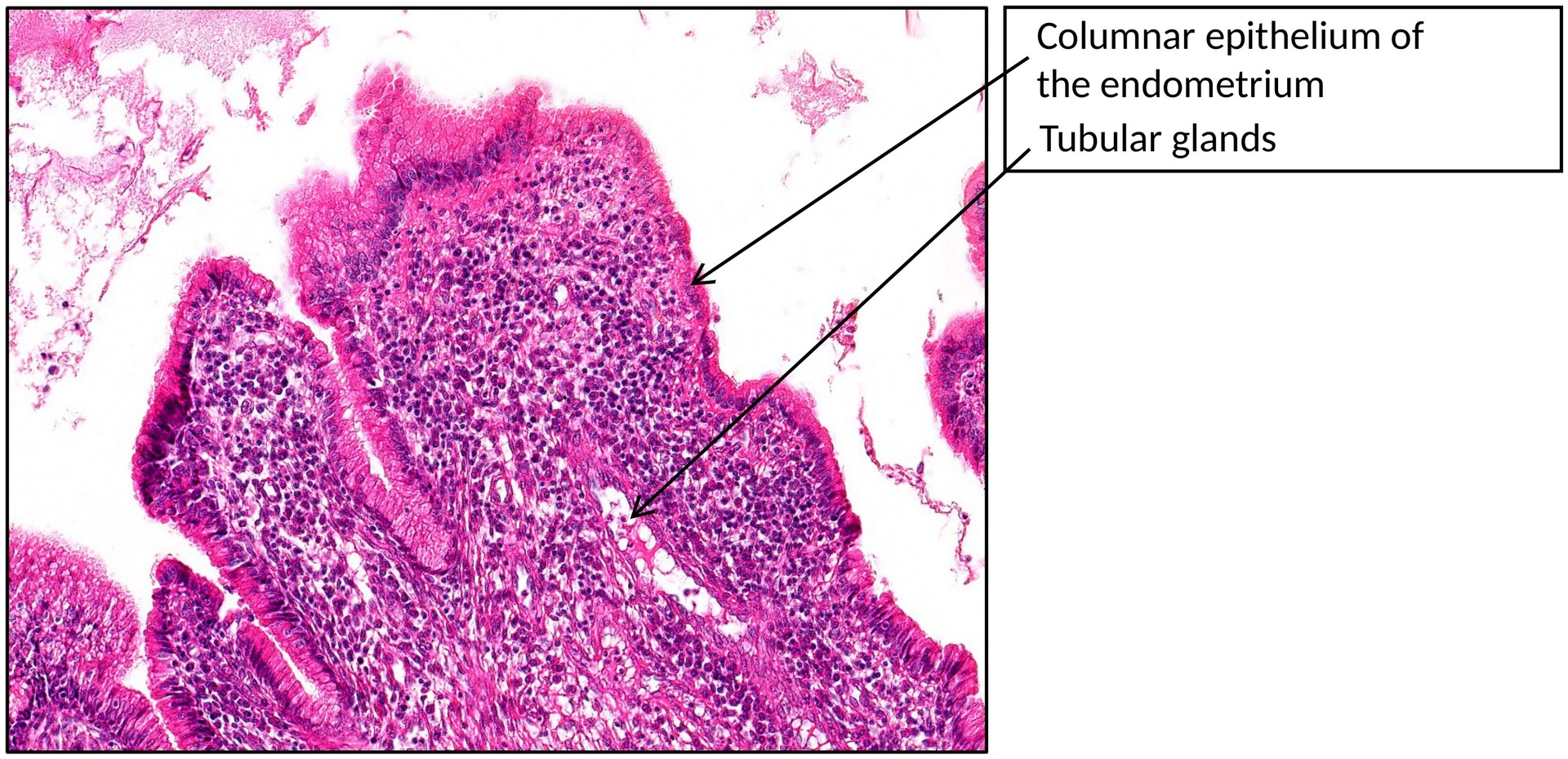
The endocervical mucosa is lined by a single-layered columnar epithelium, rich in mucus-secreting cells. The nuclei are displaced towards the basal pole, while the pale cytoplasm reflects the mucin content. Some epithelial cells bear cilia. Beneath the surface epithelium lie reserve (basal) cells, capable of regenerating the mucosa.
-
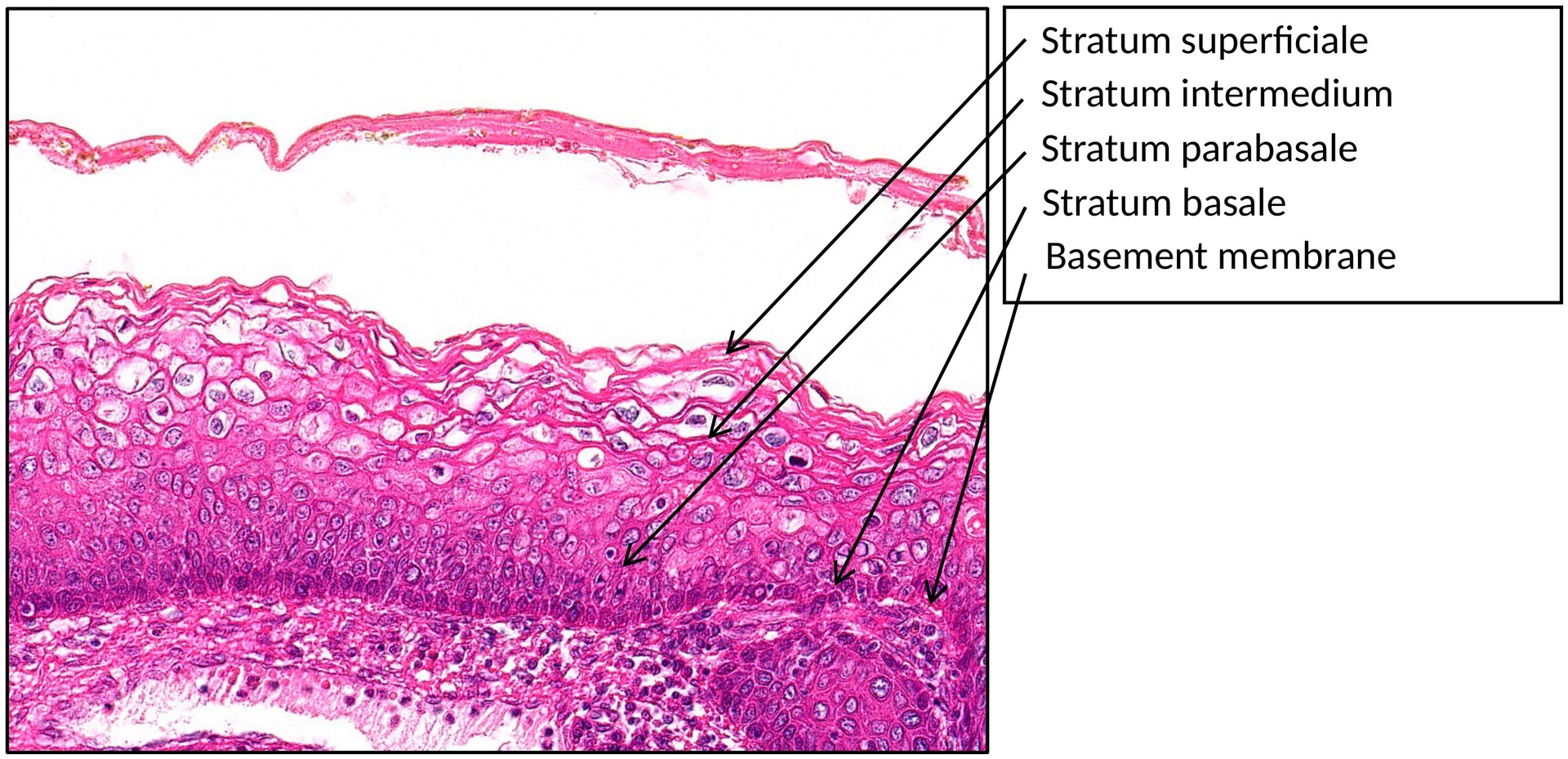
The vaginal epithelium, by contrast, consists of stratified, non-keratinized squamous epithelium. Beneath it lies the lamina propria, followed by a muscular layer interspersed with connective tissue fibres, and an outer connective tissue adventitia.
-
Cervical glands are simple tubular glands that secrete mucus into the cervical canal. When their openings become covered by the overgrowing squamous epithelium of the vagina, retention cysts—known as Nabothian follicles—form.
-
The cervical wall contains smooth muscle and dense connective tissue, continuous with the myometrium of the uterus.
TASKS:
• Identify the stratified, non-keratinized squamous epithelium of the vaginal mucosa.
• Locate the single-layered, high columnar epithelium of the endocervix.
• Observe the transition zone (squamocolumnar junction) between the two epithelia.
• Identify the cervical glands and any Nabothian follicles.
• Distinguish the smooth muscle layers of the cervix (continuous with the myometrium) and the muscularis of the vagina.
License
University of Basel