CONNECTIVE TISSUE (GENERAL HISTOLOGY)
2.2
Loose (Areolar) Connective Tissue (Mesentery)
Specimen:
Specimen Details:
Organ: Mesentery
Origin: Rat
Staining: Van Gieson/Elastin
Method and Specimen Description:
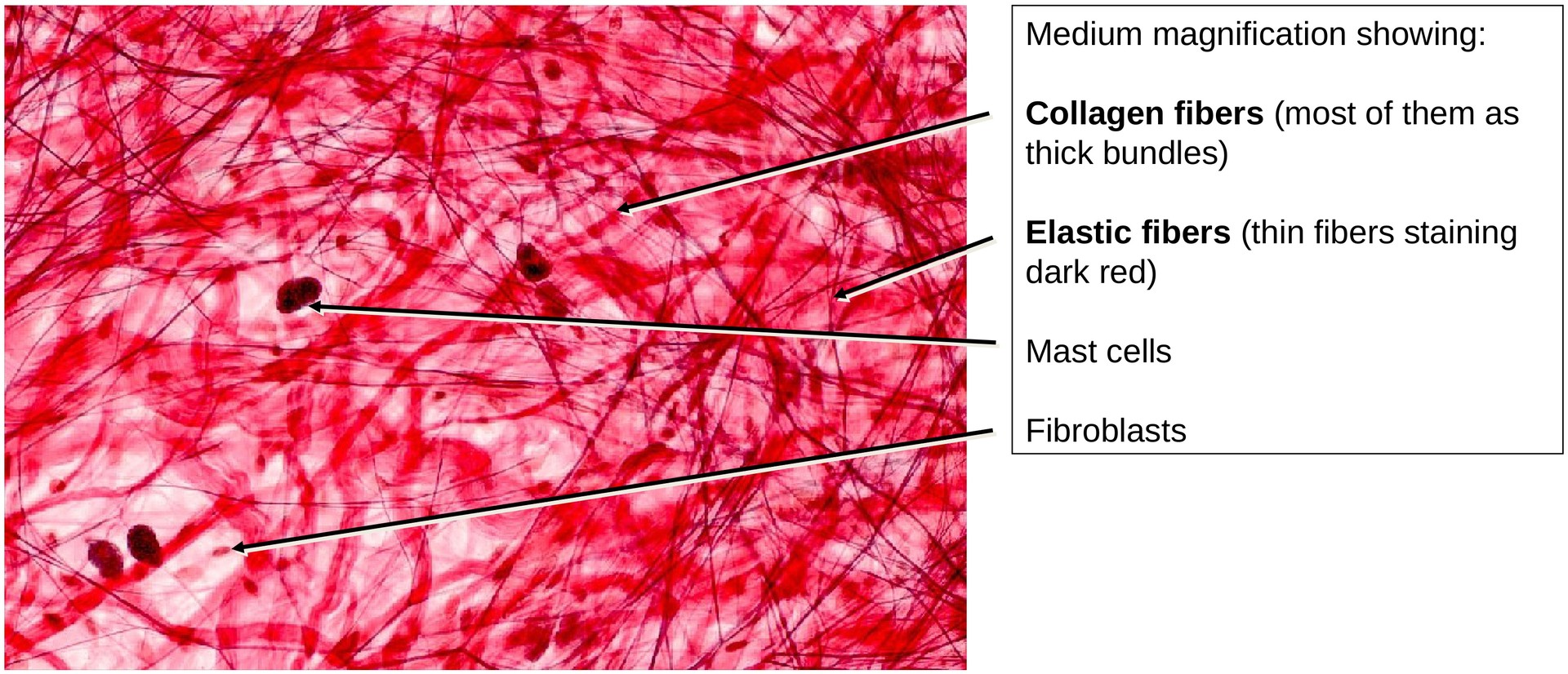
This whole-mount preparation primarily demonstrates the connective tissue located between the two layers of the peritoneum. Cell boundaries are not stained in this specimen. The staining highlights collagen fibers, elastic fibers, and cell nuclei, allowing differentiation of the principal components of loose (areolar) connective tissue.
Objective of the Examination:
-
To recognize and differentiate collagen and elastic fibers in loose connective tissue.
-
To identify mast cells and their cytoplasmic granules.
-
To understand that histological staining techniques can be used to selectively visualize specific tissue components.
Special Features of the Specimen:
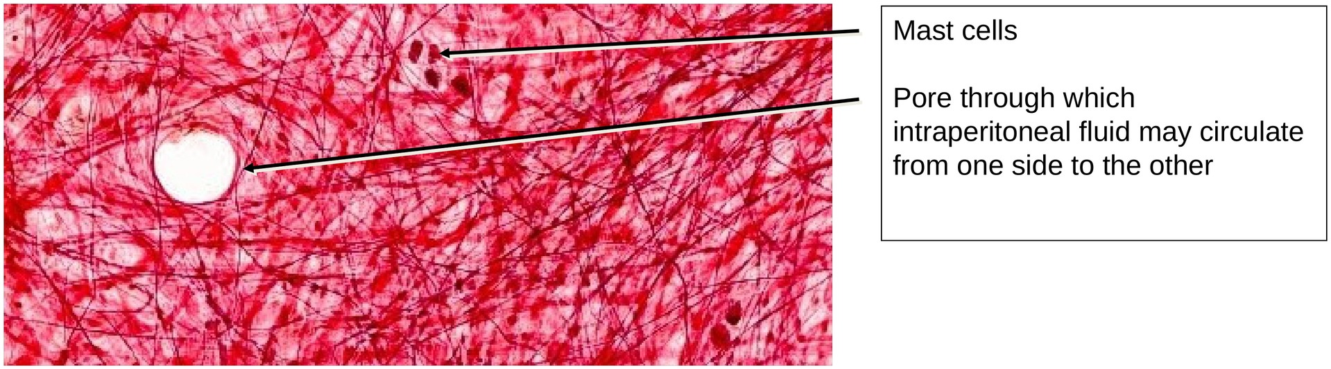
General Features (overview, low magnification): The mesentery, which serves as a suspensory fold for the intestinal tube, consists of a double layer of peritoneum enclosing loose connective tissue. Small blood vessels can be identified in some regions. Due to the thickness of the whole-mount preparation, not all tissue levels appear in focus simultaneously. The connective tissue of the peritoneum contains numerous fibers, providing both elasticity and mechanical stability.
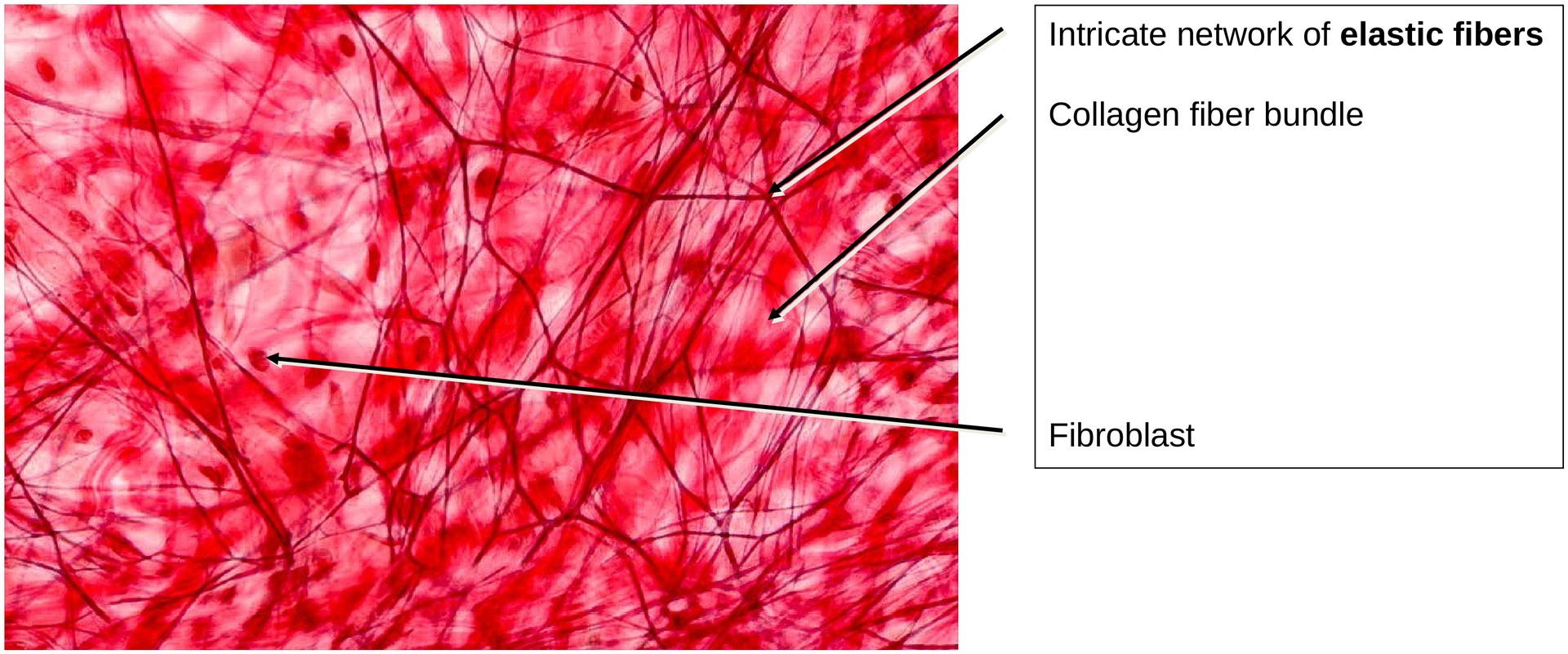
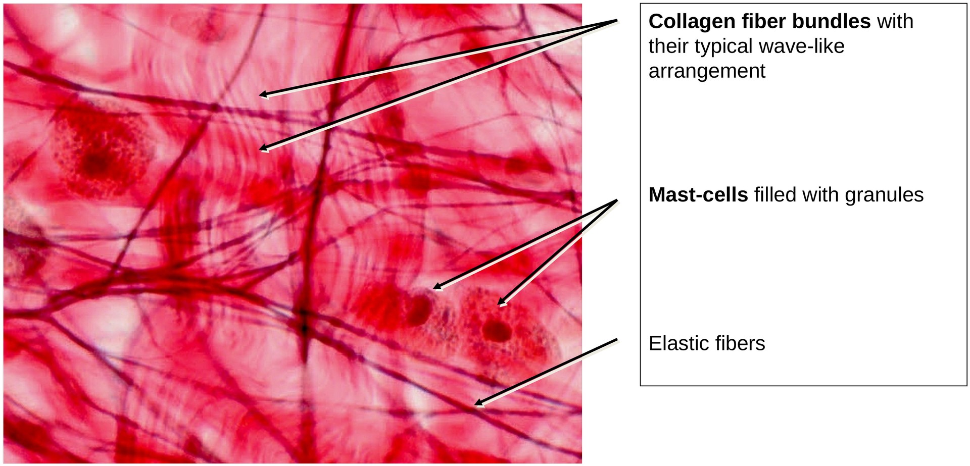
Collagen Fibers: In the mesentery, collagen fibers are loosely arranged in irregular, wavy bundles that overlap one another in a seemingly disorganized pattern. With Van Gieson staining, these fibers appear red. They are the main contributors to the tissue’s tensile strength.
Elastic Fibres: Fine networks of elastic fibers are interwoven among the collagen bundles. These fibers follow an elongated, branching course, forming true molecular-level branch points. Due to the elastin staining, they appear blue to violet. Their double contours result from the high refractive index of elastin. Elastic fibers provide the tissue with resilience and the ability to return to its original shape after stretching.
Cells of the Mesentery: The nuclei of the mesothelial cells lining the peritoneal surfaces are visible as large, oval structures. Most of the other stained nuclei belong to fibroblasts and fibrocytes, which are responsible for producing and maintaining the extracellular matrix. Scattered throughout the tissue are mast cells, easily recognized by their densely granular cytoplasm. In some areas, mast cells have ruptured during preparation, causing the granules to disperse into the surrounding tissue. These granules contain biologically active substances such as heparin and histamine, which play important roles in inflammatory and allergic reactions.
Tasks:
• Identify and distinguish collagen and elastic fibers in the preparation.
• Observe how collagen fiber bundles appear to split and merge with other bundles — an effect often described as false branching.
• Locate elastic fibers, which are thin, well-defined, and form a fine reticular network.
• Search for mast cells, identifiable by their granule-filled cytoplasm, and note areas where granules have dispersed.
• Attempt to trace the course of individual fibers, recognizing the irregular, interwoven nature of the connective tissue matrix.
License
University of Basel
Downloads