FEMALE REPRODUCTIVE ORGANS (ANATOMICAL MICROSCOPY)
10.5
Uterine body 2 (Endometrium)
Specimen:
Specimen Details:
Organ: Uterus
Origin: Human
Staining: Hematoxylin - Eosin (H&E)
Method and Specimen Description:
Routine histological preparation stained with hematoxylin and eosin (H&E), providing clear contrast between nuclei (blue–purple) and cytoplasm or extracellular material (pink).
Objective of the Examination:
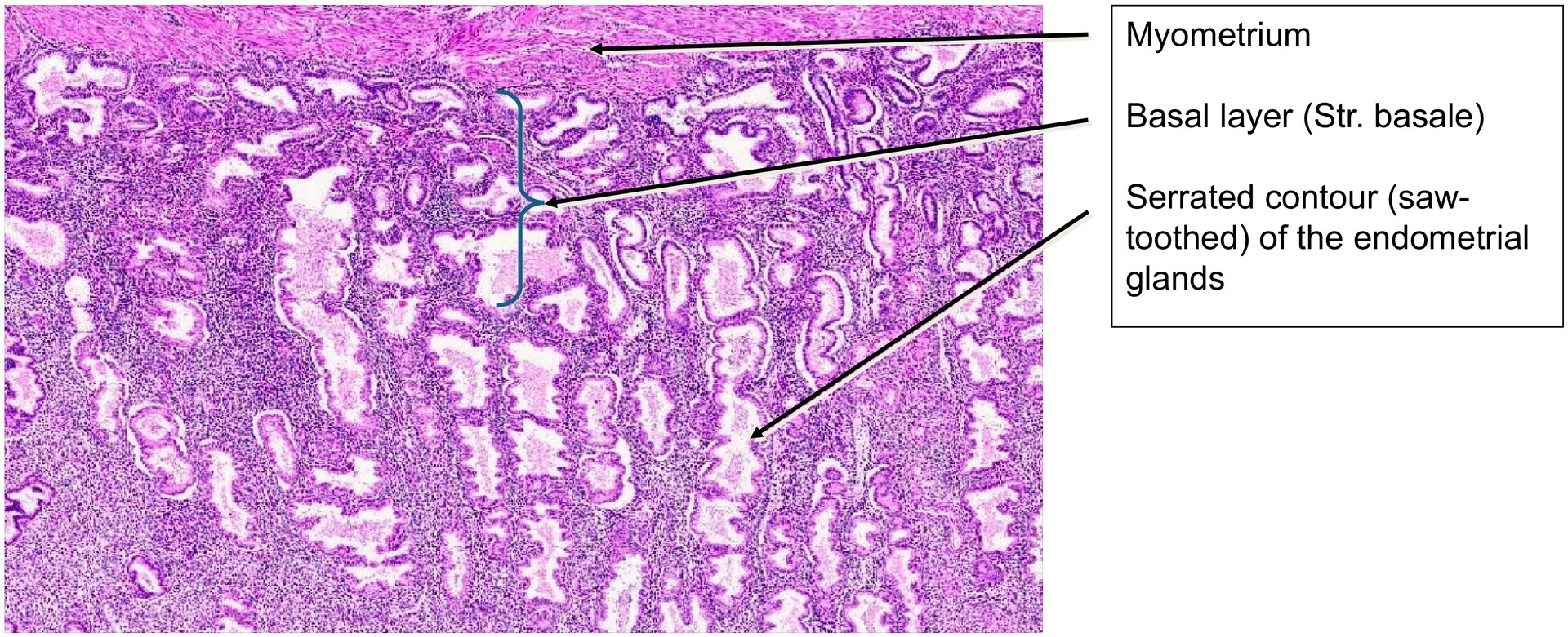
To study the endometrium during the mid to late secretory phase, particularly the glandular morphology with its characteristic saw-toothed (serrated) contours.
Special Features of the Specimen:
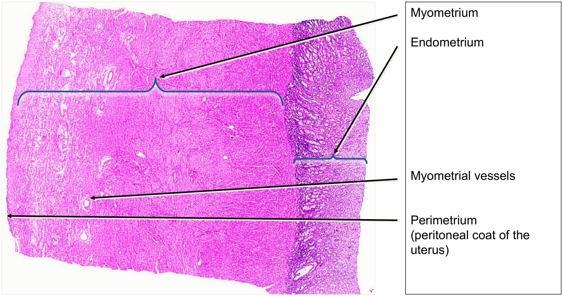
The uterus is the most powerfully muscular organ in the female body, composed predominantly of smooth muscle forming the myometrium. Although this is the major component of the wall, the focus of this specimen is the endometrium.
The section represents the mid to late secretory phase of the menstrual cycle. This stage is identified by the serrated or saw-toothed outlines of the endometrial glands, a feature that appears only after the mid-secretory phase. In the early secretory phase, basal vacuoles containing glycogen would still be visible within the glandular epithelium—these are absent in the current specimen.
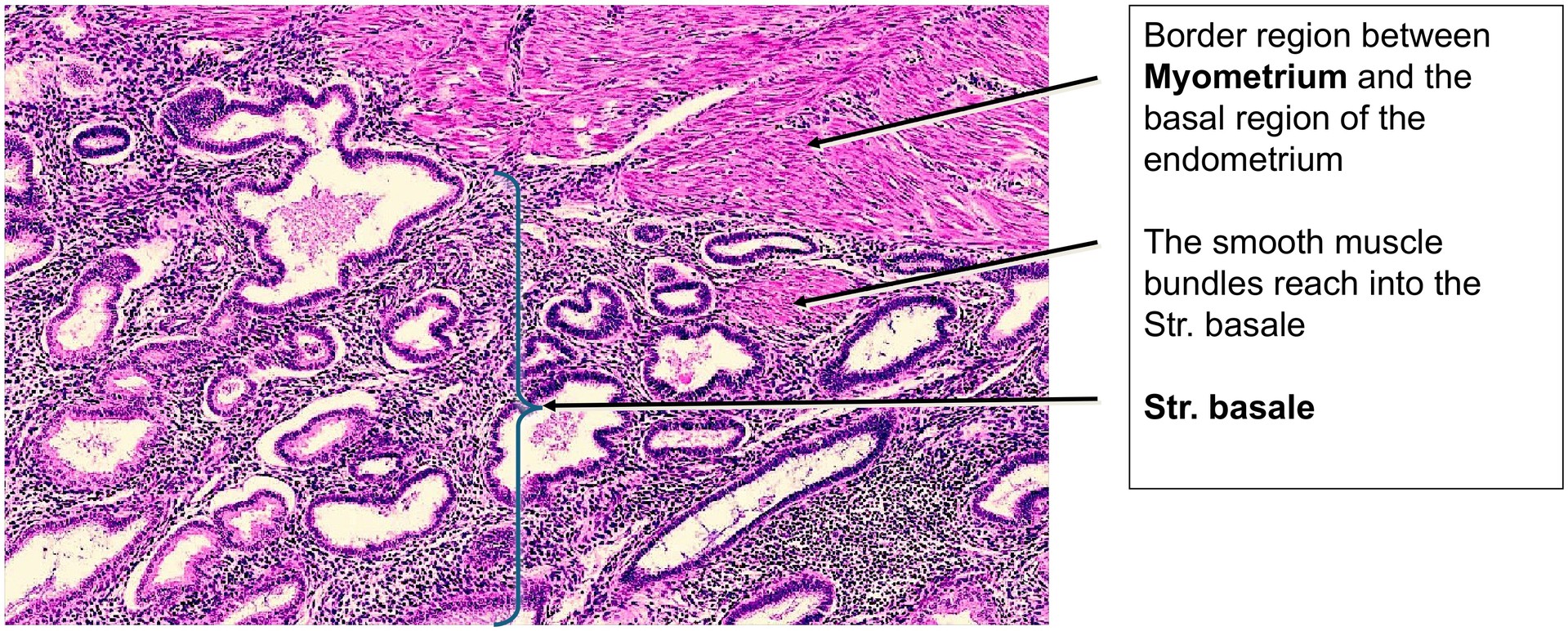
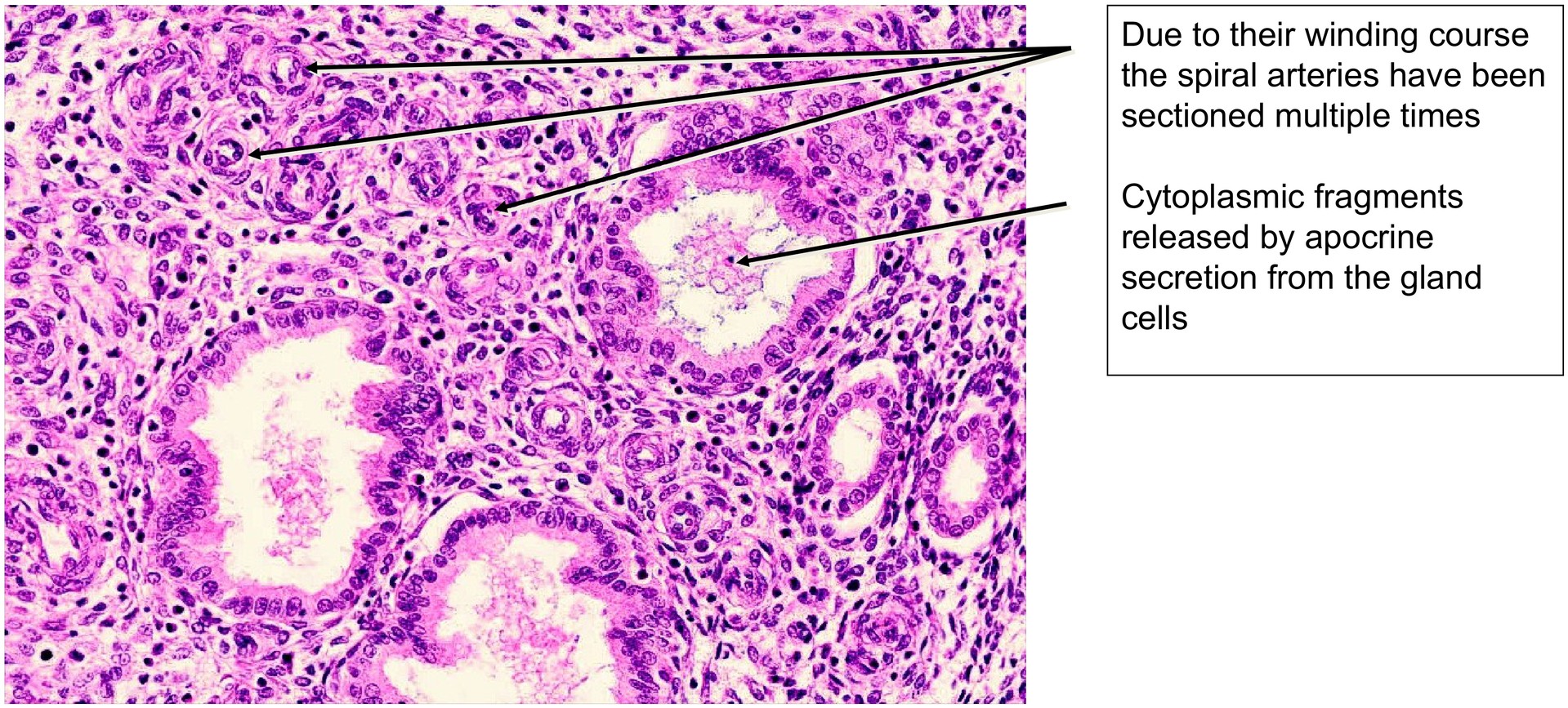
Secretions can be seen within the glandular lumina, consisting partly of cytoplasmic fragments released by apocrine secretion, rich in glycogen. The tubular glands extend throughout the height of the endometrium and may penetrate slightly into the myometrium.
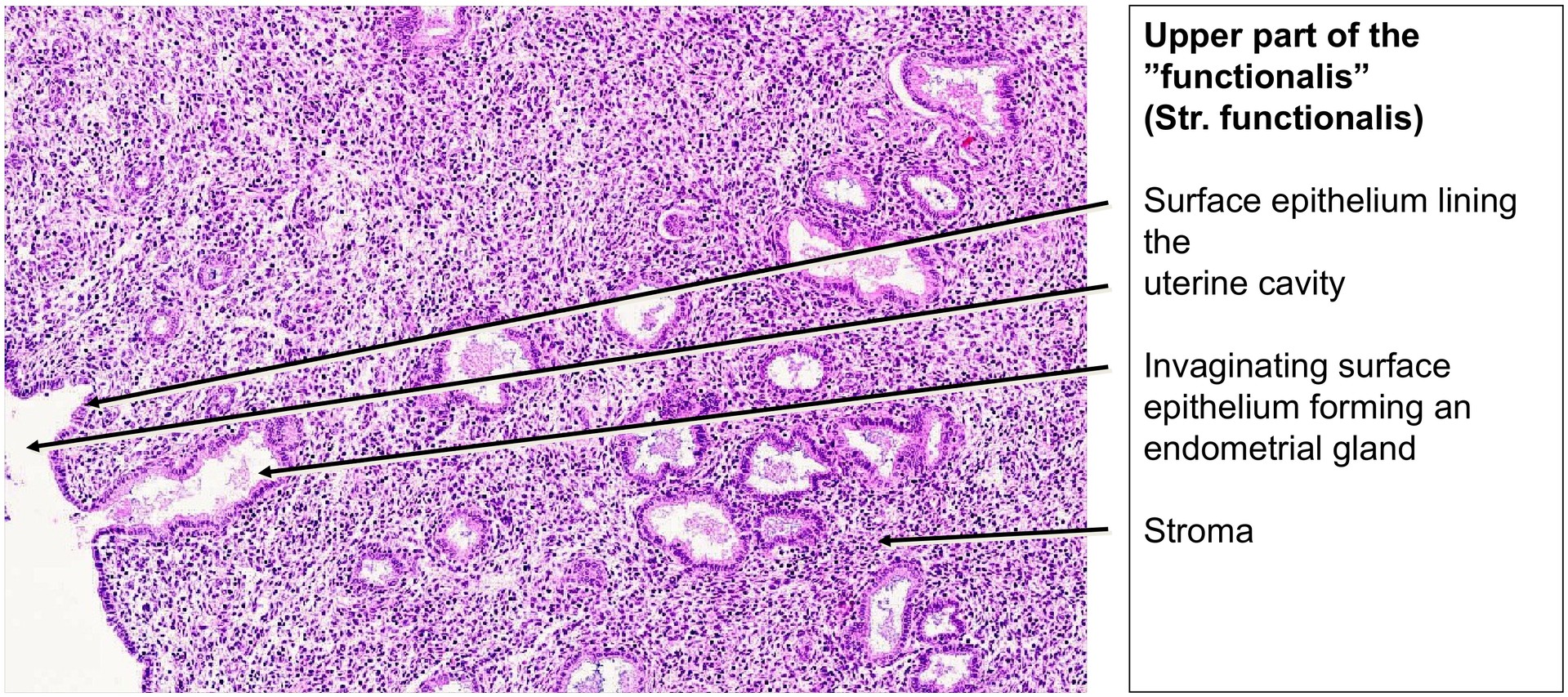
Two distinct endometrial layers are clearly visible: the stratum basalis (basal layer) and the stratum functionalis (functional layer). The basalis has a denser, darker stroma, while in the functionalis only partial differentiation into stratum spongiosum and stratum compactum is visible.
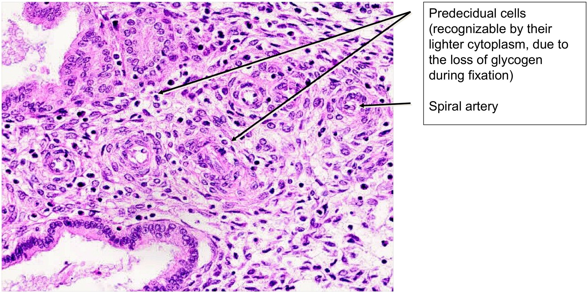
In the border region towards the basalis, the stroma becomes slightly edematous, and early predecidual cells can be identified near the uterine cavity — indicating that the tissue is not yet at the end of the secretory phase. In later stages, the spongiosa and compacta would be more distinctly separated.
Spiral arteries are readily visible and often appear in multiple cross-sections due to their coiled course within the stroma.
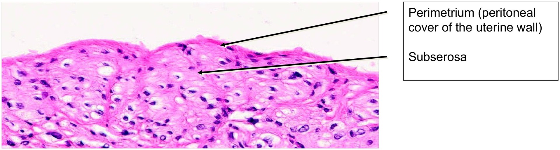
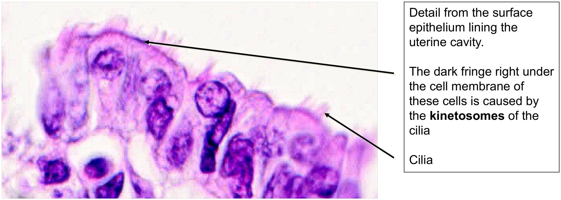
The surface epithelium is only partially preserved, likely owing to post-mortem autolysis, but ciliated epithelial cells can still be recognized in certain areas. Externally, the myometrium is covered by the peritoneal serosa, referred to as the perimetrium.
Tasks:
• Distinguish the myometrium from the endometrium at low magnification.
• Examine the deepest portions of the glands and observe how they extend into the myometrium—note that these basal regions persist after menstruation and act as the regenerative source of the endometrium.
• Describe the structure and shape of the glandular tubules.
• Determine the phase of the menstrual cycle using glandular morphology and stromal characteristics.
• Locate spiral arteries within the endometrial stroma.
• Describe the surface epithelium and note the presence of ciliated cells (kinocilia).
• Trace the arrangement and orientation of smooth muscle bundles in the myometrium.
• Identify the connective tissue septa that separate the muscle bundles.
• Identify the type of the larger vessels within the myometrium.
• Determine which structure covers the outer surface of the myometrium (the perimetrium).
• Locate vessels at the border between the endometrium and myometrium—these are typically basal arteries.
License
University of Basel
Downloads