MALE REPRODUCTIVE ORGANS (ANATOMICAL MICROSCOPY)
11.9
Prostate 1
Specimen:
Specimen Details:
Organ: Prostate
Origin: Human
Staining: Haematoxylin - Eosin (H&E)
Method and Specimen Description:
Normal histological section stained with H&E for general structural overview.
Objective of the Examination:
To study the structure of the human prostate, including its glandular epithelium and muscular components.
Specific Features of the Specimen:
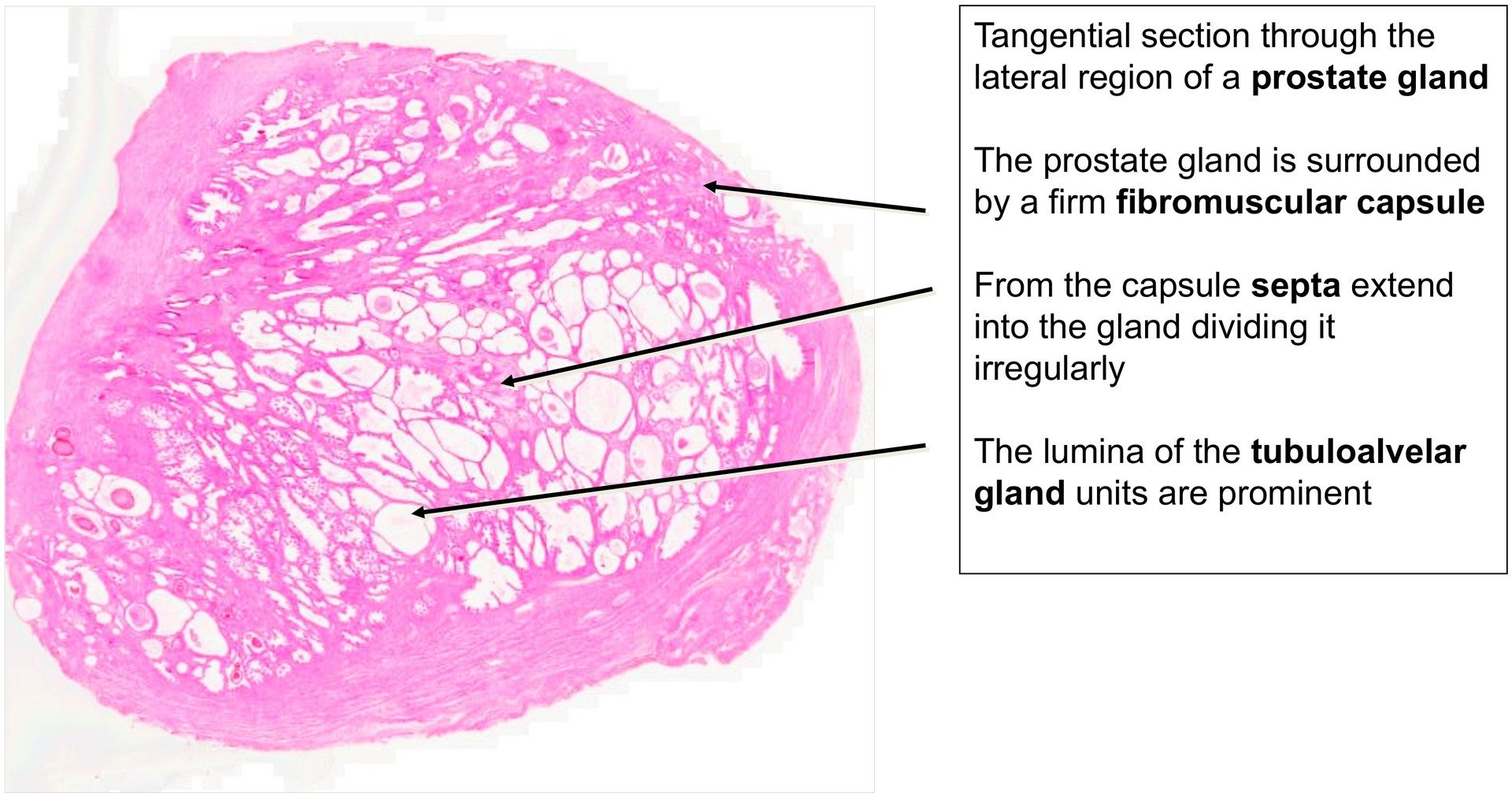
The prostate consists of approximately 30–50 tubuloalveolar glandular units, enclosed by a dense fibrous connective tissue capsule. About 15–20 excretory ducts open into the seminal colliculus within the prostatic urethra. From the capsule, septa extend into the gland, dividing the parenchyma irregularly.
This particular section is taken tangentially through the lateral region of the gland; therefore, neither the urethra nor the seminal colliculus is visible.
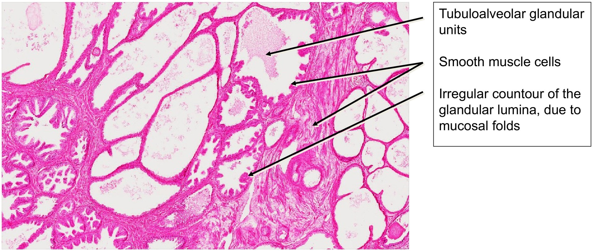
At medium magnification, the abundance of smooth muscle tissue is striking. It surrounds both the individual glands and the entire organ, forming a fibromuscular capsule. This muscular component contributes to the firm consistency of the prostate, making it easily palpable during rectal examination.
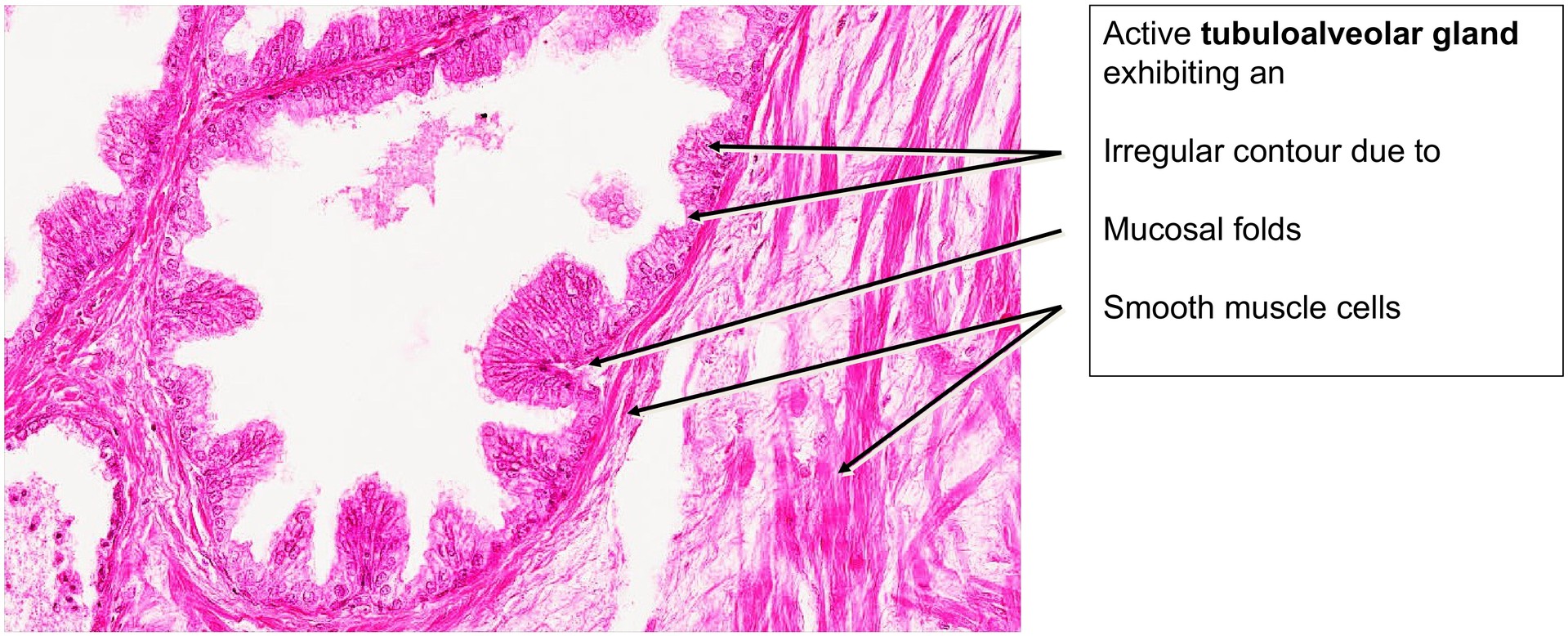
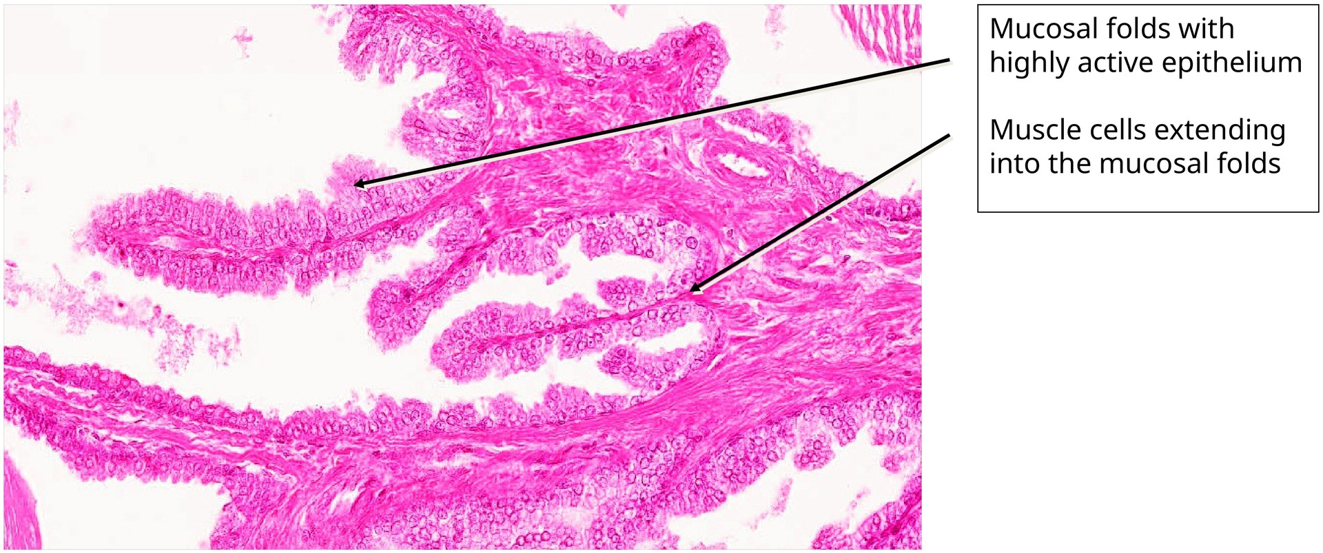
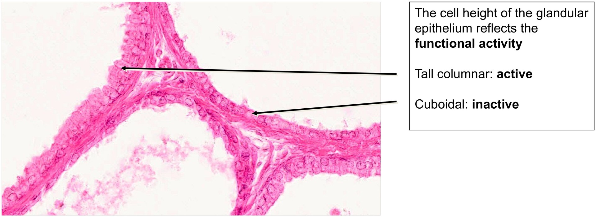
The glandular lumina exhibit an irregular contour due to mucosal folds. The lining epithelium is typically two-layered (pseudostratified), although local stratification may occur. It tends to be simple cuboidal in the troughs of folds and taller columnar at the crests. The cell height reflects the functional activity of the gland — cuboidal in inactive, columnar in active states.
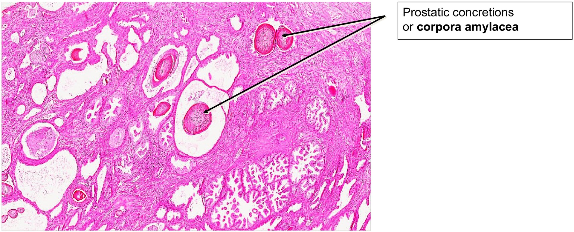
Bundles of smooth muscle cells extend into the larger mucosal folds to aid in expelling the viscous prostatic secretion during ejaculation. With age, the secretions can condense and mineralize, forming corpora amylacea (prostatic concretions or stones). These lamellated glycoprotein–mineral bodies may partially obstruct ducts and are more frequent in older men.
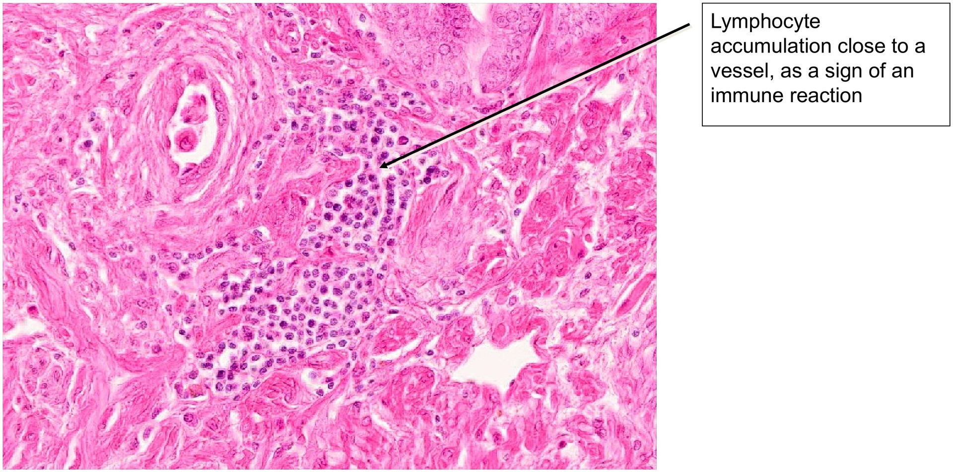
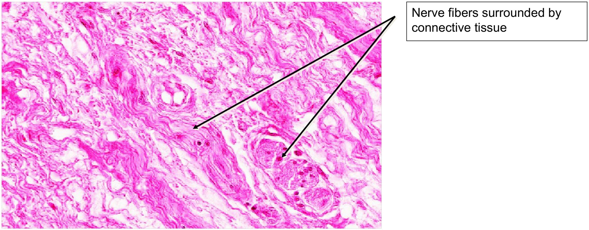
Because of the dense musculature, the gland receives a rich autonomic innervation, and nerve fibers can be identified throughout the section, particularly within the interstitial connective tissue.
Tasks:
- Gain an overview at low magnification.
- Assess the thickness and organization of the fibromuscular capsule.
- Identify smooth muscle bundles and trace their extensions into mucosal folds.
- Evaluate the epithelium — where is it simple, and where is it stratified or pseudostratified?
- Locate prostatic concretions (corpora amylacea) and consider the reason for their ring-like (lamellated) appearance.
- Identify nerve fibers within the interstitial tissue.
License
University of Basel
Downloads