DIGESTIVE ORGANS: ORAL CAVITY (ANATOMICAL MICROSCOPY)
18.12
Fungiform papillae of the tongue
Preparation:
PREPARATION DETAILS:
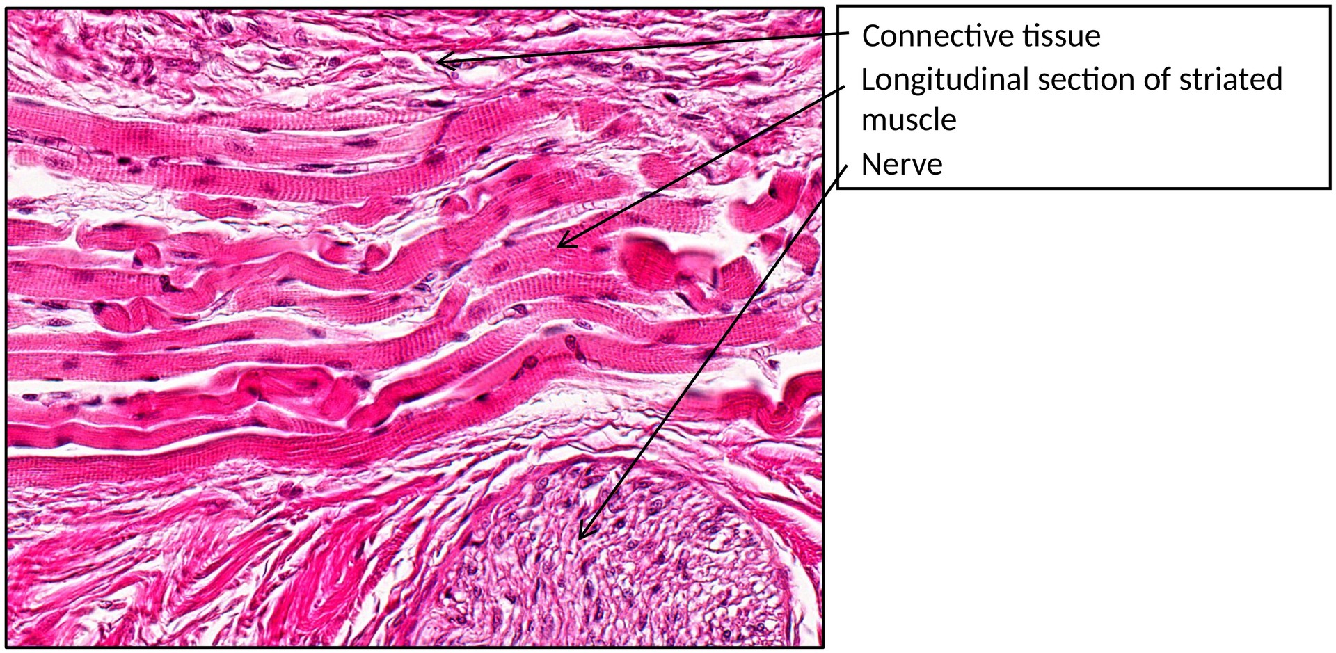
Organ: Tongue
Origin: Human
Staining: Hematoxylin Eosin (H&E)
METHOD AND SPECIMEN DESCRIPTION:
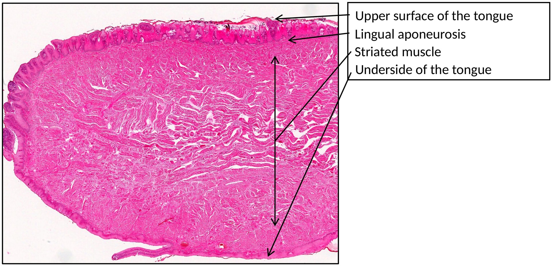
This is a normal sagittal section of a human tongue, stained with H&E for general structural visualization. The muscle fibers appear pink to red, while the connective tissue and nuclei are stained in varying shades of blue to purple.
OBJECTIVE OF THE EXAMINATION:
To study the morphology and structure of the tongue, with a specific focus on the fungiform papillae and their associated epithelial and connective tissue features.
SPECIAL FEATURES OF THE PREPARATION:
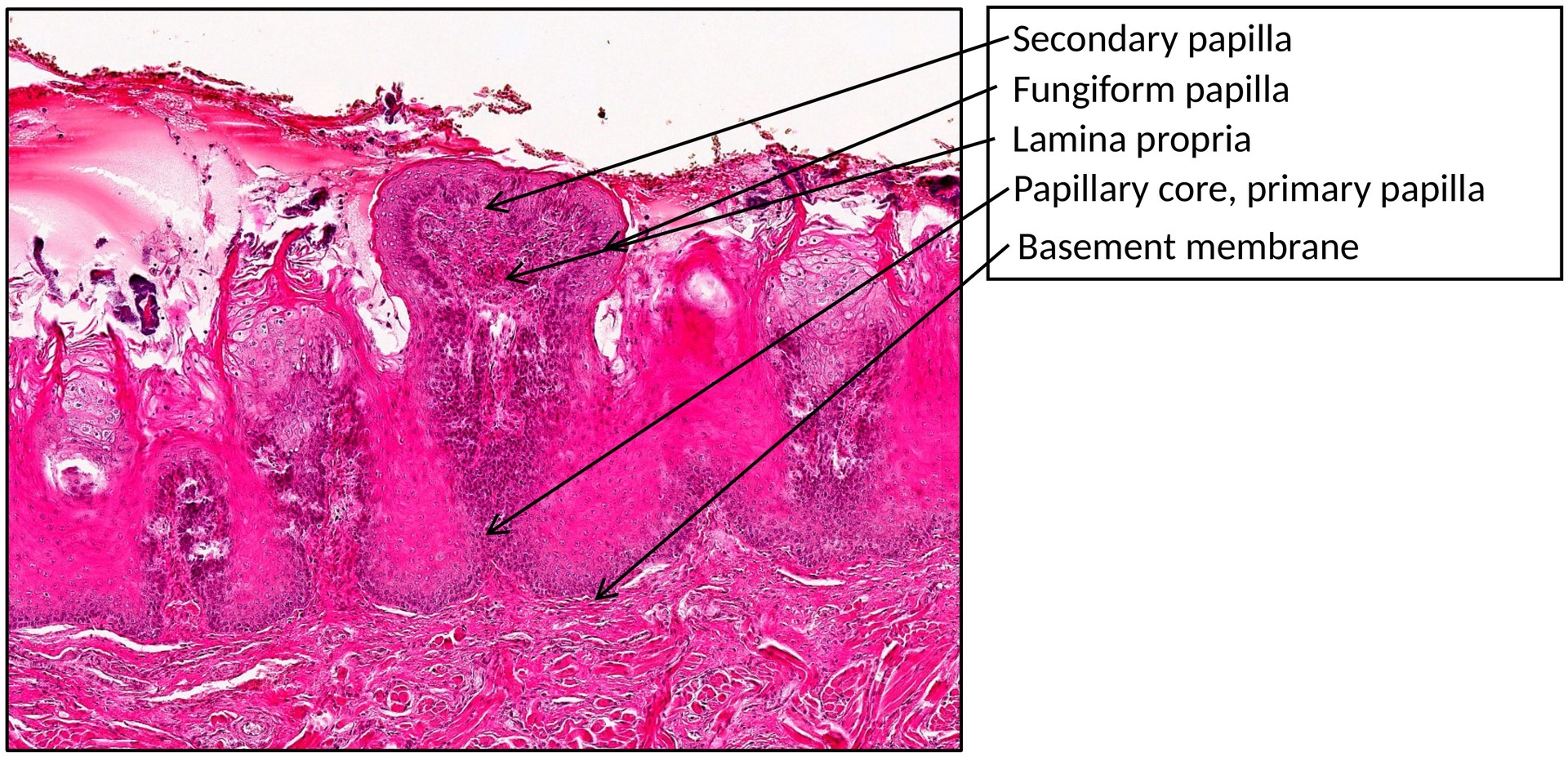
General: The fungiform papillae are mushroom-shaped projections found predominantly on the anterior two-thirds of the dorsal surface of the tongue. They are less numerous than the filiform papillae but are larger and more rounded in shape.
Each papilla consists of:
- A connective tissue core (derived from the lamina propria) that forms the papilla stalk.
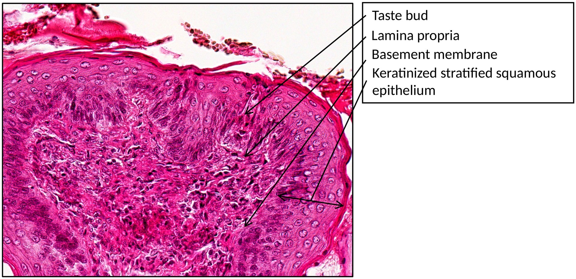
- A covering of stratified squamous epithelium, which is usually keratinized, though the degree of keratinization may vary depending on the region.
- Taste buds, which are typically embedded in the apical epithelium of the papilla and appear as pale oval structures.
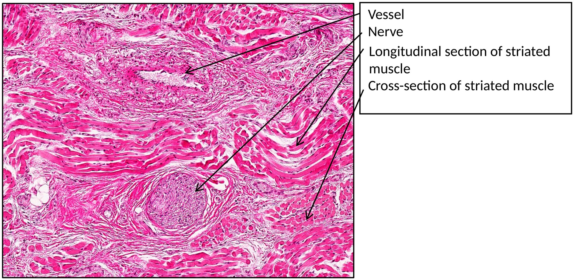
Within the connective tissue base of the papilla, numerous capillaries, nerve endings, and sensory receptors (thermo- and mechanoreceptors) are present, contributing to tactile and temperature sensation.
In adults, the number of taste buds within the fungiform papillae gradually decreases with age.
TASKS:
- Identify the dorsal and ventral surfaces of the tongue.
- Locate the fungiform papillae, lingual aponeurosis, striated muscle, blood vessels, and nerves.
- Examine the structure of the fungiform papillae:
- Identify the epithelium (keratinized stratified squamous epithelium).
- Observe the primary and secondary connective tissue papillae.
- Identify the papilla stalk and the lamina propria.
- Search for taste buds on the apical ridge of the papilla.
- Compare the structure and keratinization of the fungiform papillae with that of the filiform papillae.
License
University of Basel