DIGESTIVE ORGANS: ORAL CAVITY (ANATOMICAL MICROSCOPY)
18.8
Tooth Development I
Specimen:
Specimen Details:
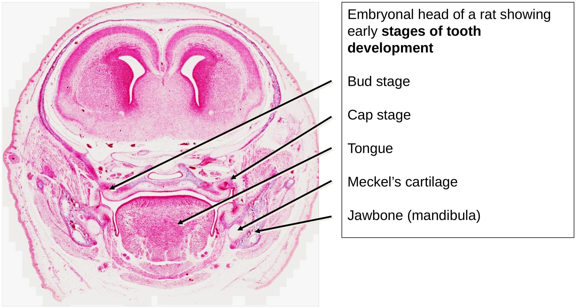
Organ: Embryonic Head
Source: Rat
Staining: Azan
Method and Specimen Description:
Normal histological section stained with Azan, which colors connective tissue blue and epithelium, muscle, and blood red. In embryonic tissue, however, due to the high water content and immaturity of organ systems, the color contrast is less pronounced.
Objective of the Examination:
To study the early stages of tooth development — specifically the bud stage and cap stage — and to understand the formation of the jaw bones and the role of Meckel’s cartilage in mandibular development.
Special Features of the Specimen:
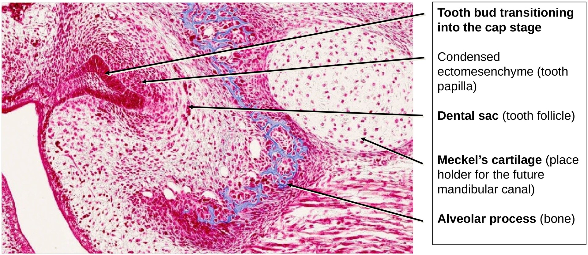
This section represents a frontal section through the developing head, slightly oblique in orientation. This is evident from the finding that one side of the jaw displays the bud stage, while the opposite side shows the cap stage of tooth development.
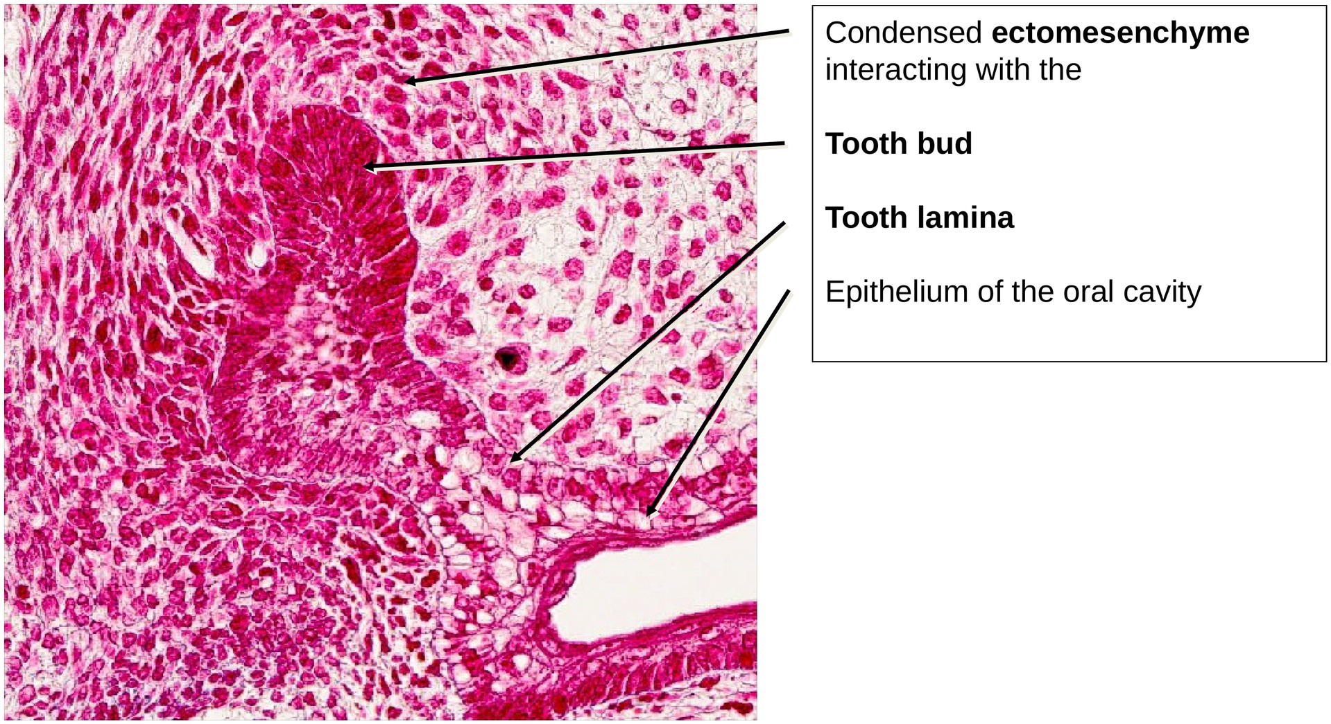
Bud Stage:
At this stage, there is a distinct thickening of the epithelial tissue that has grown downwards from the oral epithelium into the underlying ectomesenchyme. The surrounding mesenchyme condenses to form the tooth follicle, which will later give rise to the tooth-supporting structures:
- Alveolar bone
- Desmodontium (periodontal ligament)
- Cementum
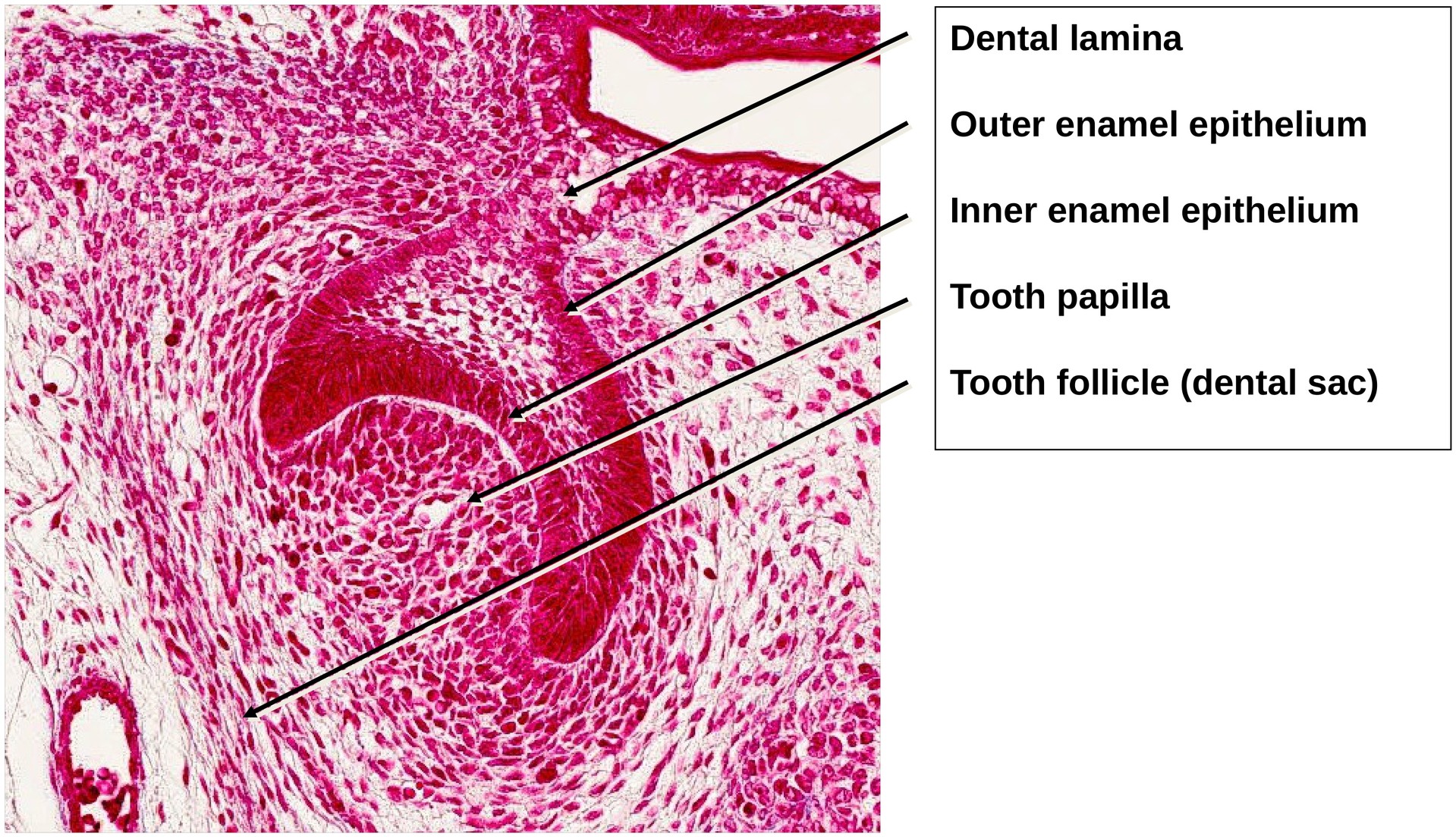
The tooth lamina remains in connection with the oral epithelium, serving as the developmental link between the tooth germ and the oral cavity.
Cap Stage:
On the opposite side of the mandible, the developing tooth germ shows clear proliferation and differentiation of the enamel organ into:
- Inner enamel epithelium
- Outer enamel epithelium
Between these layers, the enamel organ covers the tooth papilla, which will later form the dentine and pulp. Surrounding the enamel organ is the tooth follicle (dental sac), which contributes to the formation of the supporting tooth apparatus.
The light blue-stained structures represent developing bone, particularly in the mandible, where an emerging tooth alveolus (future socket) can be identified.
Adjacent to the developing mandible lies the oval Meckel’s cartilage, which serves as a temporary cartilaginous scaffold (or placeholder) for the future mandibular canal and plays an important role in the spatial organization of jaw development.
Tasks:
Using the slide, identify and describe the following structures and developmental features:
- Identify the two developmental stages — bud and cap.
- In the cap stage, locate:
- Tooth papilla
- Inner enamel epithelium
- Outer enamel epithelium
- Tooth follicle (dental sac)
- Tooth lamina
- Locate Meckel’s cartilage within the developing mandible.
- Identify the forming tooth alveolus.
- Examine the tongue in the section and identify the three muscle fibre orientations of the tongue musculature (longitudinal, transverse, and vertical).
License
University of Basel
Downloads