NERVE TISSUE (GENERAL HISTOLOGY)
7.2
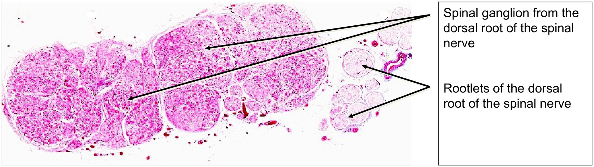
Spinal ganglion 1
Specimen:
Specimen Details:
Organ: Spinal Ganglion
Origin: Bovine
Staining: Azan
Method and Specimen Description:
Normal histological specimen stained with Azan, where connective tissue appears blue, and erythrocytes and epithelium stain red.
Objective of the Examination:
To understand the structure of a spinal ganglion, including its pseudo-unipolar neurons and the rootlets of the dorsal root of a spinal nerve.
## Special Features of the Specimen:
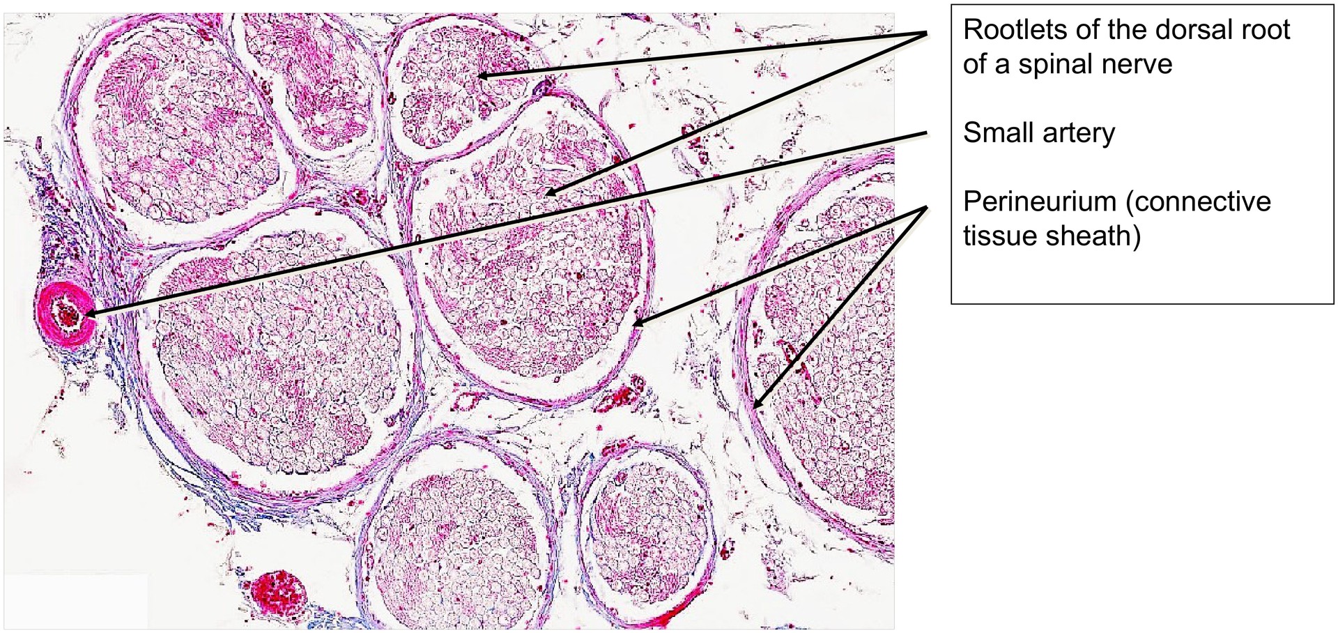
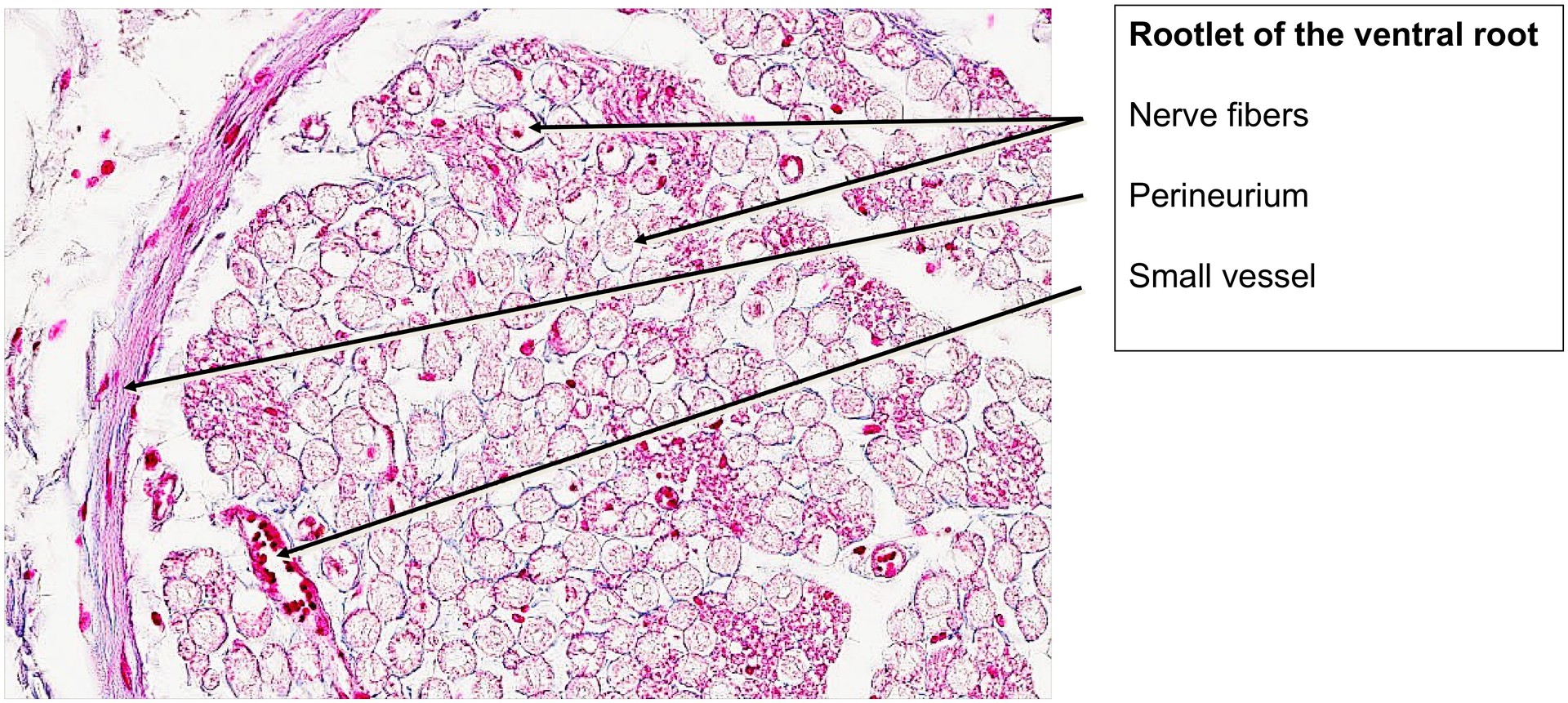
The spinal ganglion (sensory ganglion) lies at the junction between the peripheral nervous system (PNS) and the central nervous system (CNS). It forms part of the dorsal root of the spinal nerve. Alongside the dorsal root ganglion, the afferent (sensory) fibers of the spinal nerve enter the spinal cord. The ventral root, in contrast, contains only efferent (motor) fibers, some of which can also be observed in this specimen.
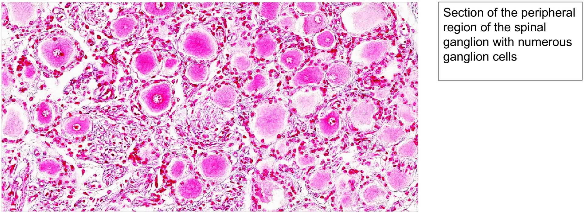
A large proportion of the neuronal nuclei are located towards the periphery of the ganglion. The neurons are pseudo-unipolar, meaning that a single process leaves the perikaryon, dividing shortly afterwards into an axonal (neuritic) and a dendritic branch. The dendritic process conducts impulses from peripheral sensory receptors (e.g. tactile corpuscles in the skin), while the neuritic process continues centrally towards the spinal cord, where it forms synapses.
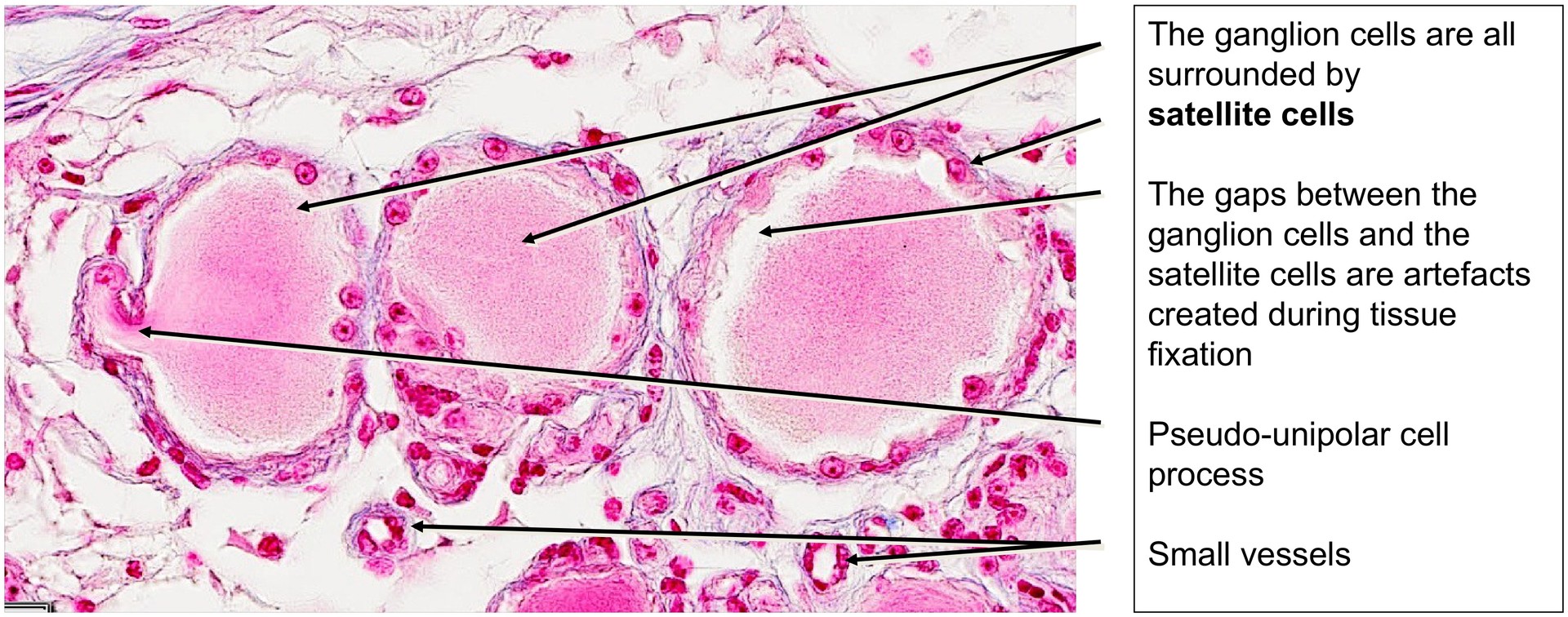
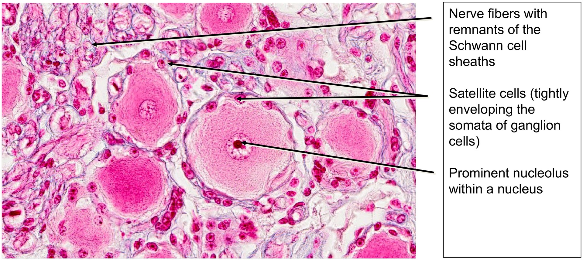
Each ganglion cell is surrounded by a layer of satellite cells, which may appear separated by small gaps — artefacts created during tissue fixation. Between the ganglion cells, particularly in the central area of the ganglion, afferent nerve fibers can be seen, though their sheaths (such as myelin) are often poorly preserved. After fixation, remnants of the Schwann cell sheaths appear as “neurokeratin”, a term unrelated to true keratin.
The nuclei of the ganglion cells are notably large, and in some cells, the nucleolus is clearly visible.
At the level of the spinal ganglion, the CNS meninges merge with the connective tissue sheaths of the peripheral nerves (endo-, peri-, and epineurium). However, the coverings of both the ganglion and the ventral motor root are structurally more similar to those of the PNS.
Tasks:
• Orient yourself using the overview and determine which structures belong to the dorsal root and which to the ventral root of the spinal nerve.
• Locate the spinal ganglion within the dorsal root and compare the sizes of the ganglion cells.
• Identify the satellite cells surrounding each ganglion cell.
• Search for perikarya with a distinct nucleus and nucleolus. Identify the afferent nerve fibers within the ganglion.
• Find the origin of a pseudo-unipolar process from a ganglion cell.
• Examine the ventral (efferent) root of the spinal nerve and consider how the bundling of nerve fibers is organized.
• Identify Schwann cells within the ventral root and observe the appearance of neurokeratin.
License
University of Basel
Downloads