SENSORY ORGANS (ANATOMICAL MICROSCOPY)
17.4
Optic nerve
Specimen:
Specimen Details:
Organ: Optic Nerve
Origin: Human
Staining: Hematoxylin - Eosin (H&E)
Method and Specimen Description:
Normal histological section stained with an overview staining (H&E).
Objective of the Examination:
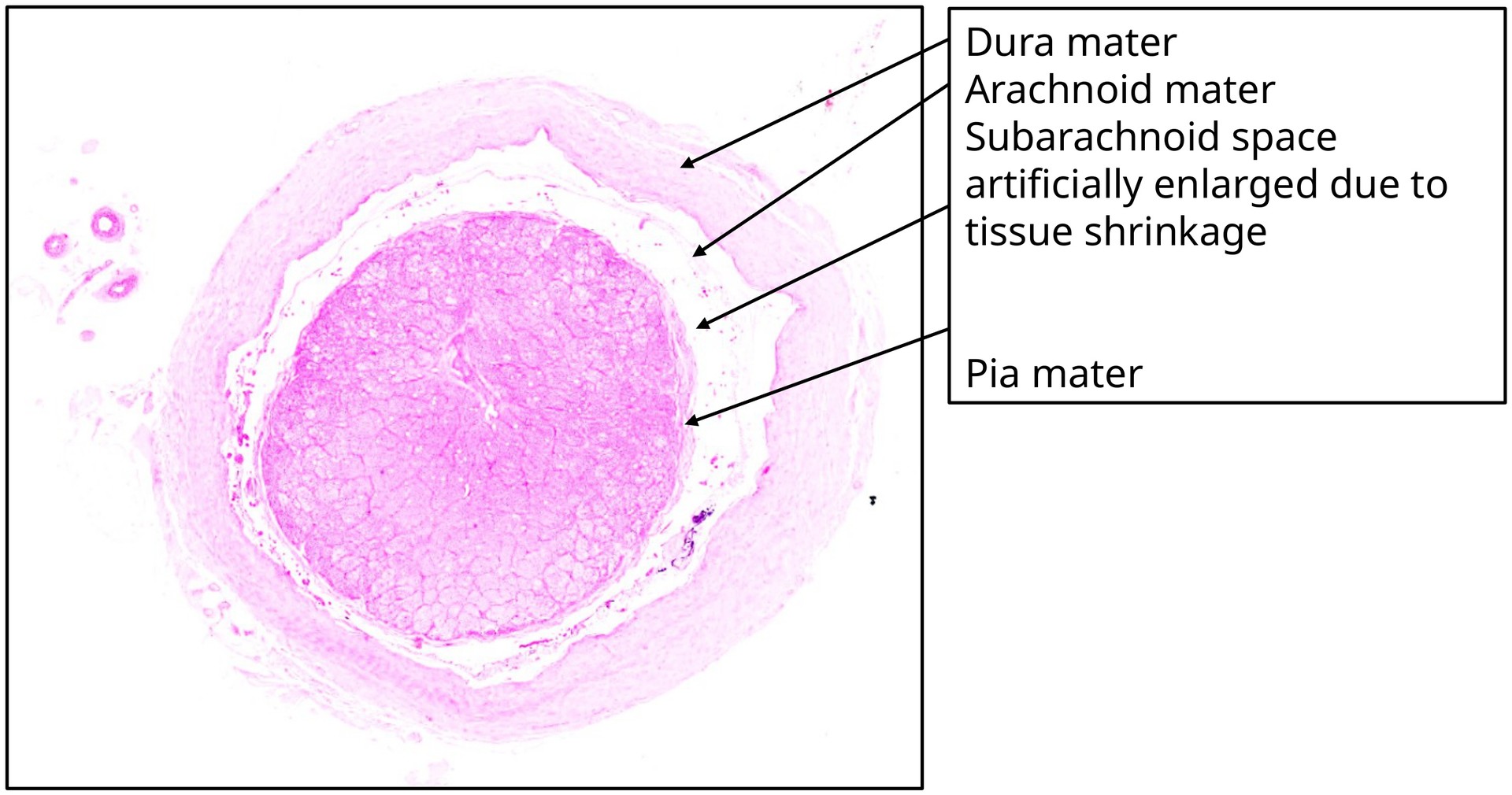
To identify the optic nerve and its meningeal sheaths (dura mater, arachnoid mater, and pia mater), and to recognize their relationship to the central retinal vessels.
Specifics of the Specimen:
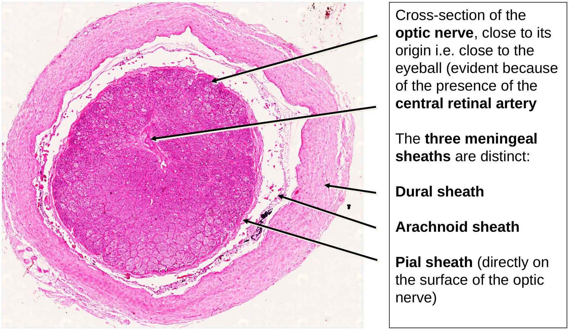
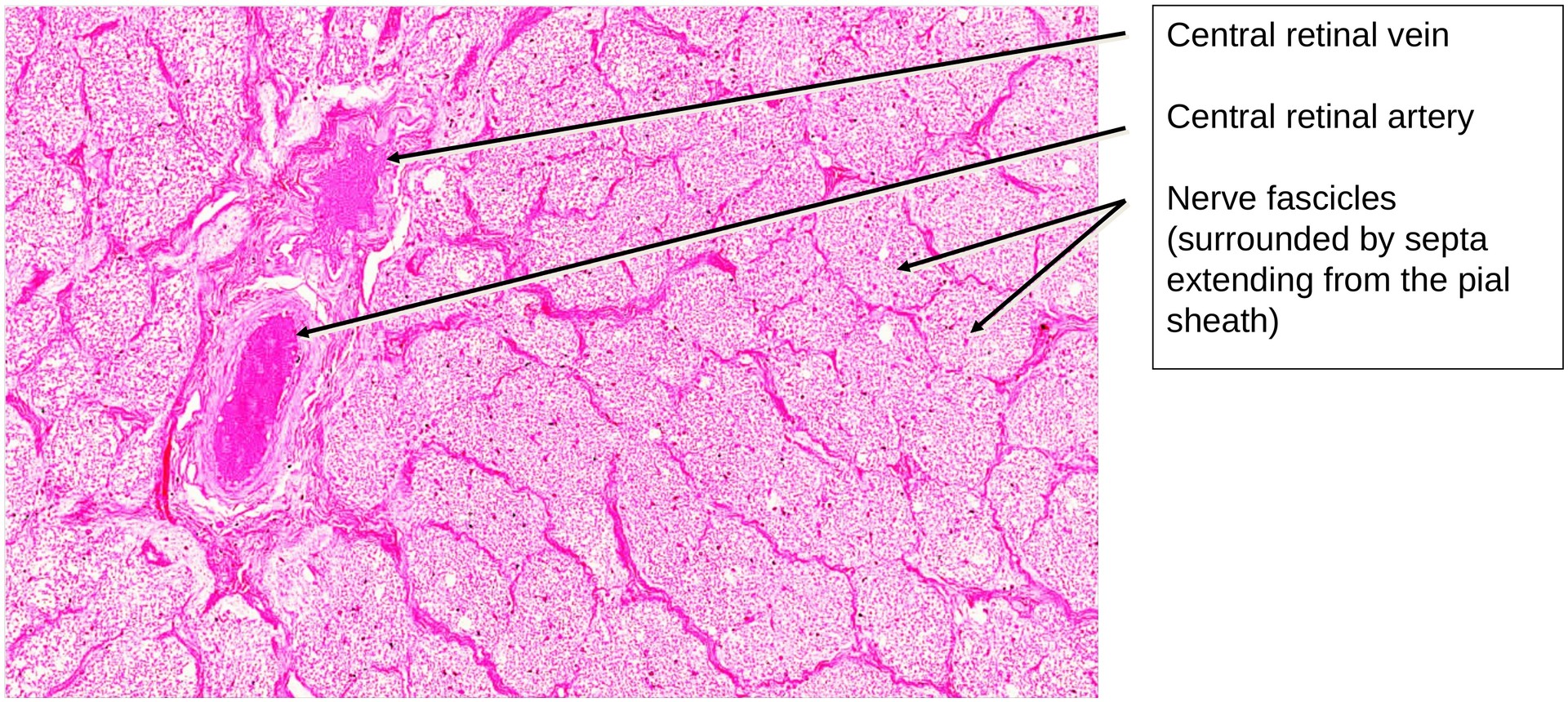
This specimen was taken close to the eyeball, which is evident from the presence of the central retinal artery, a vessel that usually enters the optic nerve only shortly before it reaches the globe.
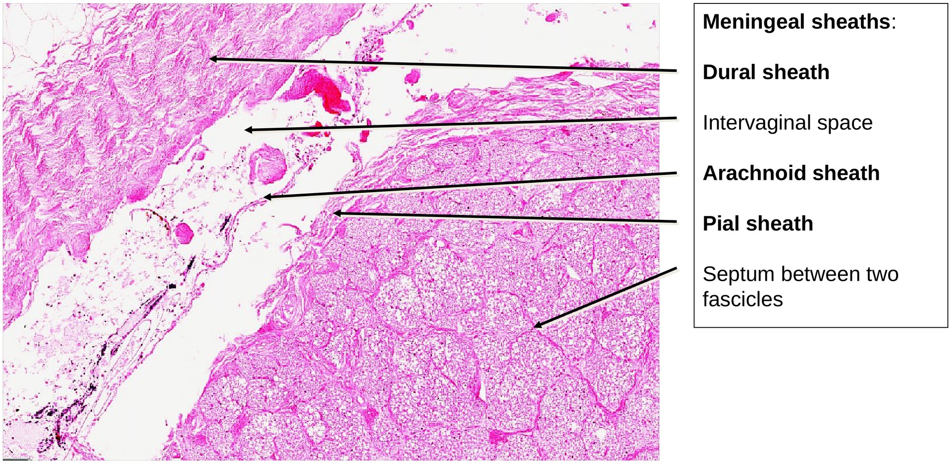
The optic nerve is characterized by its distinct meningeal sheaths, which are continuations of the meninges of the brain:
- Dural sheath (outermost layer): A tough connective tissue covering that forms the external sheath of the optic nerve.
- Arachnoid sheath: Lies internal to the dura mater. Under physiological conditions, the arachnoid adheres closely to the dura; however, in this preparation, it has detached due to fixation artefacts.
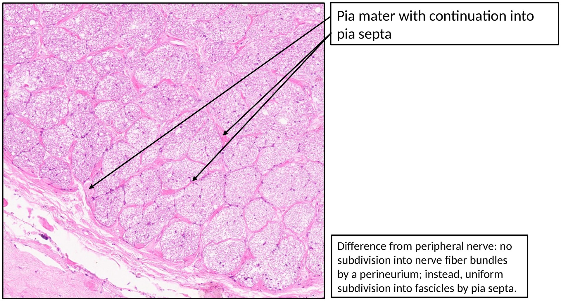
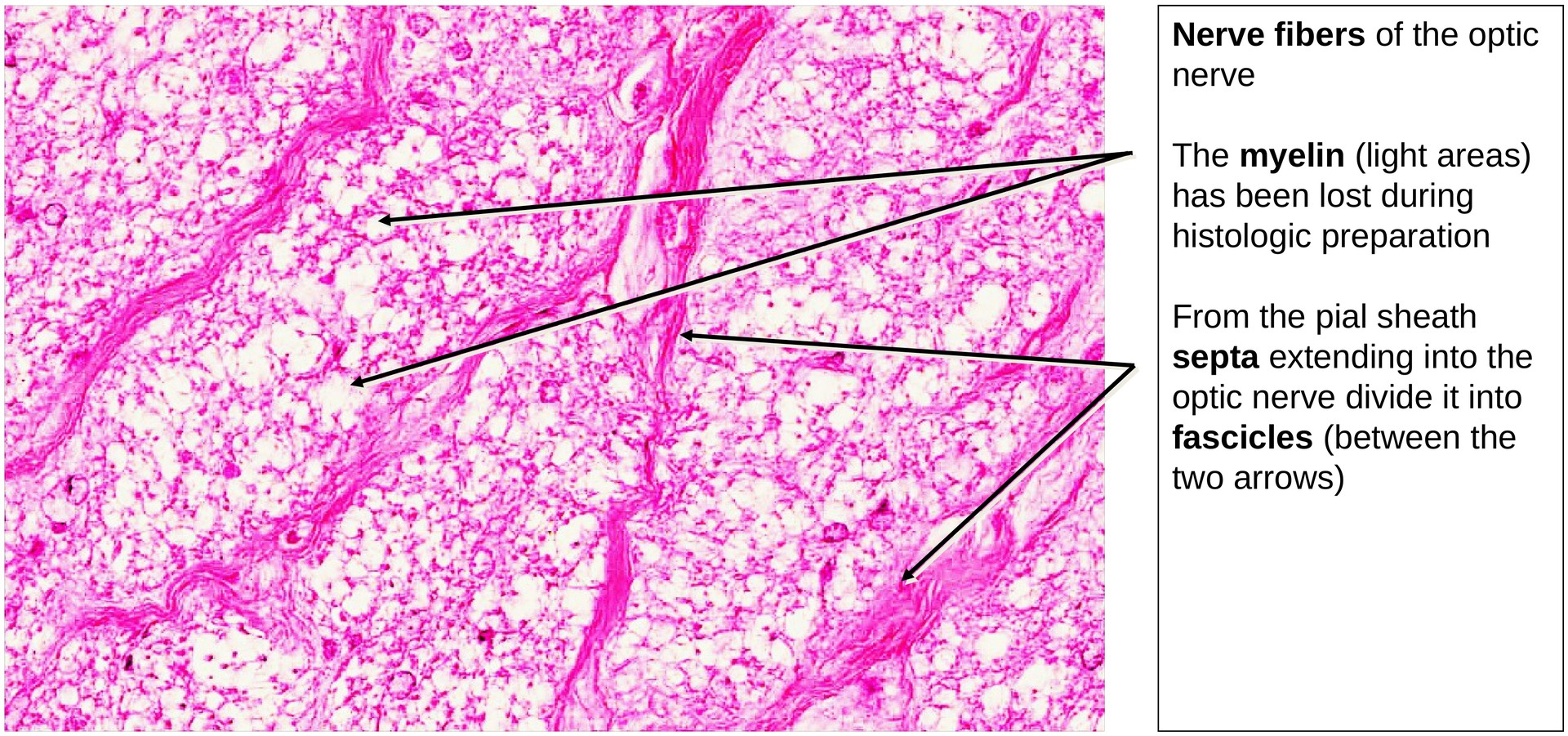
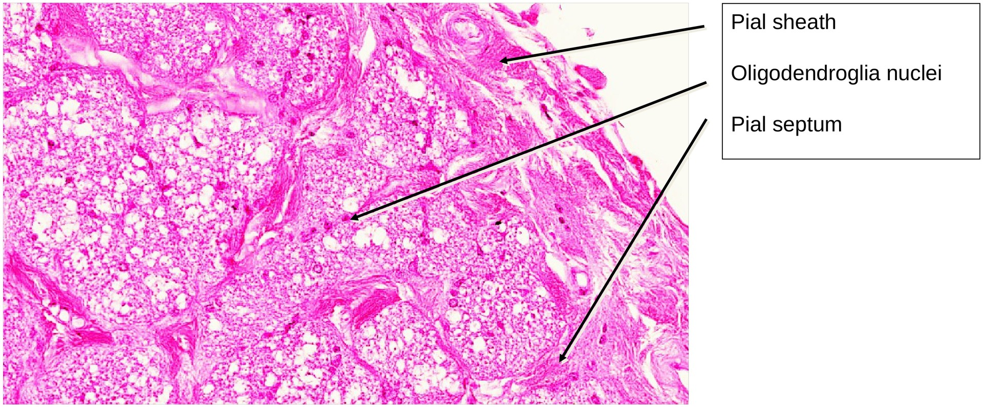
- Pial sheath (innermost layer): Originates from the pia mater and lies directly on the surface of the optic nerve. From it, pial septa extend into the interior of the nerve, dividing it into fascicles.
Between the dural and arachnoid sheaths lies the intervaginal space, which is the continuation of the subdural space of the meninges.
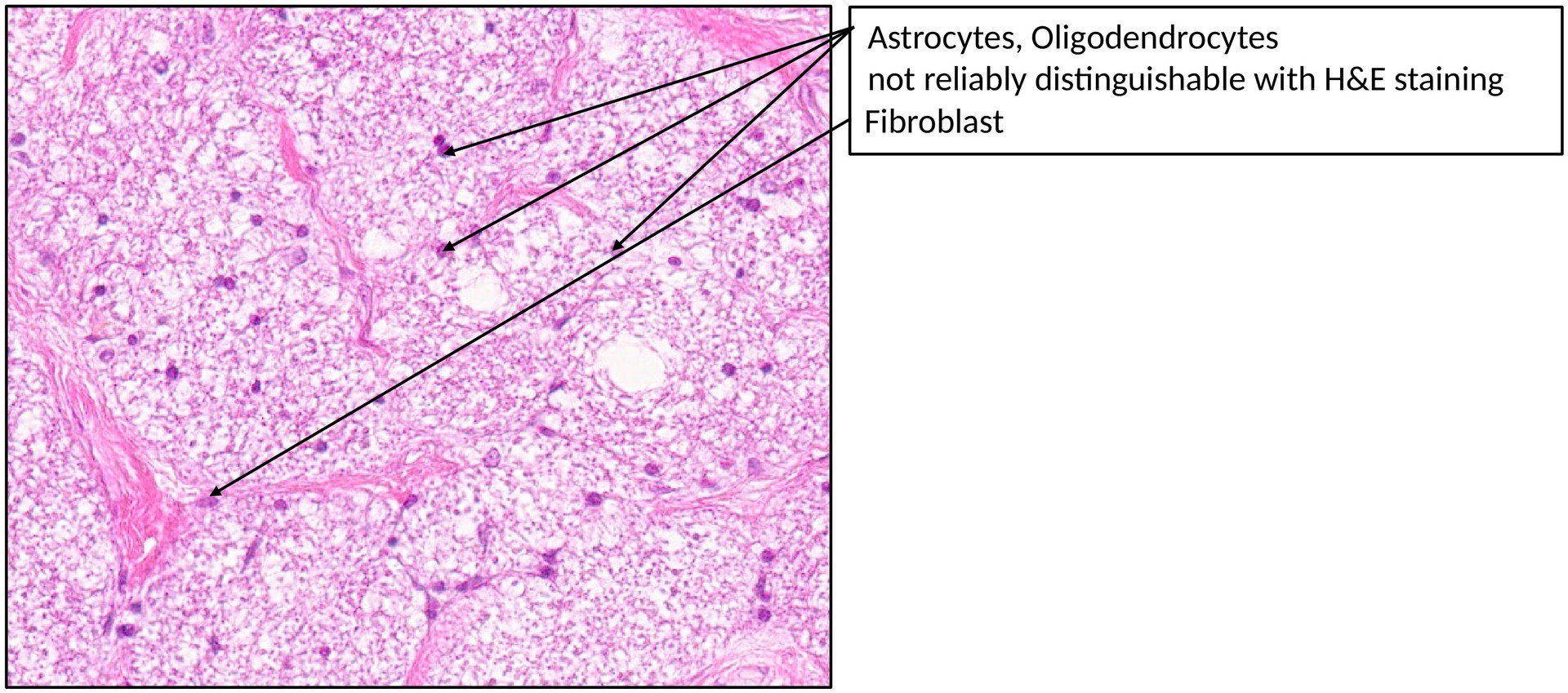
Within the optic nerve, nerve fibers are surrounded by oligodendroglial cells, which provide myelination within the central nervous system.
The central retinal artery and vein can be observed near the center of the nerve. Their presence confirms the section’s proximity to the eye.
Tasks:
Identify the following structures and answer the questions based on the specimen:
- Identify the meningeal sheaths of the optic nerve: dural sheath, arachnoid sheath, and pial sheath.
- Trace the pial septa projecting into the interior of the nerve.
- Locate the central retinal artery and vein.
- To what cell type do the nuclei located near the nerve fibers belong?
- Define the boundaries of the intervaginal space.
License
University of Basel
Downloads