NERVOUS SYSTEM (ANATOMICAL MICROSCOPY)
16.3
Brain 3 (Calcarine sulcus)
Specimen:
SPECIMEN DETAILS:
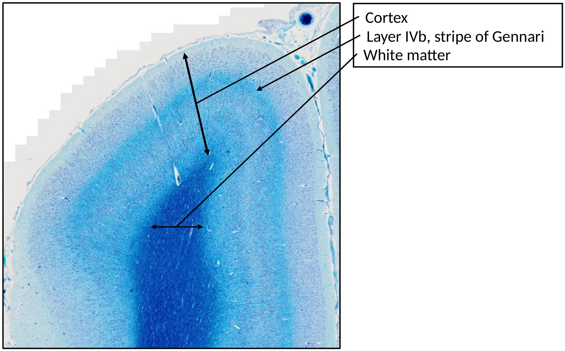
Organ: Brain Calcarine Sulcus (Primary Visual Cortex, V1)
Origin: Human
Staining: Luxol Fast Blue/Cresyl Violet
METHOD AND SPECIMEN DESCRIPTION:
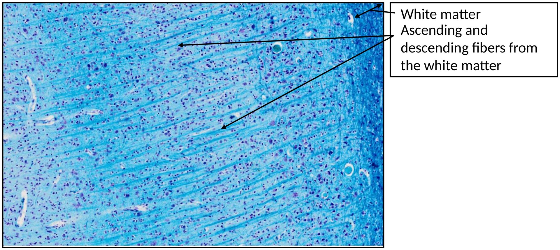
Cresyl violet is a basic dye that binds to acidic structures such as RNA and DNA, staining the chromatin, nucleolus, and rough endoplasmic reticulum (Nissl substance) blue–violet. Luxol Fast Blue selectively stains myelin sheaths, coloring the white matter and myelinated fibers in blue.
This combined staining therefore allows the simultaneous visualization of neuronal cell bodies (grey matter) and myelinated fiber tracts (white matter).
OBJECTIVE OF THE EXAMINATION:
To study the course of the fibers in the grey and white matter of the cerebral cortex, with particular attention to the primary visual cortex (V1) and the Gennari stripe.
SPECIFIC FEATURES OF THE SPECIMEN:
The laminar structure of the cortex corresponds to that of specimen 16.2 (Brain – Calcarine Sulcus). The Gennari stripe, located in layer IVb, appears as a distinct light blue band due to the densely packed tangentially running myelinated fibers.
At several points, blue-stained fiber bundles from the lateral geniculate nucleus (LGN) can be seen traversing the deeper cortical layers before terminating in layer IVc, which is characteristic of the visual cortex.
The dark-stained elongated structures visible throughout the section represent capillaries filled with blood.
TASKS:
- From which thalamic nucleus do the clearly visible fibers terminating in layer IVc originate?
- Identify the Gennari stripe within layer IVb.
- To which class of cortical fibers do the tangentially running fibers in the Gennari stripe belong?
License
University of Basel