BONES (GENERAL HISTOLOGY)
5.3
Ossification, endochondral (Epiphyseal plate, rat)
Preparation:
Preparation Details:
Organ: Tibia (Long bone)
Origin: Rat
Staining: Hematoxylin - Eosin (H&E)
Method and Specimen Description:
In order to section this long bone, which is already partially ossified, it first had to be demineralized. This process is achieved using acids or chelating agents. The prepared sections were subsequently stained with a general overview stain.
Objective of the Examination:
To gain detailed knowledge of endochondral ossification, including the interaction between chondroclasts and osteoblasts, and to understand the structure of articular cartilage.
Special Features of the Preparation:
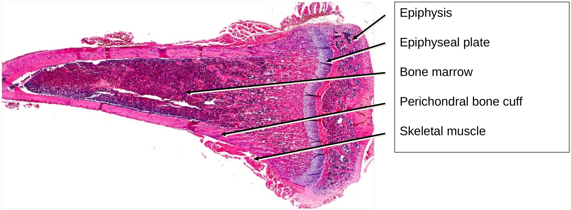
Even at low magnification, the distinct regions of the long bone can be identified: epiphysis, epiphyseal plate, and diaphysis.
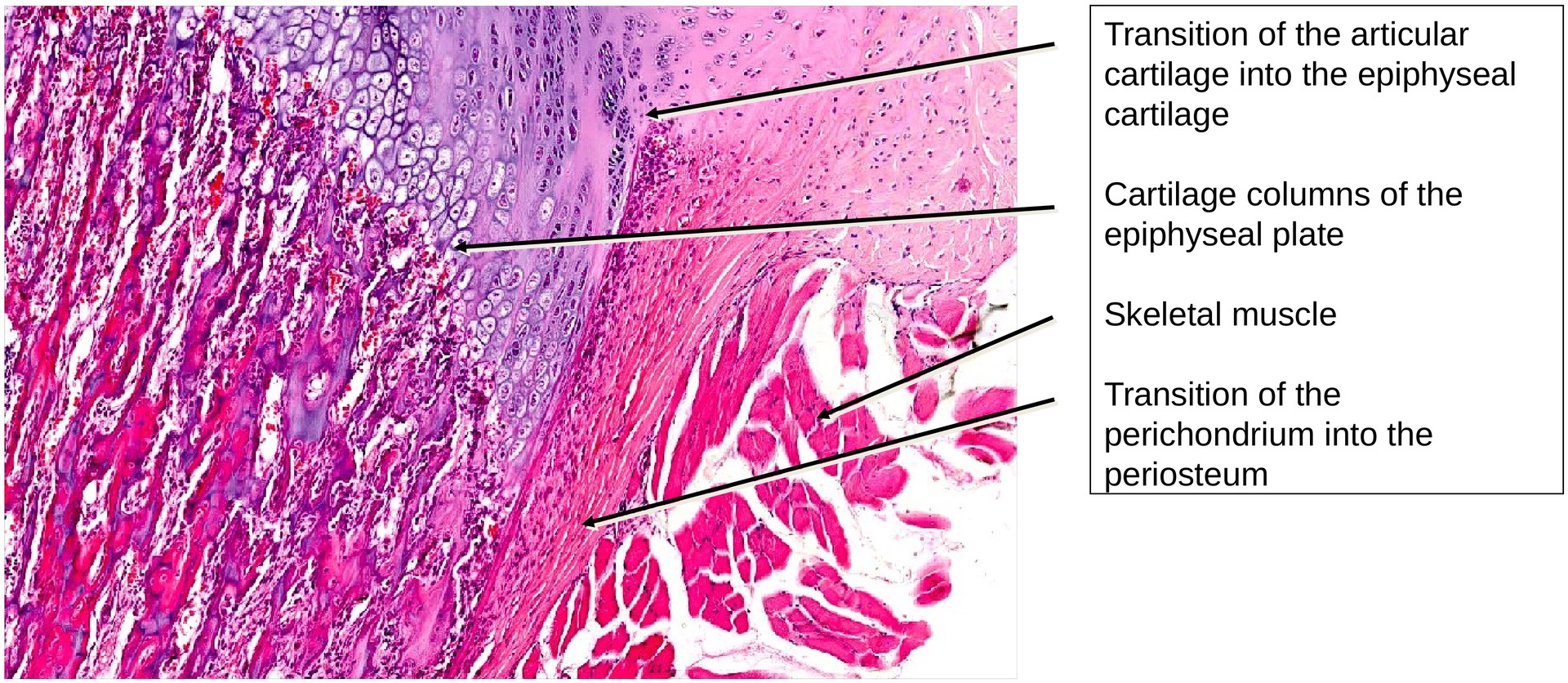
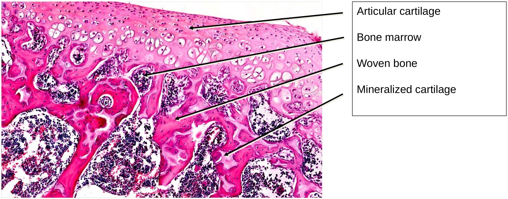
The epiphysis is covered by articular cartilage, which transitions into the epiphyseal cartilage within the epiphyseal plate.
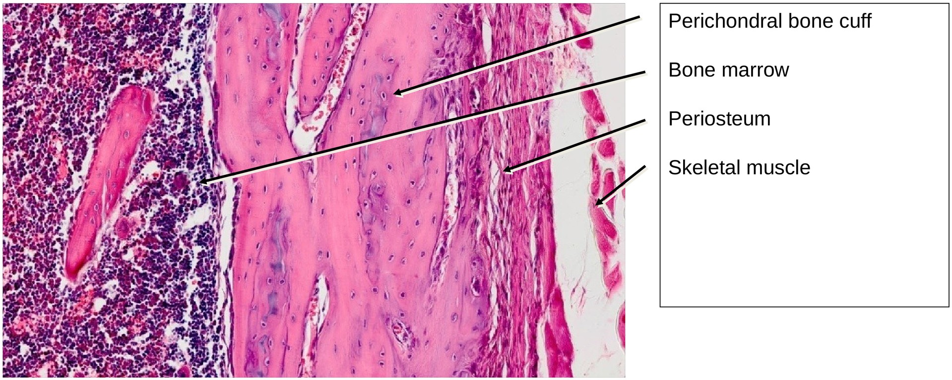
The joint capsule is partially preserved in this section and merges into the perichondrium, or, in the area of the bone, into the periosteum.
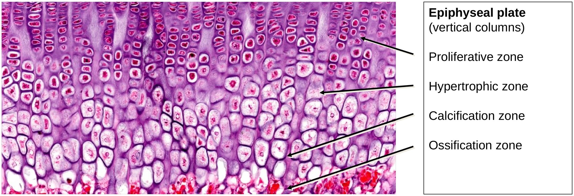
The epiphyseal plate consists of hyaline cartilage arranged in vertical columns. Within these cartilage columns, the following zones can be distinguished: the resting zone, proliferative zone, hypertrophic zone, calcification zone, and ossification zone (as described in standard histological literature).
Calcification Zone:
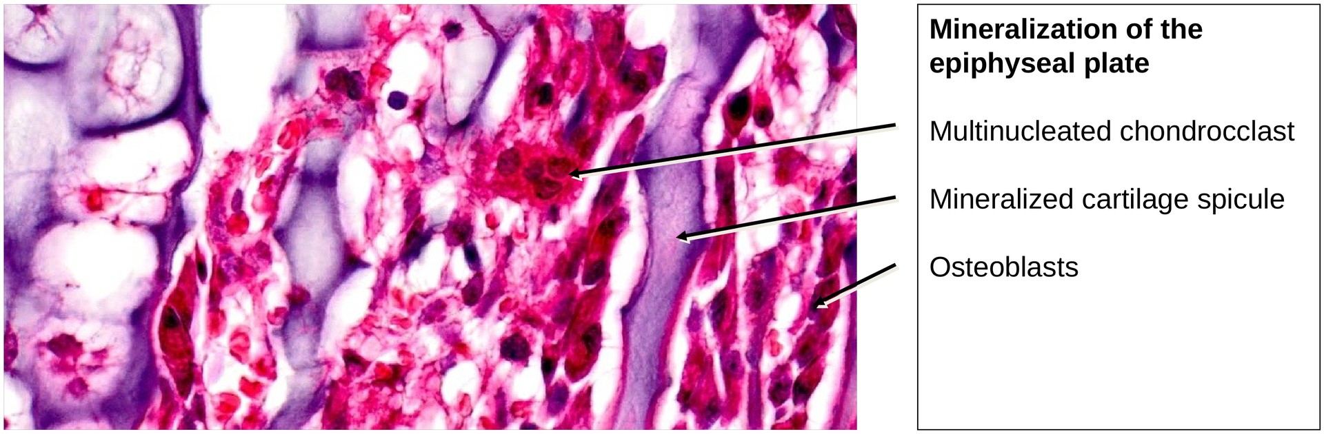
In this zone, part of the cartilaginous intercellular matrix is resorbed by chondroclasts — large, multinucleated cells similar in structure and function to osteoclasts. Their resorptive activity opens the cartilage lacunae, allowing capillaries and associated cells, such as osteoblasts, to invade the area.
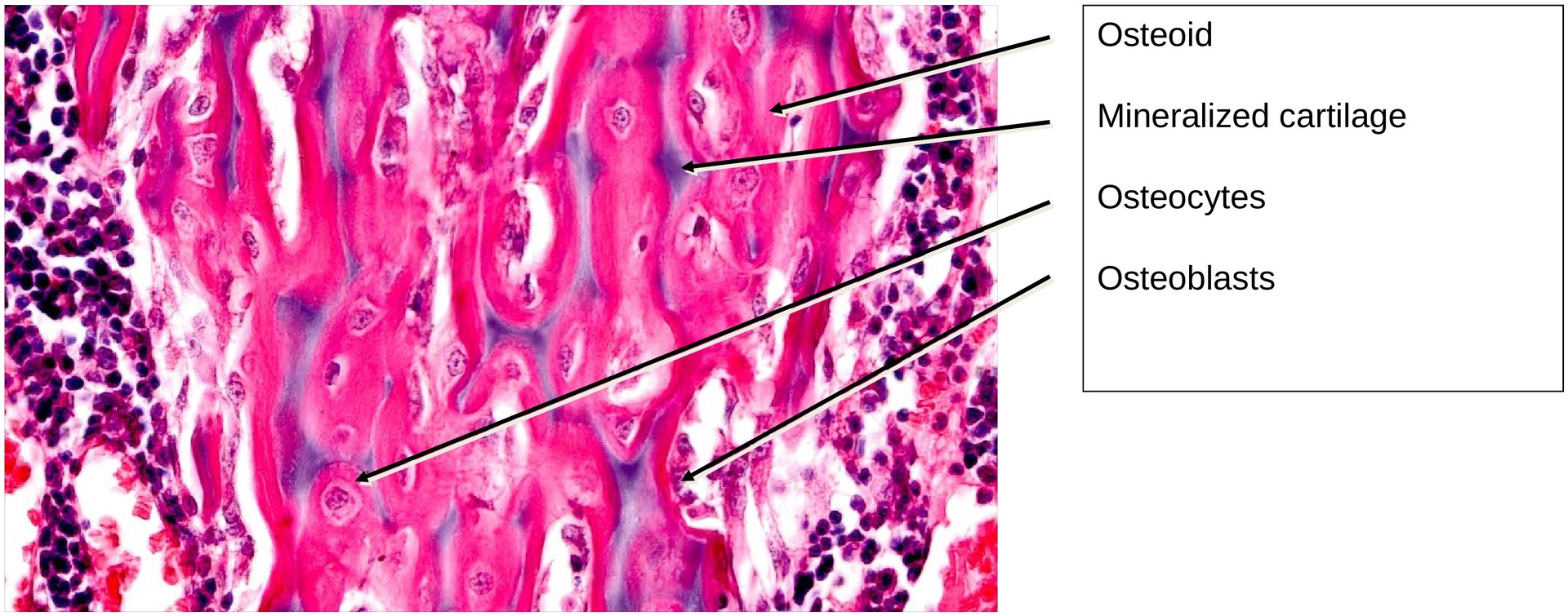
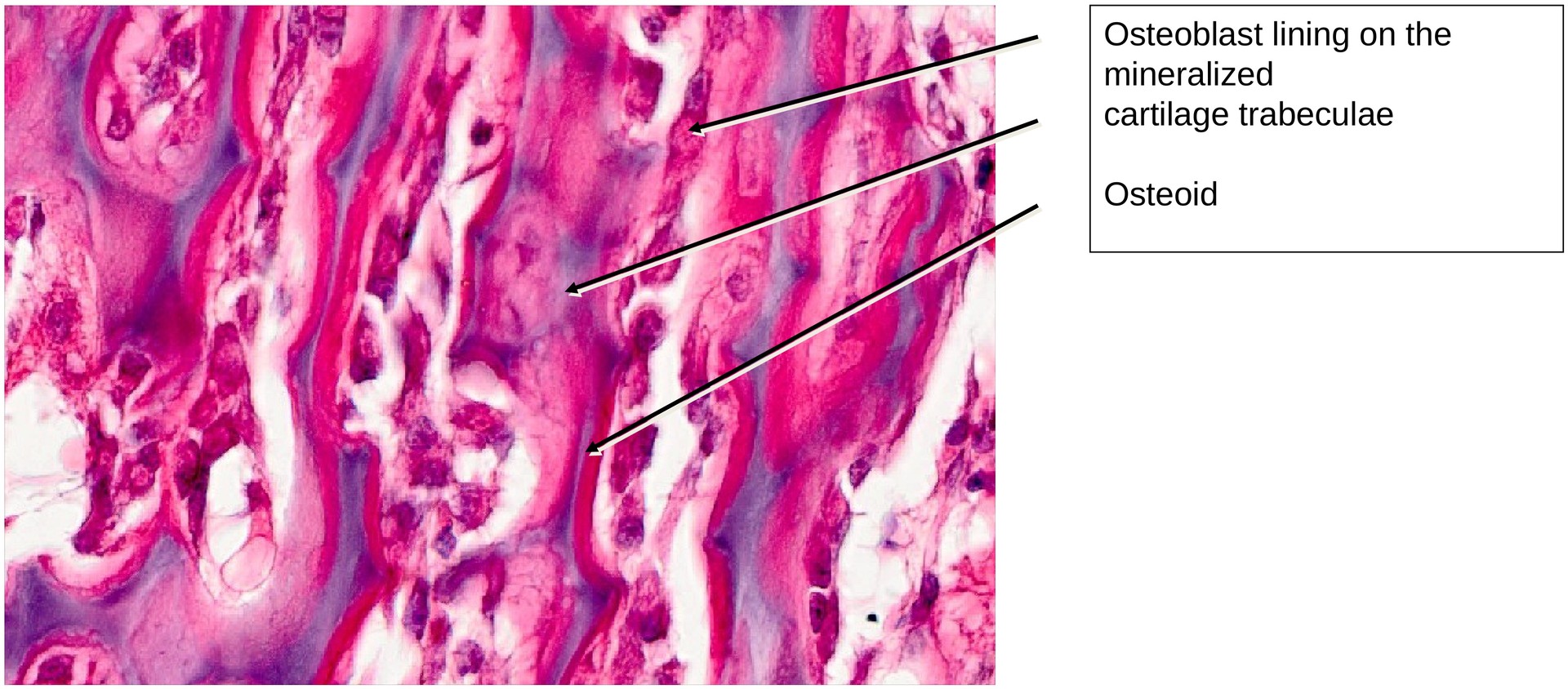
Ossification Zone:
In this region, the remaining mineralized cartilage trabeculae (stained blue) are lined by an osteoblastic layer. These osteoblasts secrete osteoid (stained red), which then undergoes secondary mineralization to form bone. The transition from cartilage columns to bone trabeculae can be clearly followed from the calcification zone towards the developing bone marrow.
Bone Marrow:
Near the epiphysis, primary bone marrow can be observed, consisting of mesenchymal cells, osteoblasts, and blood vessels.
Towards the diaphysis, the primary bone marrow transitions into secondary bone marrow, which is haematopoietically active and also known as red bone marrow.
Tertiary bone marrow, which has been converted to fatty marrow, is not yet present in this specimen.
Tasks:
• Identify the following components of the long bone: epiphysis, diaphysis, and epiphyseal plate.
• Note the already ossified regions — the bone within the epiphysis and the perichondral bone cuff in the diaphyseal area. What type of bone is this?
• Identify the distinct zones in the cartilage columns of the epiphyseal plate: resting zone, proliferative zone, hypertrophic zone, and calcification zone.
• Locate a chondroclast in the calcification zone.
• Identify the osteoblasts (osteoblastic layer) along the mineralised cartilage trabeculae.
• Observe the blood vessels that have invaded the calcification zone.
• Identify the periosteum. Which structure surrounds the bone on both sides, external to the periosteum?
License
University of Basel
Downloads