DIGESTIVE ORGANS: LIVER, GALLBLADDER, PANCREAS (ANATOMICAL MICROSCOPY)
20.3
Liver, Pig
Specimen:
Specimen Details:
Organ: Liver
Origin: Pig
Staining: Van Gieson
Method and Specimen Description:
Normal histological section stained with Van Gieson, a connective tissue stain that highlights the lobular structure of the pig liver particularly well. In this staining, hepatocytes appear yellow and connective tissue red.
Objective of the Examination:
To study the microscopic architecture of the liver, with emphasis on its distinct lobulation and the arrangement of the supplying and draining vessels.
Special Features of the Specimen:
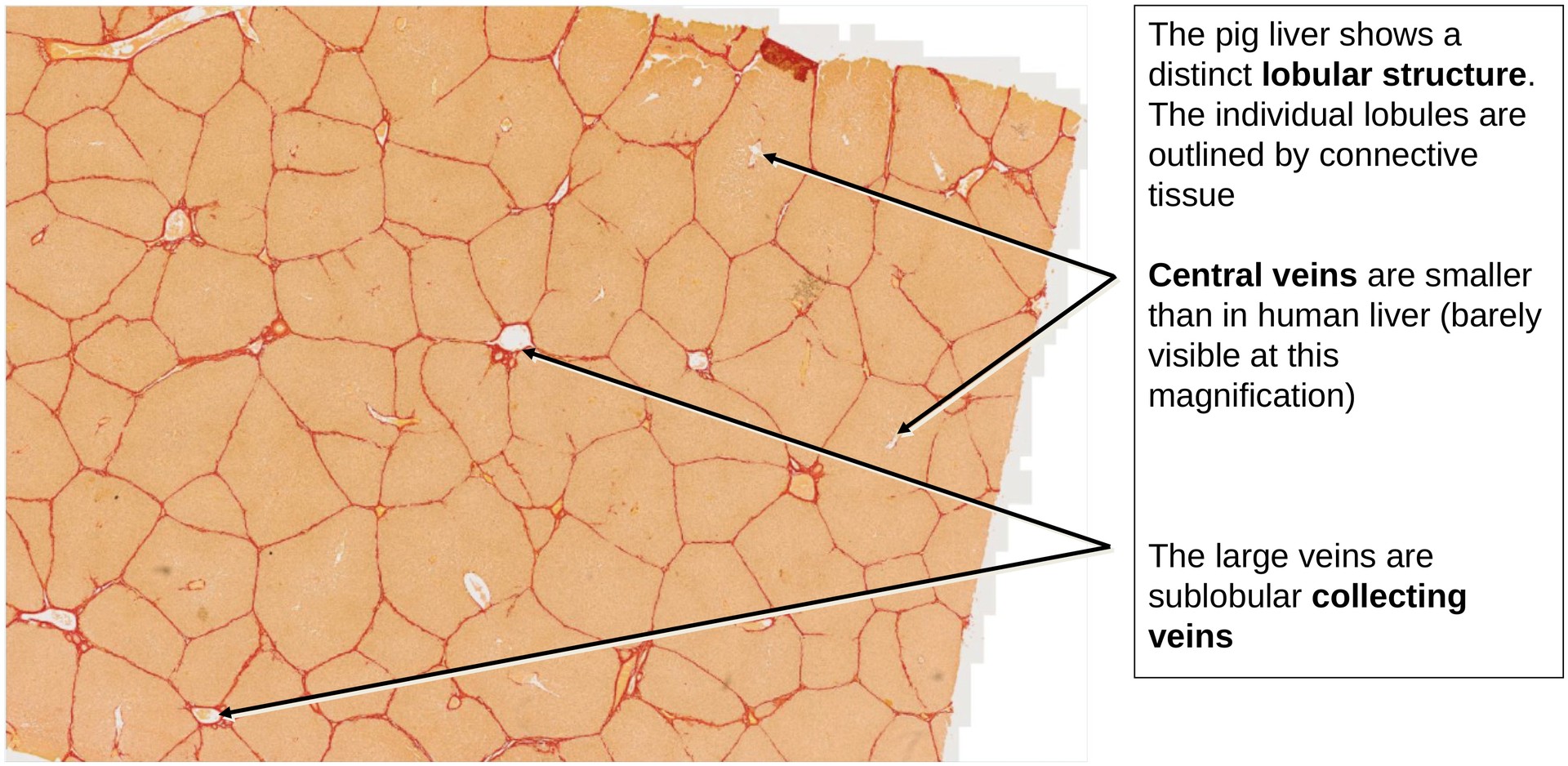
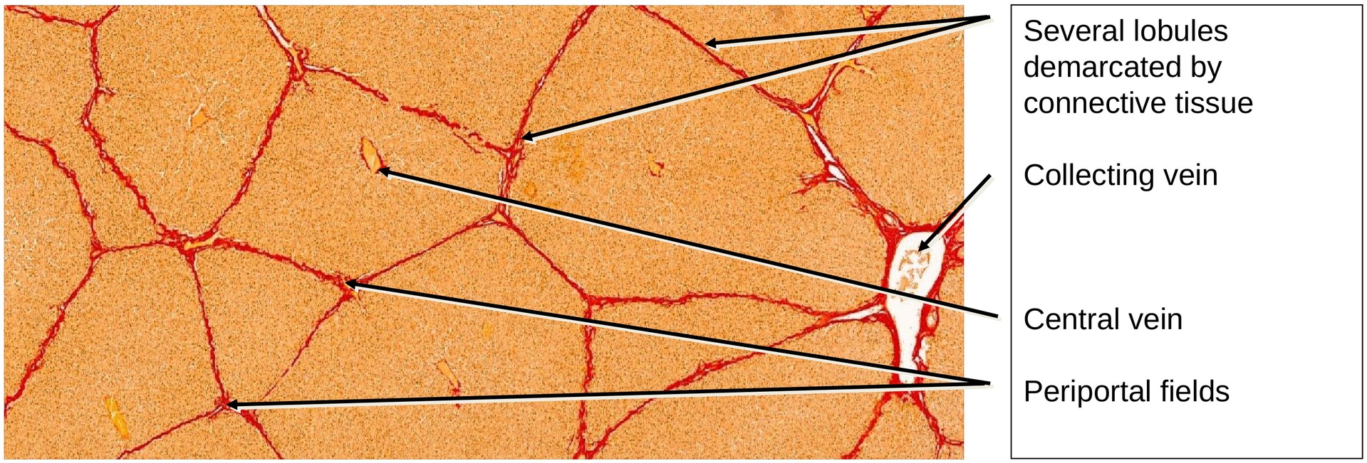
The pig liver exhibits a clearly demarcated lobular structure, in contrast to the human liver. This distinct appearance is due to the abundant interlobular connective tissue, which sharply delineates each liver lobule.
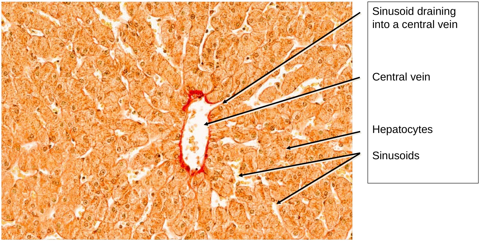
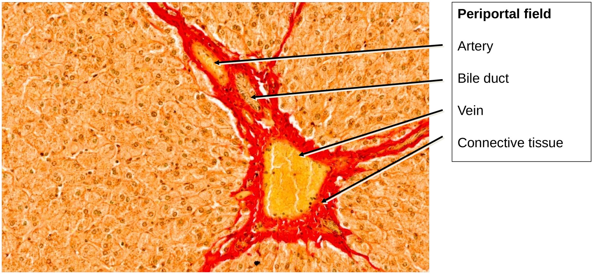
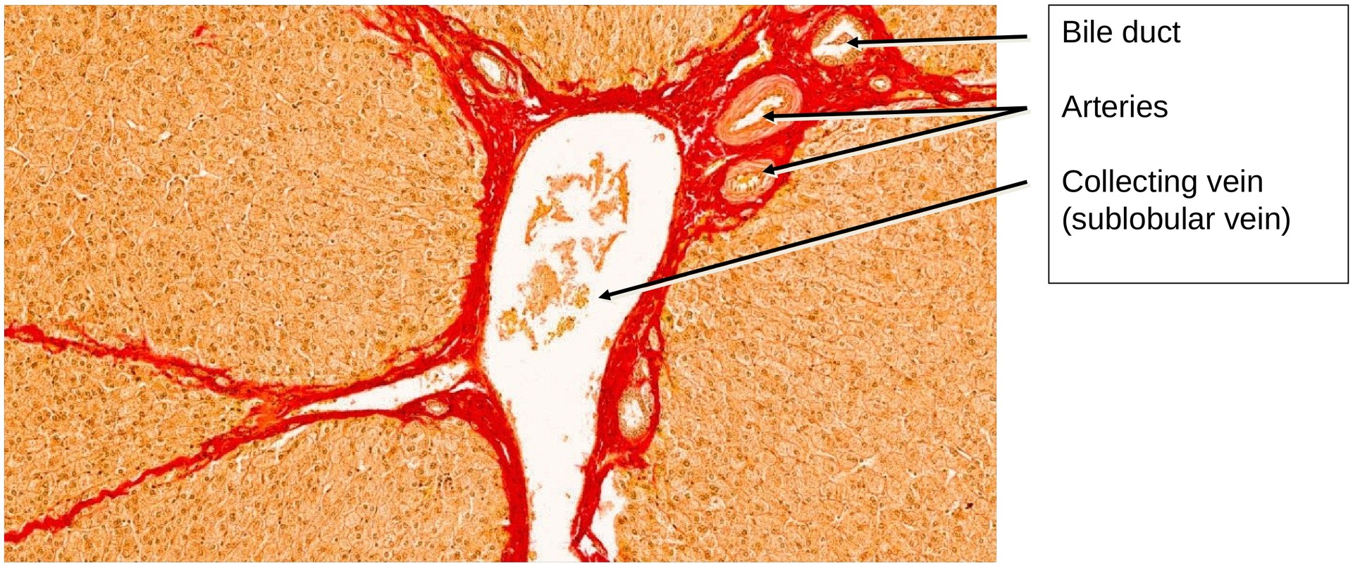
Each lobule is more or less polygonal in outline and arranged around a central vein, located at its center. At the corners of adjacent lobules, surrounded by connective tissue, lie the periportal fields (portal tracts or Glisson’s triads). These typically contain:
- a branch of the portal vein (afferent venous vessel),
- a branch of the hepatic artery (afferent arterial vessel), and
- a bile duct (efferent duct).
The connective tissue not only separates lobules but also carries larger collecting veins, which drain blood from the central veins as sublobular veins. These merge into the hepatic veins, which ultimately empty into the inferior vena cava.
In some areas, the hepatic sinusoids around the central veins have remained open, rather than collapsed during fixation. This makes it possible to observe the transition of the sinusoids into the central veins, which is normally less apparent in fixed material.
Tasks:
- Observe the lobular organization of the pig liver and note the polygonal outline of the individual lobules.
- Identify the central veins within several lobules (not always well stained).
- Trace the sinusoids, where visible, and observe their opening into the central veins.
- Locate a periportal field and consider why it is described as periportal.
- Differentiate between the venous and arterial branches within the periportal field, based on their wall structure and lumen size.
- Identify a bile duct in the periportal field — note its cuboidal epithelium and clear lumen.
License
University of Basel
Downloads