LYMPHATIC ORGANS (ANATOMICAL MICROSCOPY)
15.8
Thymus, child
Specimen Details:
Specimen Details:
Organ: Thymus
Origin: Human (Child)
Staining: Hematoxylin - Eosin (H&E)
Method and Specimen Description:
Standard histological section stained with H&E for general morphological assessment.
Objective of the Examination:
To study the thymus in childhood, in which both cortex and medulla are still fully developed. The specimen serves as a reference for comparison with other lymphatic organs and with the involuted adult thymus.
Special Features of the Specimen:
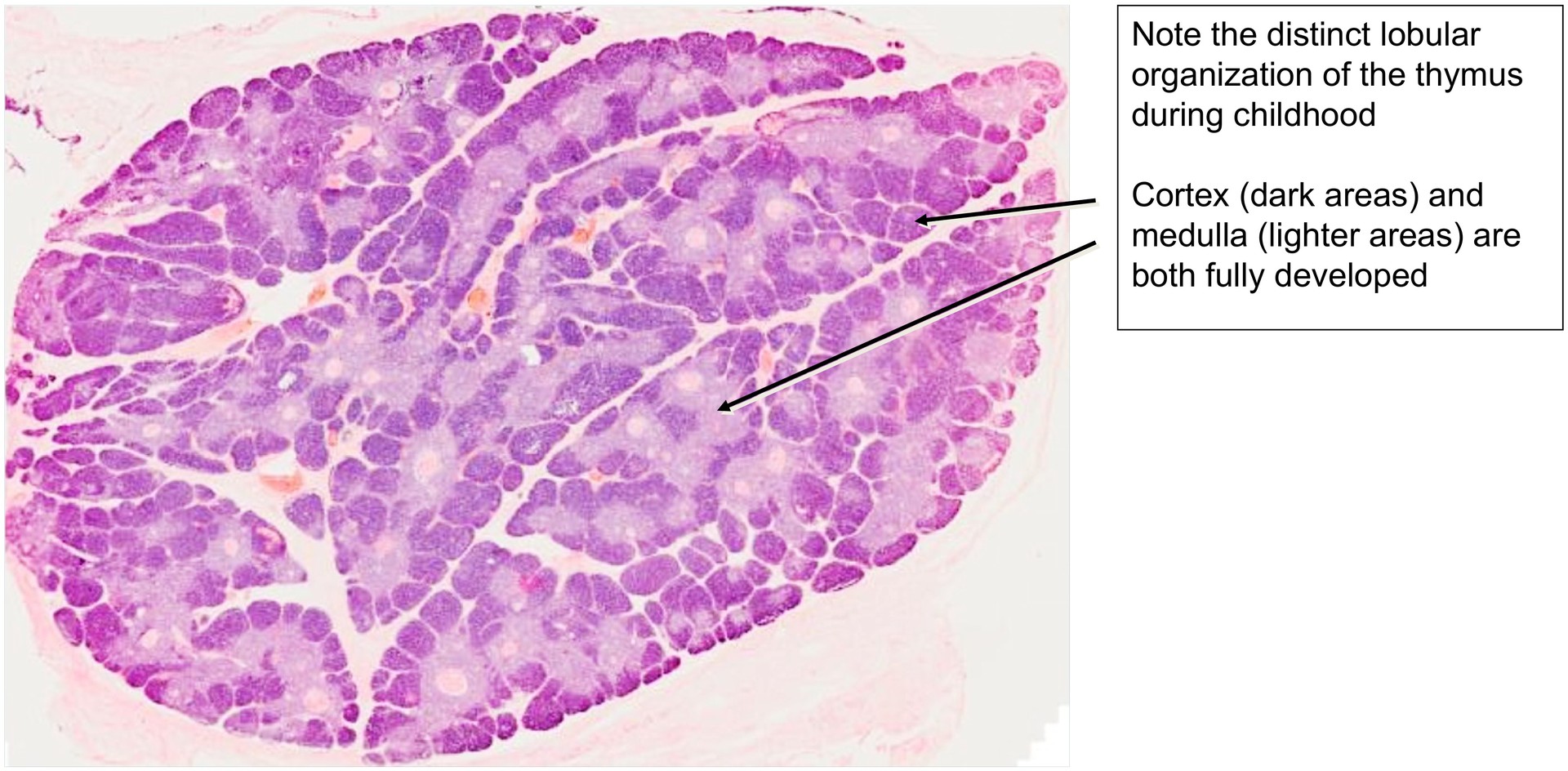
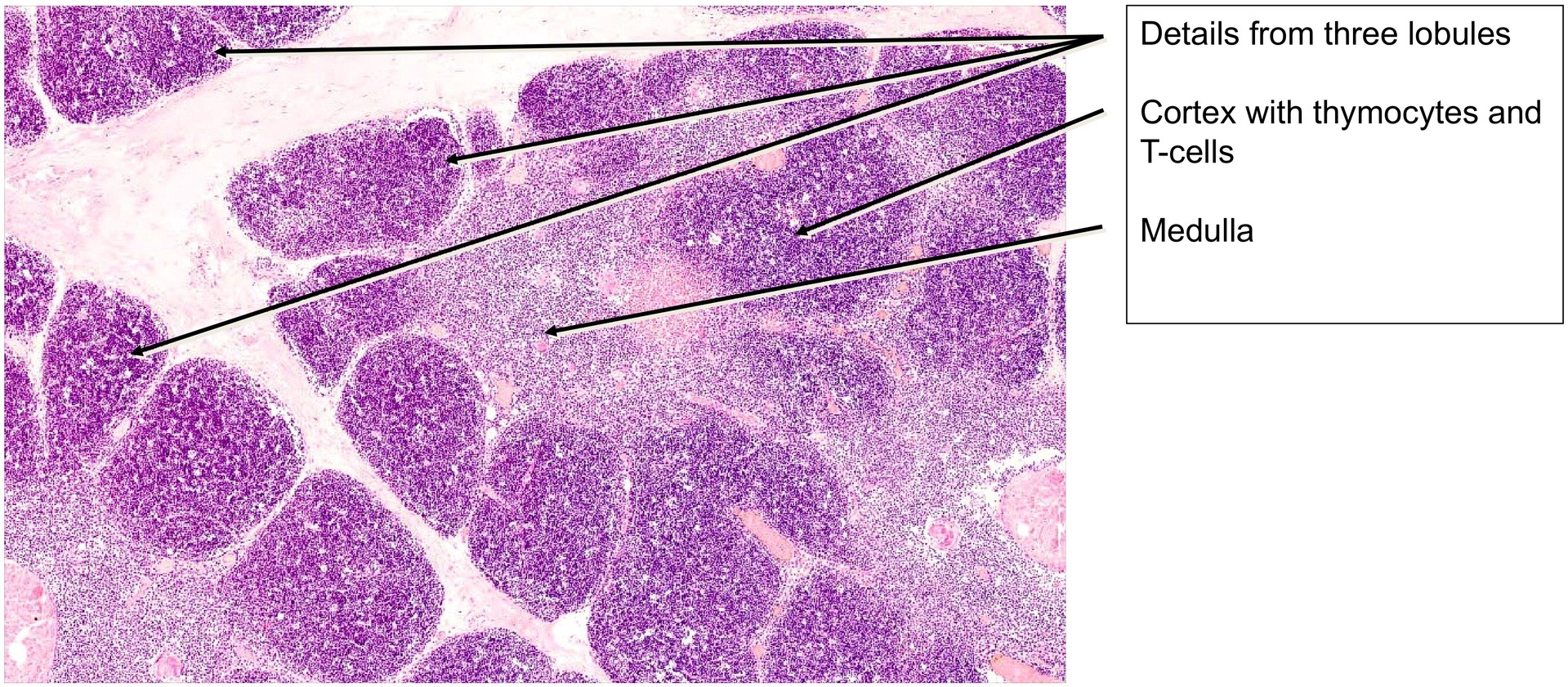
At low magnification, the child’s thymus displays a distinct lobular organization, with each lobule showing a darker-stained cortex and a lighter-stained medulla.
The dense appearance of the cortex results from the high cellularity of thymocytes, which possess relatively little cytoplasm and closely packed nuclei. These thymocytes originate from the bone marrow and migrate towards the medulla during maturation, from where they later populate other lymphatic organs.
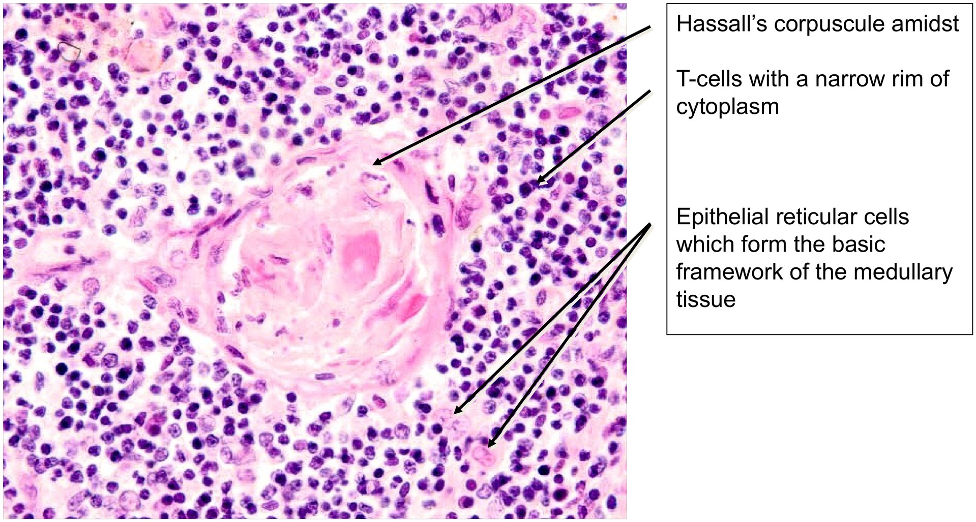
The medulla, by contrast, appears paler, as it is composed mainly of epithelial reticular cells (derived from the endoderm of the 3rd and 4th pharyngeal pouches), which have larger nuclei and broader cytoplasmic extensions. In addition, dendritic cells and macrophages are present within the medulla, though they cannot be distinctly identified in standard H&E sections.
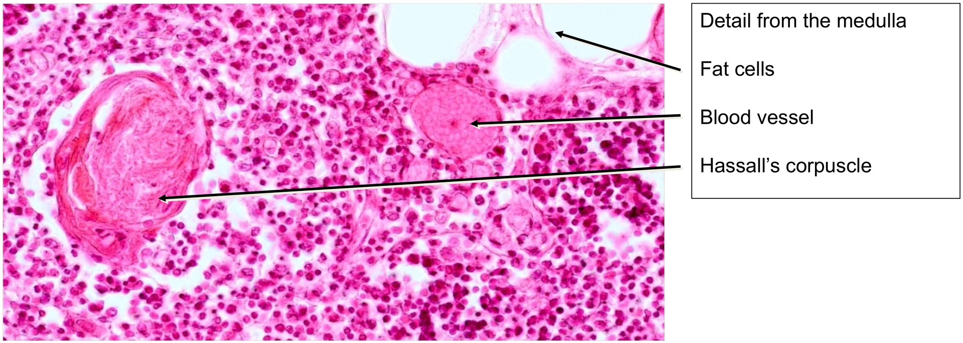
A characteristic feature of the medulla are the Hassall’s corpuscles, which display a concentric, onion-skin-like structure. These corpuscles contain cytokeratin filaments and keratohyalin granules, and are formed from epithelial reticular cells undergoing transformation. In this specimen, some exceptionally large Hassall’s corpuscles can be observed.
Tasks:
- At low magnification, observe the lobular structure of the thymus and distinguish clearly between cortex and medulla.
- At higher magnification, identify thymocytes and developing T-cells, and distinguish them from the epithelial reticular cells of the thymic framework (the latter exhibit larger nuclei and broader cytoplasm).
- Compare the relative proportions of cortex and medulla as a reference for evaluating thymic involution in the adult specimen.
- In the medulla, note the heterogeneous cell population, but do not attempt detailed classification.
- Locate and study Hassall’s corpuscles in the medulla — some are very large in this preparation — and compare their structure with that of nearby blood vessels.
Search for any fat cells present (relatively few, in contrast to the adult thymus).
License
University of Basel
Downloads