SKIN AND APPENDAGES (ANATOMICAL MICROSCOPY)
13.2
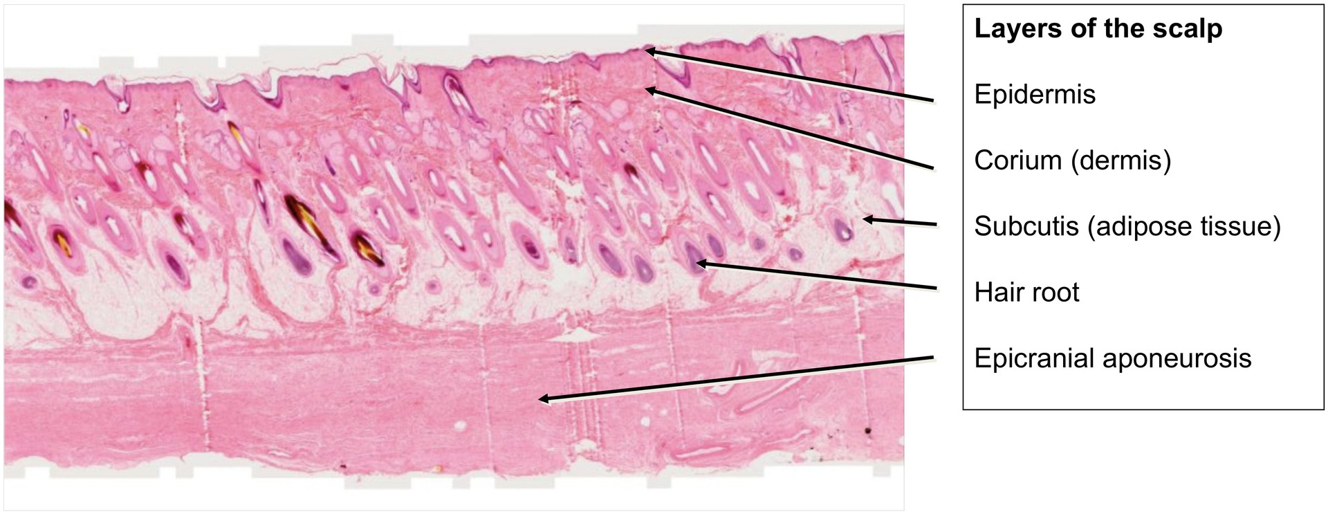
Scalp, longitudinal cross section
Specimen:
Specimen Details:
Organ: Scalp
Origin: Human
Staining: Hematoxylin - Eosin (H&E)
Method and Specimen Description:
Normal histological section of the human scalp. The section was made more or less parallel to the obliquely embedded hair shafts within the scalp, allowing an extended view of the hair follicles and associated structures in their full length.
Objective of the Examination:
To study the formation and structure of hair, including its specific layers, the hair shaft, and the hair bulb.
Special Features of the Specimen:
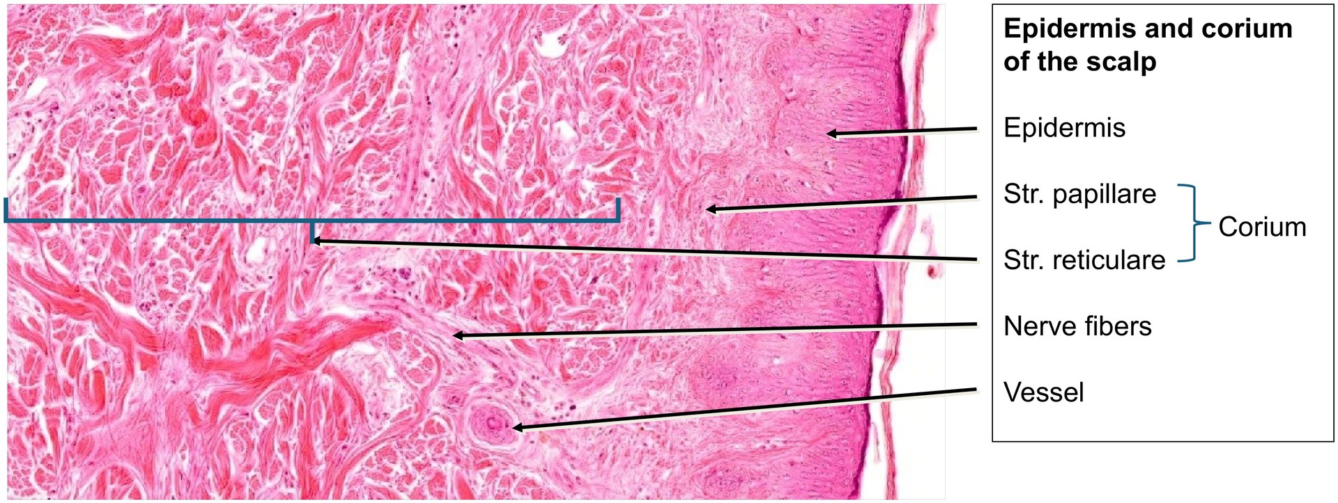
This specimen clearly demonstrates the layered architecture of the scalp, extending from the epidermis, through the corium (dermis) and subcutis, to the epicranial aponeurosis.
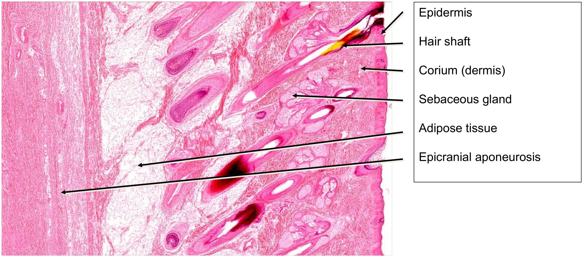
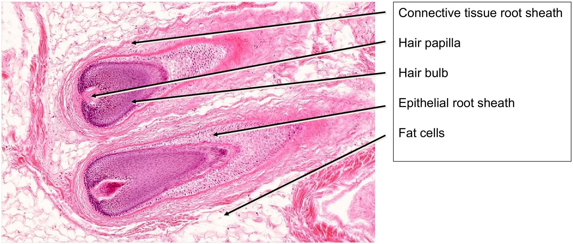
Once the hairs have emerged through the epidermis, they are referred to as hairs. Within the hair follicle, the developing structure is known as the hair shaft, surrounded by the hair root sheaths:
- the fibrous root sheath,
- the outer epithelial root sheath, and
- the inner epithelial root sheath.
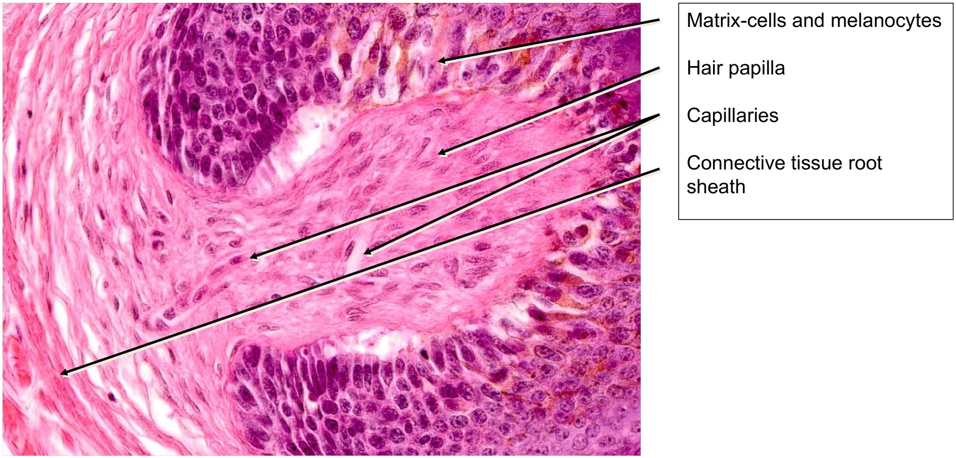
In the subcutaneous layer, the hair bulbs are visible, each containing a hair papilla that provides vascular and nutritive support. The hair papilla typically contains capillaries (though these may collapse during fixation and are not always visible). The hair matrix cells surrounding the papilla give rise to both the hair shaft and the epithelial root sheaths.
Once keratinization is complete, the hair is held in place only by the sheath cuticle, meaning the actual hair shaft is sometimes lost during histological preparation — resulting in an apparently “empty” root sheath.
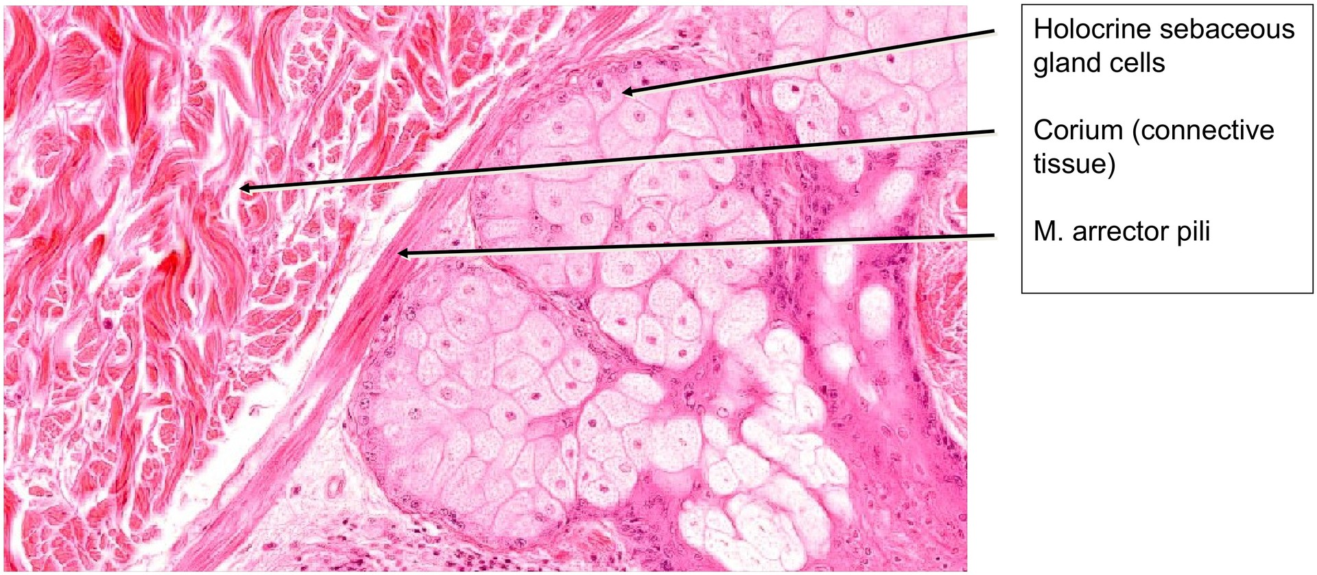
Openings of the sebaceous glands into the hair follicles are particularly well visible in this specimen. The sebaceous glands exhibit a clear maturation gradient — from basal cells to fully sebum-filled cells near the duct opening.
The arrector pili muscle (hair erector) can also be identified in several areas, running directly beneath the sebaceous glands. This smooth muscle aids in the release of sebum through contraction and is responsible for the familiar “goosebumps” effect in vivo.
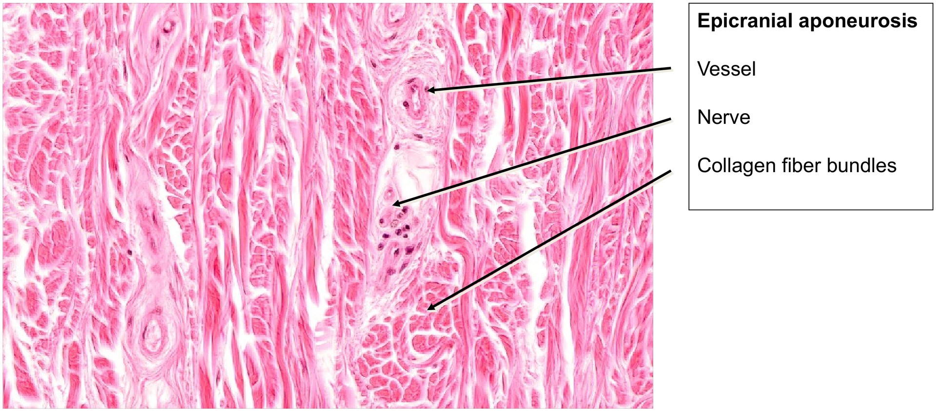
Beneath the subcutis lies the dense, fibrous epicranial aponeurosis, traversed by blood vessels and nerves, forming the tough connective layer characteristic of the scalp.
Tasks:
- Orient yourself within the specimen: identify the epidermis, corium (dermis), and subcutis.
- Locate the hair bulbs — in which layer of the skin are they found?
- Search for a centrally sectioned hair bulb to examine the hair papilla.
- Identify melanocytes in the matrix region.
- Identify the sebaceous glands.
- Locate an arrector pili muscle — where is it positioned relative to the hair follicle and sebaceous gland?
- Determine the type of gland the sebaceous gland represents (apocrine, holocrine, or merocrine).
- Review the layers of the epidermis — which are relatively thin or poorly developed in the scalp?
- Describe the predominant structure in the region of the epicranial aponeurosis.
License
University of Basel
Downloads