CARTILAGE (GENERAL HISTOLOGY)
4.3
Hyaline cartilage (rib)
Specimen Details:
Specimen Details:
Organ: Costal cartilage
Origin: Human
Staining: Hematoxylin - Eosin (H&E)
Method and Specimen Description:
Histological section, stained with a general H&E stain to demonstrate the structural features of hyaline cartilage.
Objective of the Examination:
To study the structure and growth mechanisms of hyaline cartilage.
Special Features of the Specimen:
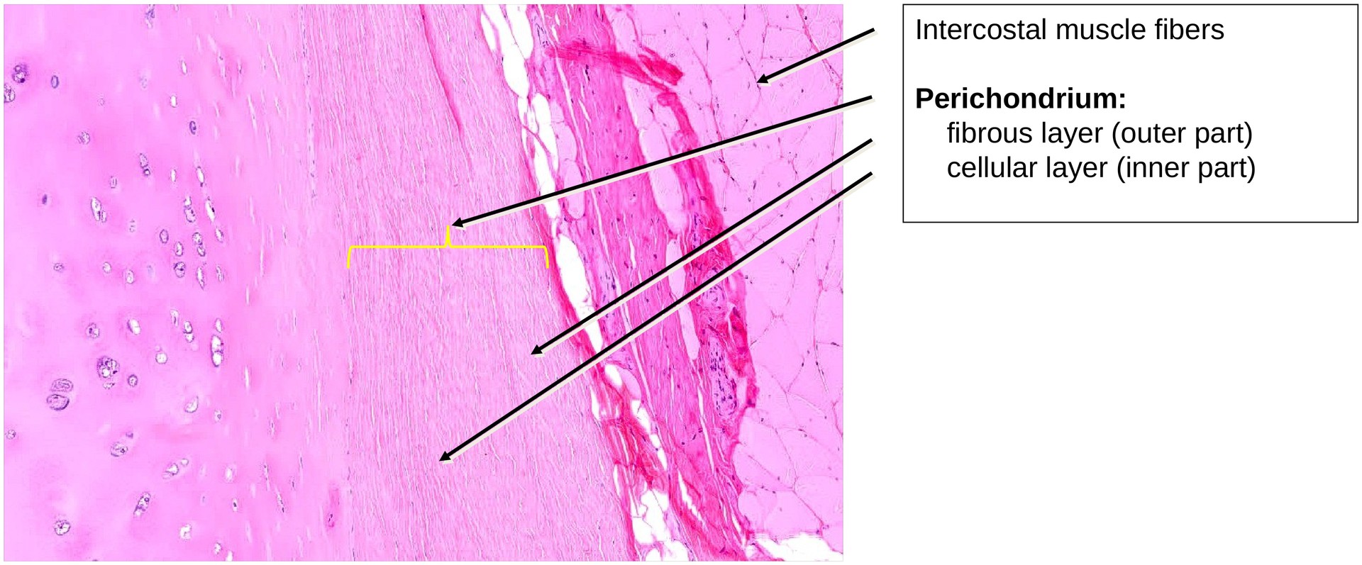
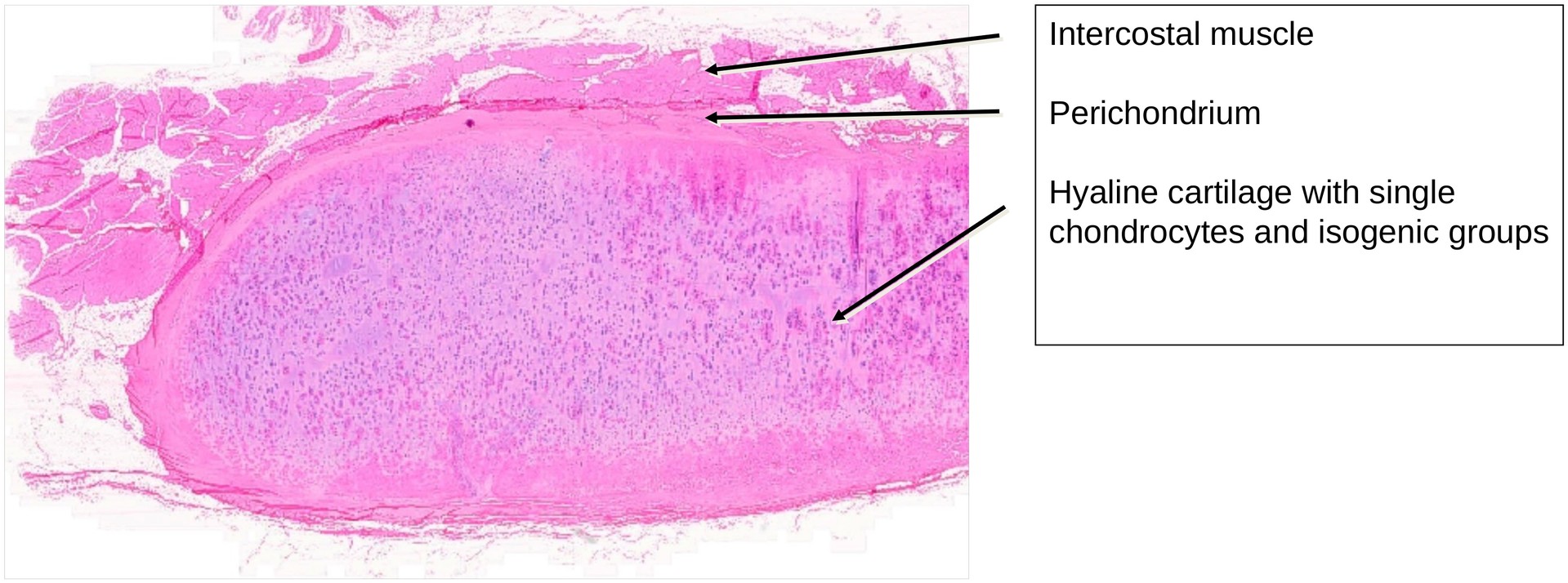
In this cross-section through costal cartilage, the tissue is covered externally by the perichondrium.
The perichondrium consists of two layers:
-
an outer fibrous layer (stratum fibrosum), and
-
an inner cellular layer (stratum cellulosum).
From the cellular layer, the cartilage undergoes appositional growth, contributing to cartilage thickening and limited regeneration.
Cartilage growth occurs in two ways:
-
Appositional growth — from the perichondrium, where undifferentiated cells differentiate into chondroblasts, and later chondrocytes.
-
Interstitial growth — by division of existing chondrocytes within the cartilage matrix.
The perichondrium consists of collagenous connective tissue fibers and fibroblasts. Beneath it lies a layer of flattened undifferentiated connective tissue cells, capable of developing into chondroblasts.
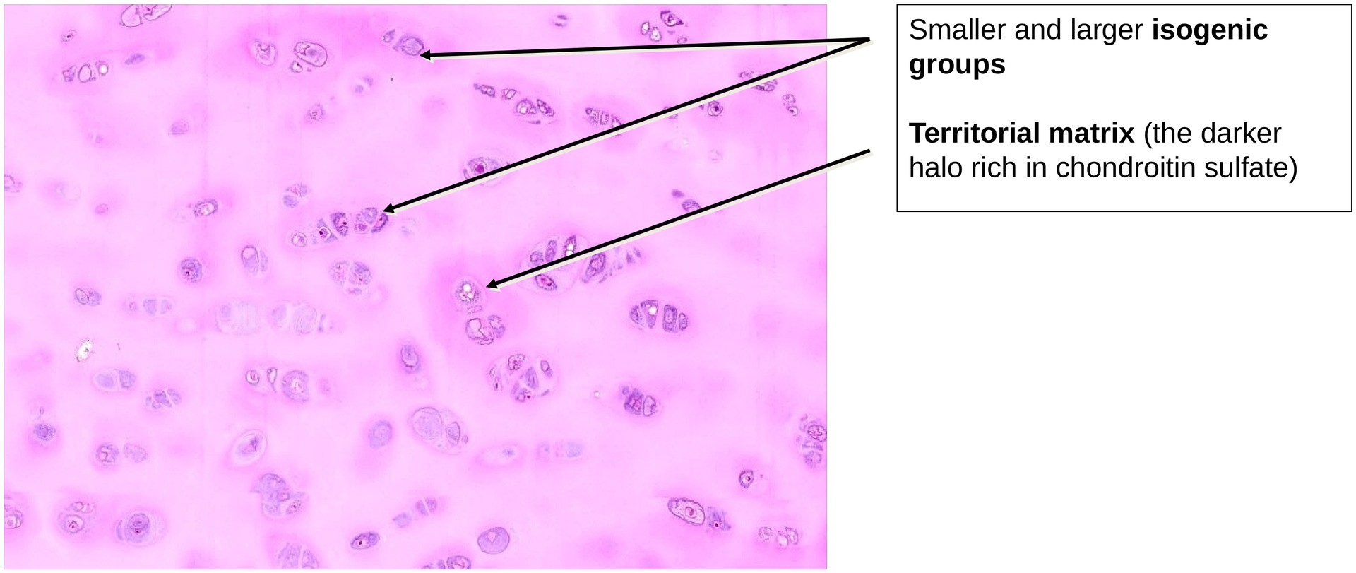
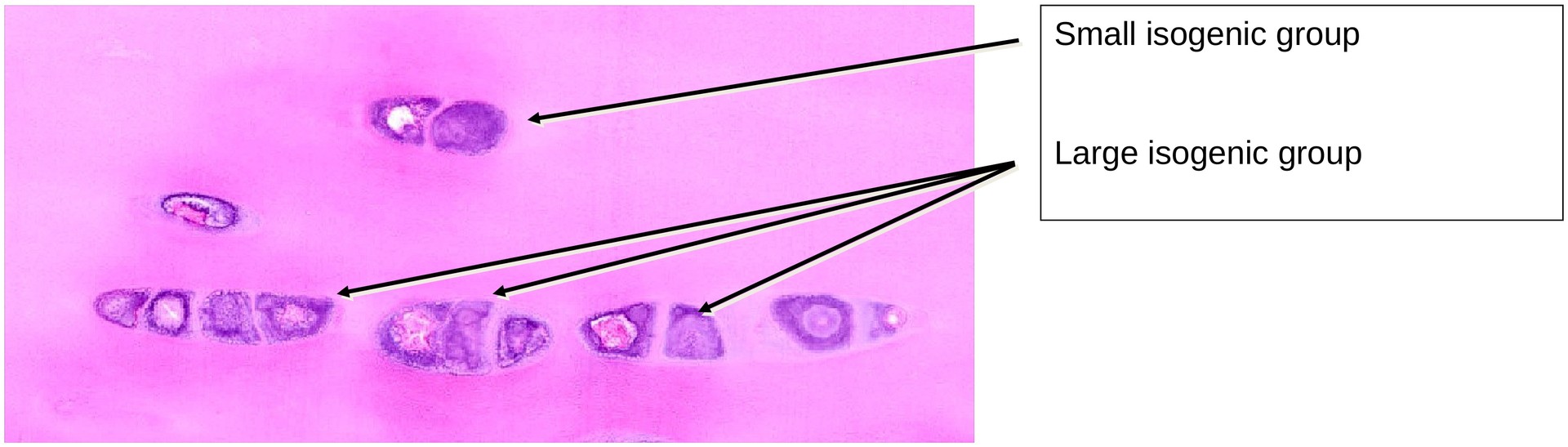
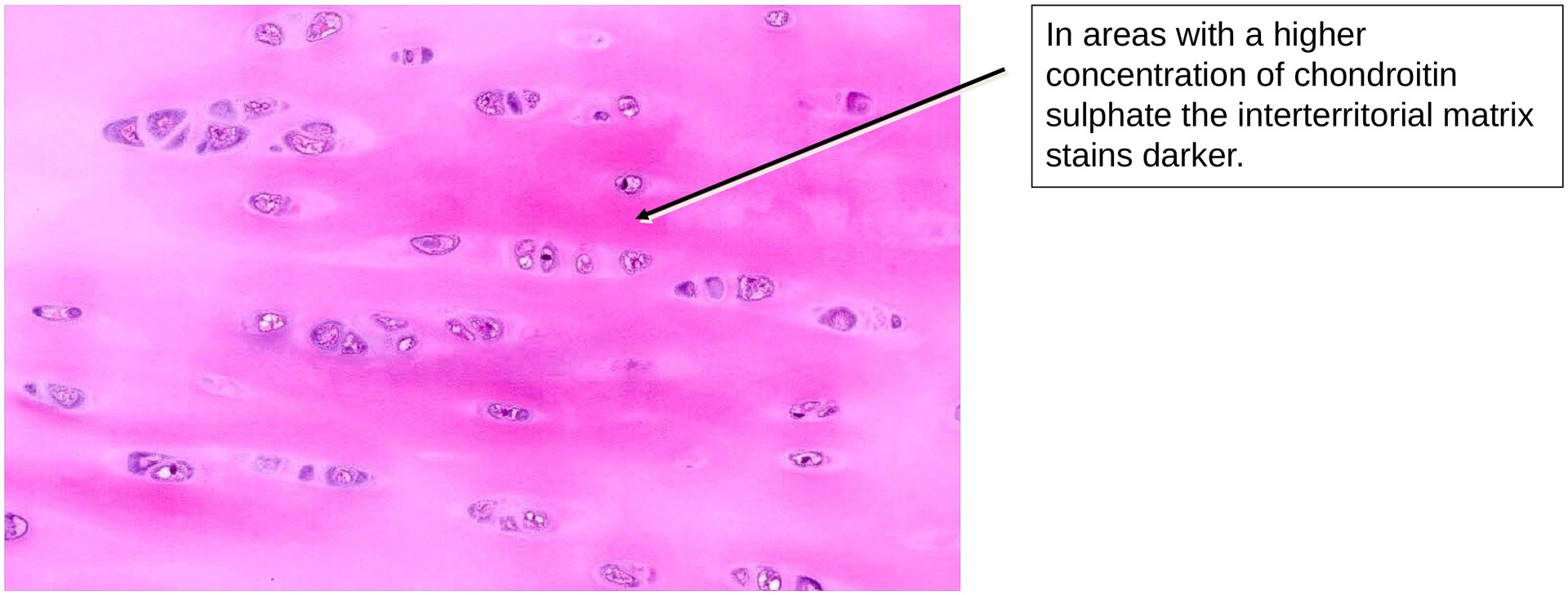
Chondrocytes are found in lacunae, each surrounded by a wall known as the cartilage capsule. When several chondrocytes derived from the same parent cell occupy a single lacuna, they form an isogenic group (or chondron). The darker-stained halo surrounding the chondron represents a region rich in chondroitin sulphate, forming the territorial matrix. The areas between these territories form the interterritorial matrix.
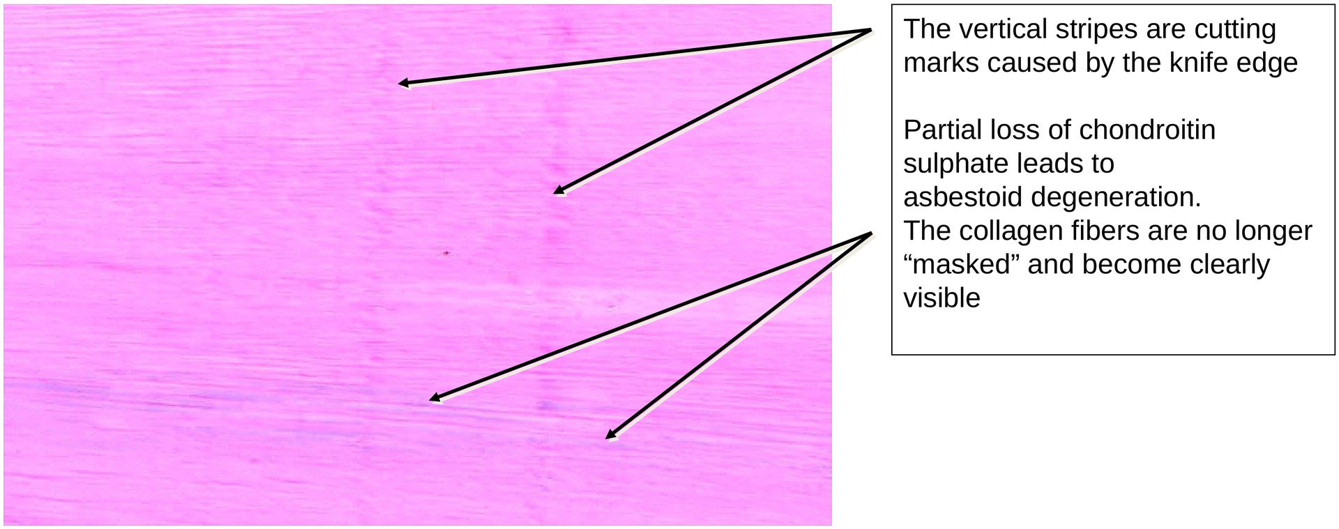
Occasional striated regions indicate age-related degenerative changes, beginning as early as adulthood. This process is termed asbestoid degeneration, resulting from the demasking of collagen fibrils due to the loss of chondroitin sulphate.
In general, collagen fibers are not visible in hyaline cartilage, as they share the same refractive index as chondroitin sulphate, rendering them optically masked.
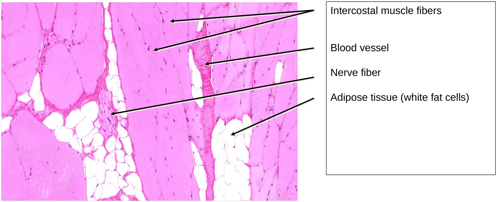
Because the ribs are moved by intercostal muscles during respiration, it is not uncommon to find skeletal muscle, adipose tissue, nerves, and blood vessels in this specimen.
Due to the different consistencies of cartilage, perichondrium, and adjacent intercostal muscle, sectioning artefacts may occur, where different tissue layers appear superimposed at the cartilage edge.
Tasks:
-
Examine the different sizes of the isogenic groups (chondrons) and consider how this variation can be explained.
-
Attempt to identify collagen fibers in this specimen. Where can they be seen clearly, and where are they practically invisible? What accounts for this difference?
-
Locate areas showing early asbestoid degeneration. Describe the underlying cause.
-
Identify, where possible, the cellular and fibrous layers of the perichondrium.
-
Locate skeletal muscle cut in cross-section. How can you still recognize it as skeletal muscle tissue?
License
University of Basel
Downloads