BONES (GENERAL HISTOLOGY)
5.2
Ossification, perichondral (Finger)
Specimen Details:
Specimen Details:
Organ: Finger (Long bone)
Origin: Human, Newborn
Staining: Goldner trichrome
Method and Specimen Description:
Paraffin sections of bone tissue are only possible after demineralization. However, this specimen was not demineralized and was instead embedded directly in plastic (methyl methacrylate) following fixation and dehydration. Sections were cut to a thickness of 5 μm using a tungsten knife.
This preparation method avoids the extraction or loss of mineral components that typically occurs during acid treatment for demineralization.
Objective of the Examination:
To study the ossification process in a long bone and to recognize the structural organization of the finger.
Special Features of the Specimen:
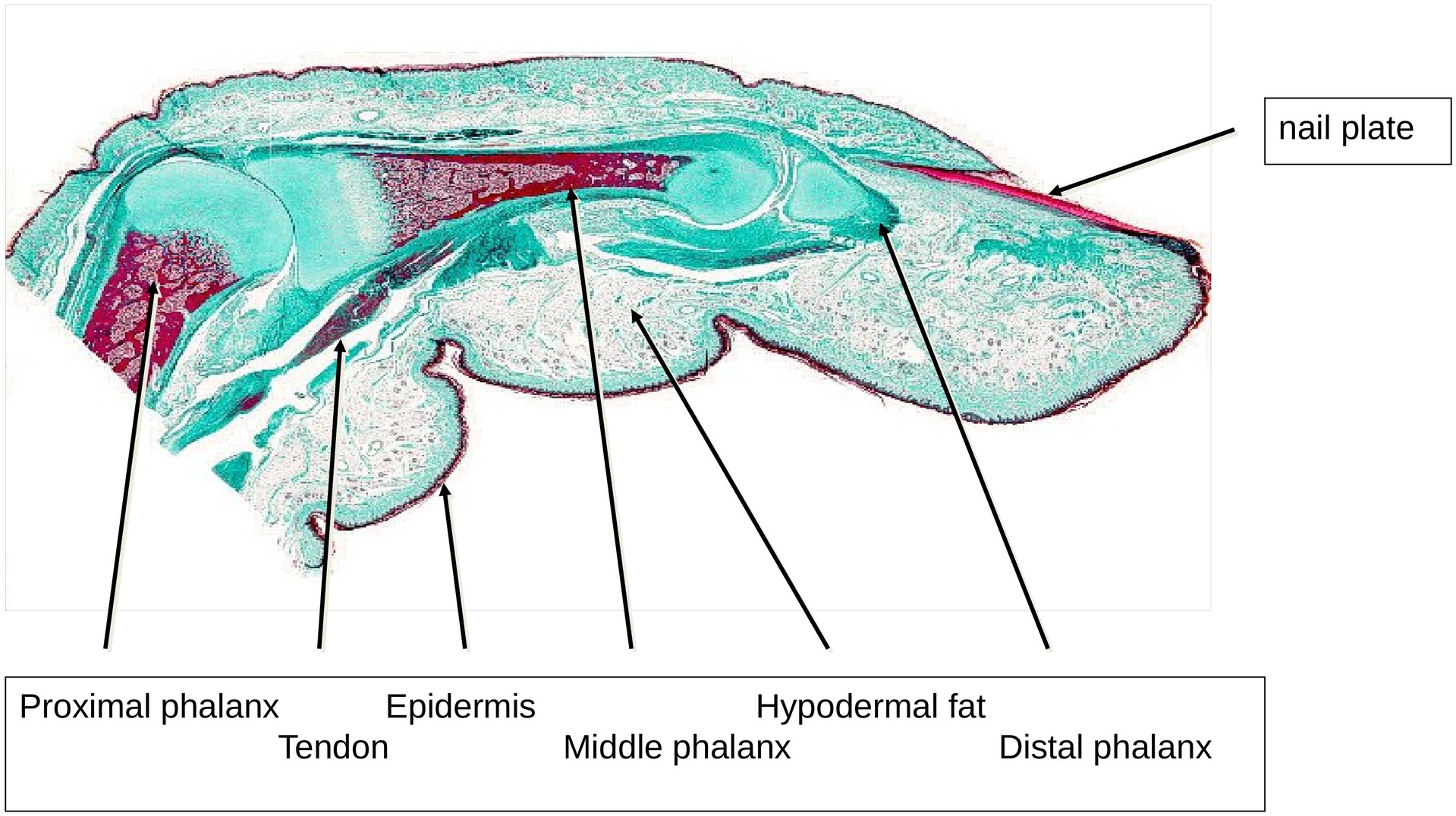
This longitudinal section through a finger allows identification of the dorsal and palmar aspects, the nail plate, fingertip, and the distal, middle, and proximal phalanges.
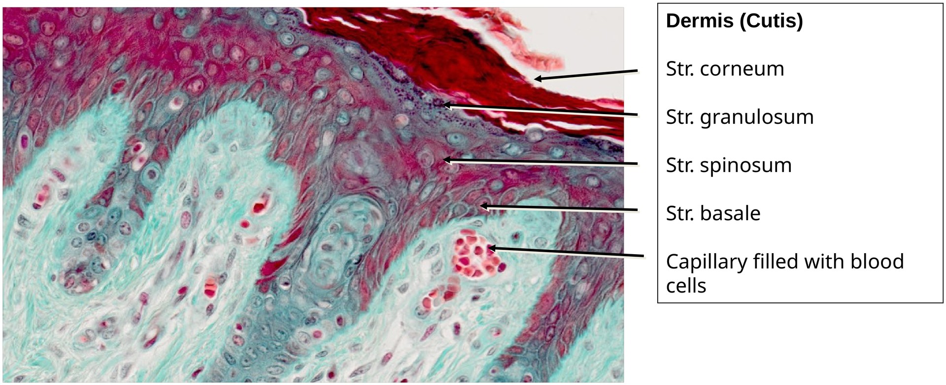
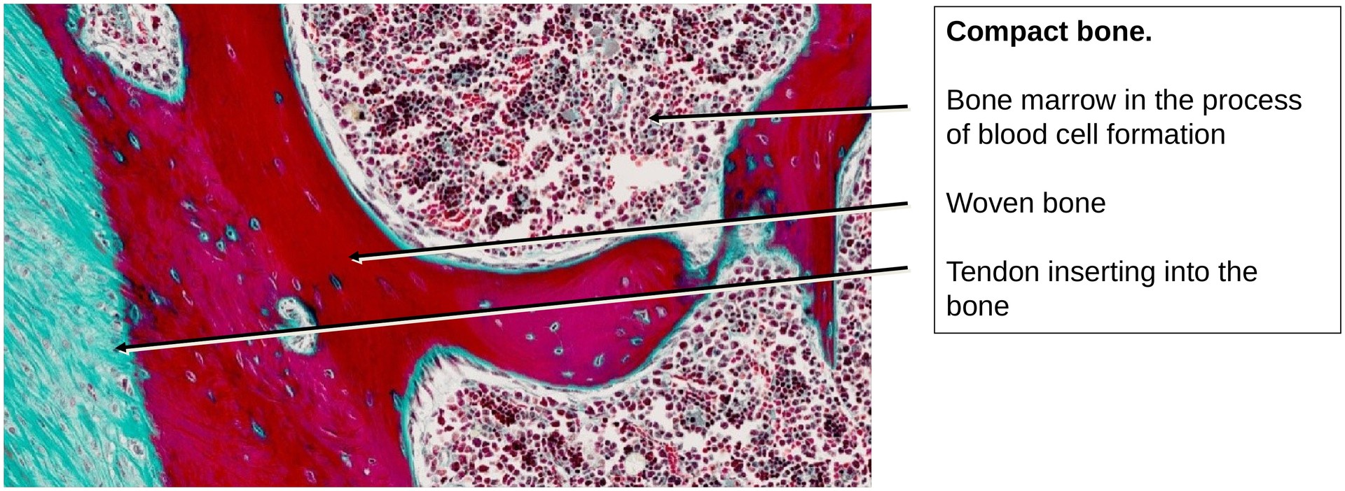
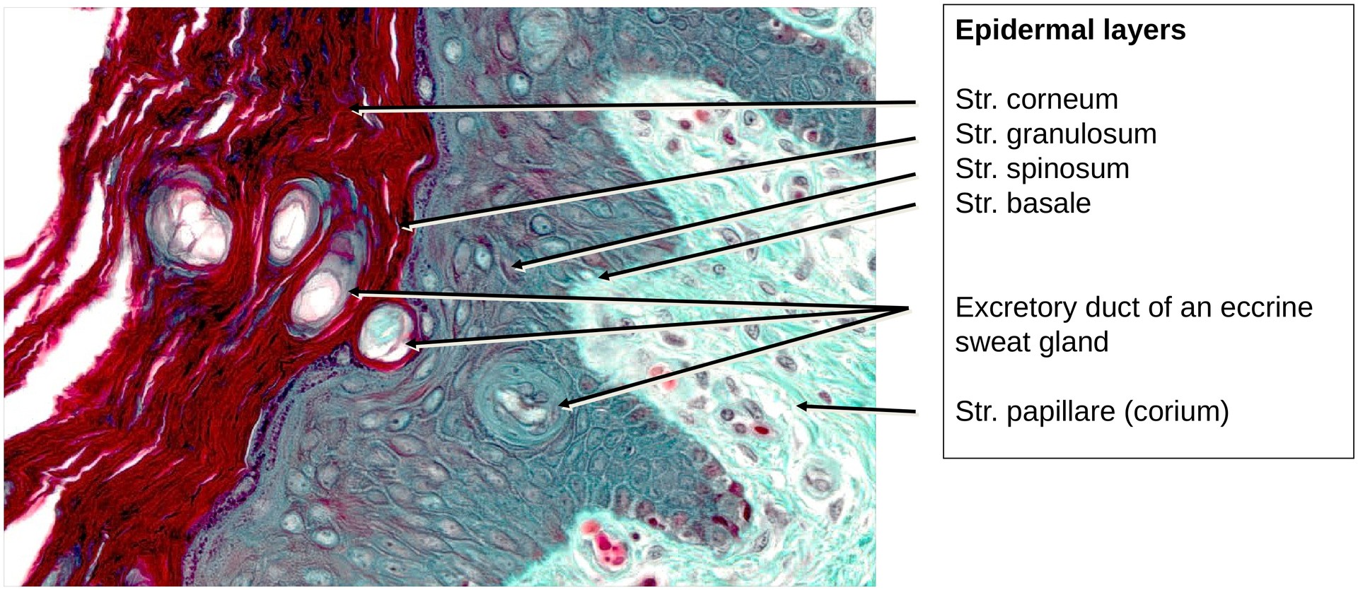
The finger is externally covered by a moderately keratinized epidermis. Within the digit lie the preformed cartilaginous phalanges, where the shaft (diaphysis) is already undergoing perichondral ossification. The bone produced at this stage is still woven bone.
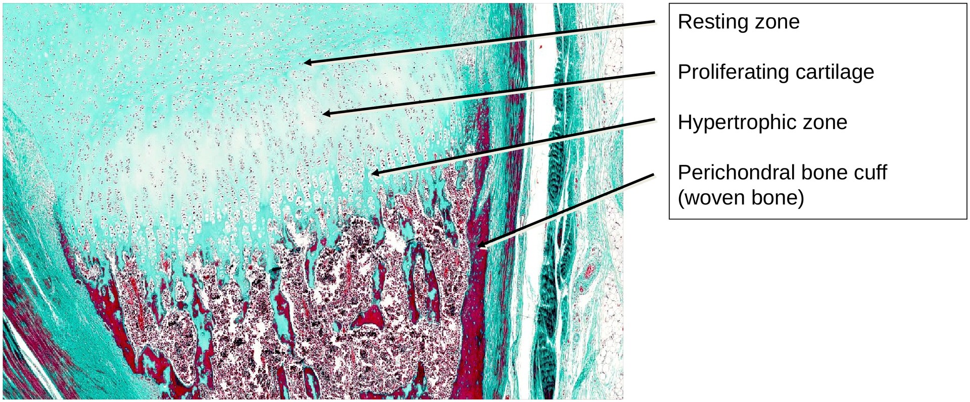
At the articular ends (epiphyses), endochondral ossification is occurring. Here, the classical zones of cartilage transformation are clearly recognizable:
-
Resting zone
-
Proliferation zone
-
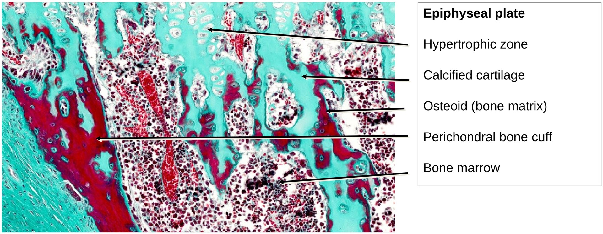
Hypertrophic zone
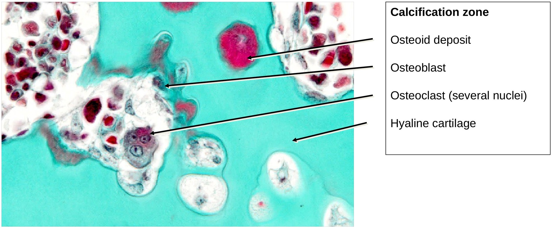
followed by the zone of ossification, which contains primary bone trabeculae. These trabeculae still contain remnants of mineralized cartilage, which will later be remodeled — first into woven bone, and subsequently into lamellar bone.
The proximal end of the middle phalanx provides the best view of the ossification process.
Within the diaphyseal region, a perichondral bone cuff of woven bone (stained red in this preparation) surrounds the cartilage. The epiphyses consist of hyaline cartilage, while red bone marrow is present within the marrow cavity.
Compact bone of the diaphysis:
The outer surface of the compact bone is covered by the periosteum, a fibrous membrane containing fibroblasts and collagen fibers.\
At the epiphyseal region, the periosteum transitions into the joint capsule.
The articular cartilage itself lacks a perichondrium.
The inner cellular layer of the periosteum contains osteoblasts and blood vessels, which are responsible for appositional bone growth forming the perichondral bone cuff.
The newly formed bone is homogeneously stained and contains osteocytes, although vascular channels and canaliculi are usually absent at this early stage.
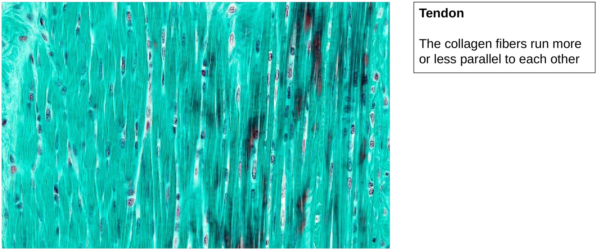
Since there are no muscles within the fingers, only tendons are present, inserting into the phalanges. Dense collagenous tendons are clearly visible on both the dorsal and palmar sides.
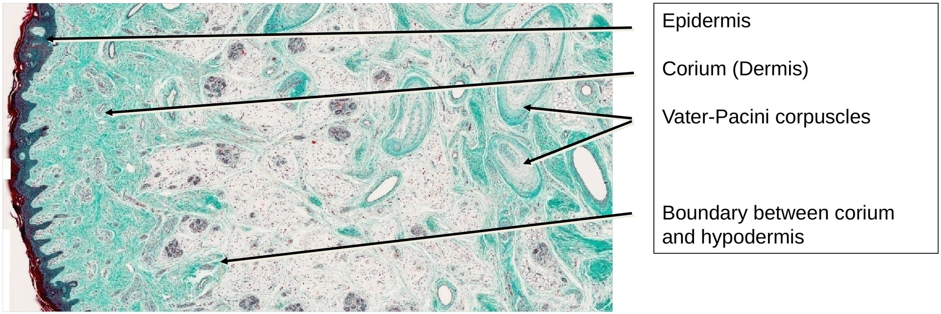
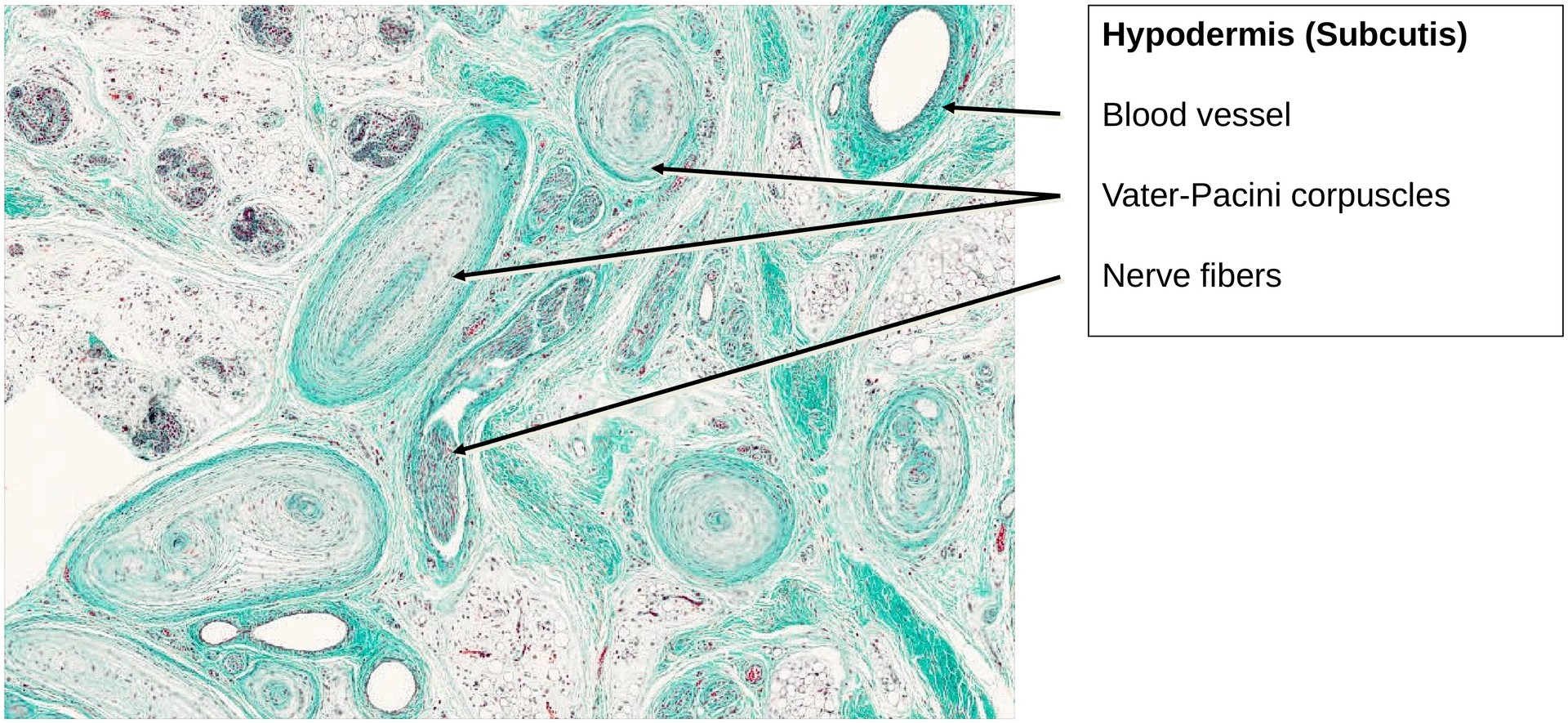
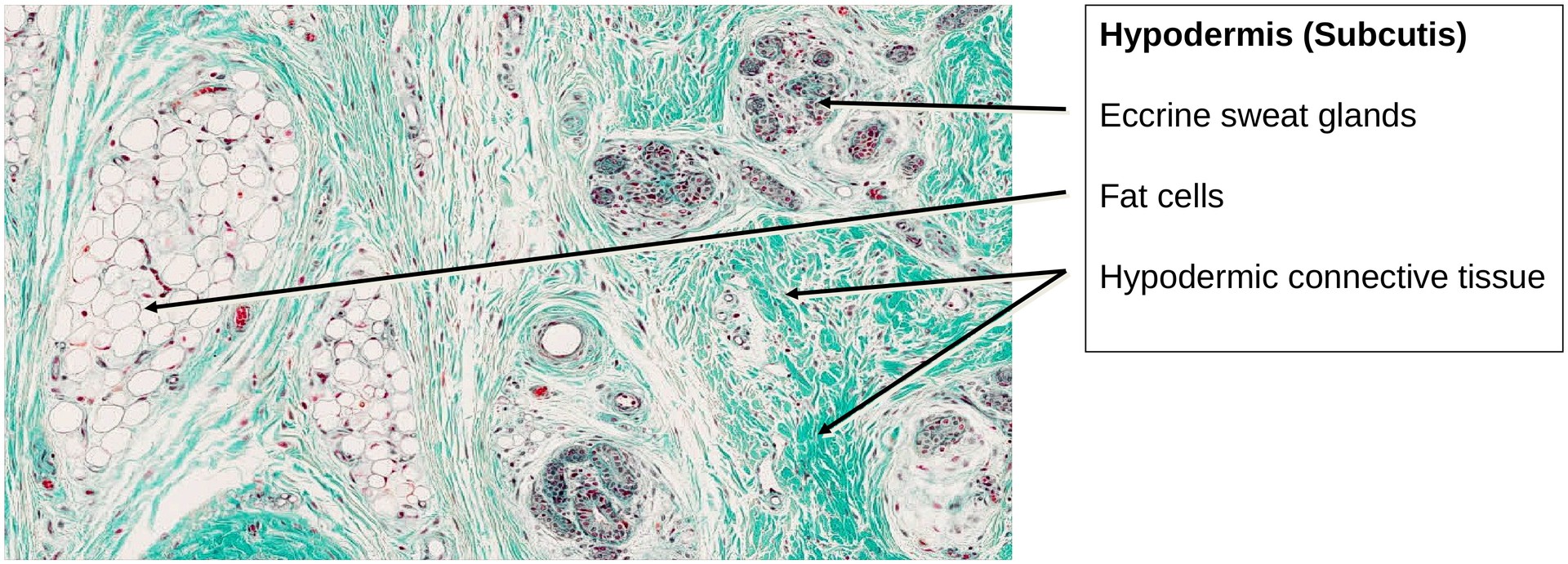
On the palmar aspect, adipose tissue accumulates within the subcutaneous layer (subcutis), supported by dense connective tissue (collagen). Here, Vater–Pacini corpuscles (responsible for vibration sensation) can be identified.
Developing eccrine sweat glands are visible in both the dorsal and palmar skin.
On the dorsal side of the distal phalanx, the nail plate (deep red staining) can be seen resting on the nail bed, which represents a specialized part of the epidermis.
Tasks:
• Orientation: Identify the individual phalanges and the general layout of the specimen.
• Identify the layers of the skin and compare the thickness of each.
• Locate the tendons inserting into the developing bones and examine their structure and attachment.
• Study the ossification zone at the proximal epiphysis of the middle phalanx.
• Identify the individual zones of the cartilage column.
• Locate osteoclasts and osteoblasts and note their distribution.
• Examine the cartilage trabeculae and interpret the differences in staining intensity.
• Find Vater–Pacini corpuscles — in which layer are they located?
• Describe the structure of the perichondral bone cuff. What type of bone is it?
• Search for nerves in both the corium and the subcutis.
License
University of Basel
Downloads