MUSCULATURE (GENERAL HISTOLOGY)
6.6
Skeletal muscle, longitudinal section (hyoglossus muscle)
Preparation:
Preparation Details:
Organ: Hyoglossus muscle
Origin: Cow
Staining: Azan
Method and Specimen Description:
This is a routine histological section stained with Azan, which stains muscle fibers and blood in varying shades of red (depending on section thickness and stain differentiation, occasionally slightly bluish) and connective tissue blue.
Objective of the Examination:
To recognize skeletal muscle fibers and understand the light-microscopic structure of myofibrils.
Additional aim: identification of connective tissue, vessels, and nerves within the perimysium.
Special Features of the Preparation:
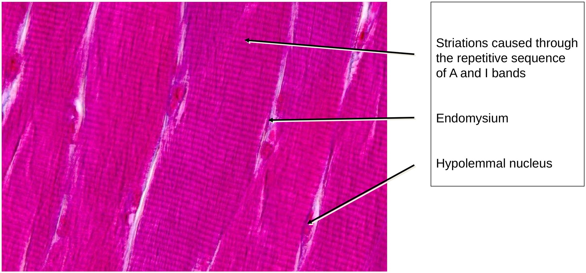
The preparation consists of bundles of individual muscle fibers, each surrounded by delicate connective tissue known as the endomysium. Some fibers are cut longitudinally, others transversely.
Structure of the Skeletal Muscle Fiber:
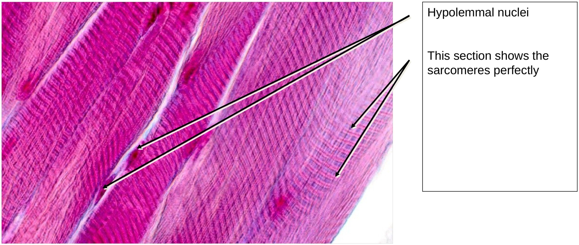
At high magnification, the sarcolemma (comprising the cell membrane, basal lamina, and reticular fiber sheath) is visible as a distinct boundary. Each fiber contains numerous elongated, peripheral nuclei located directly beneath the sarcolemma. The muscle fiber is a multinucleated cytoplasmic tube bounded by the sarcolemma.
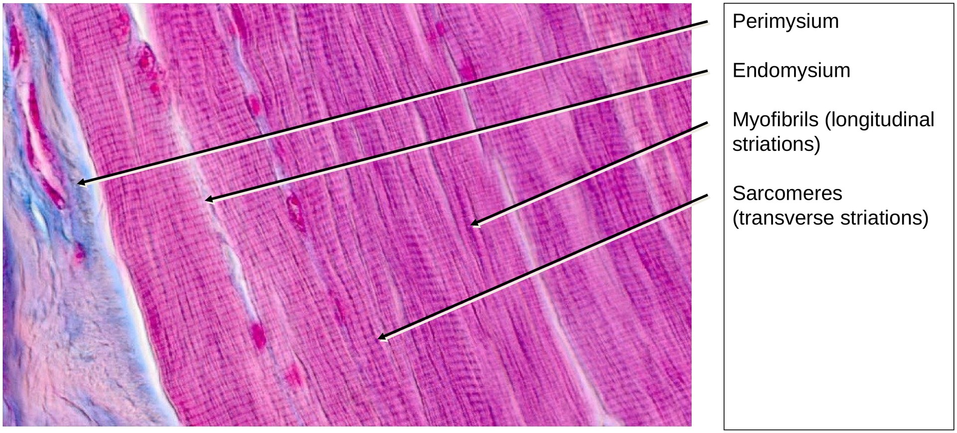
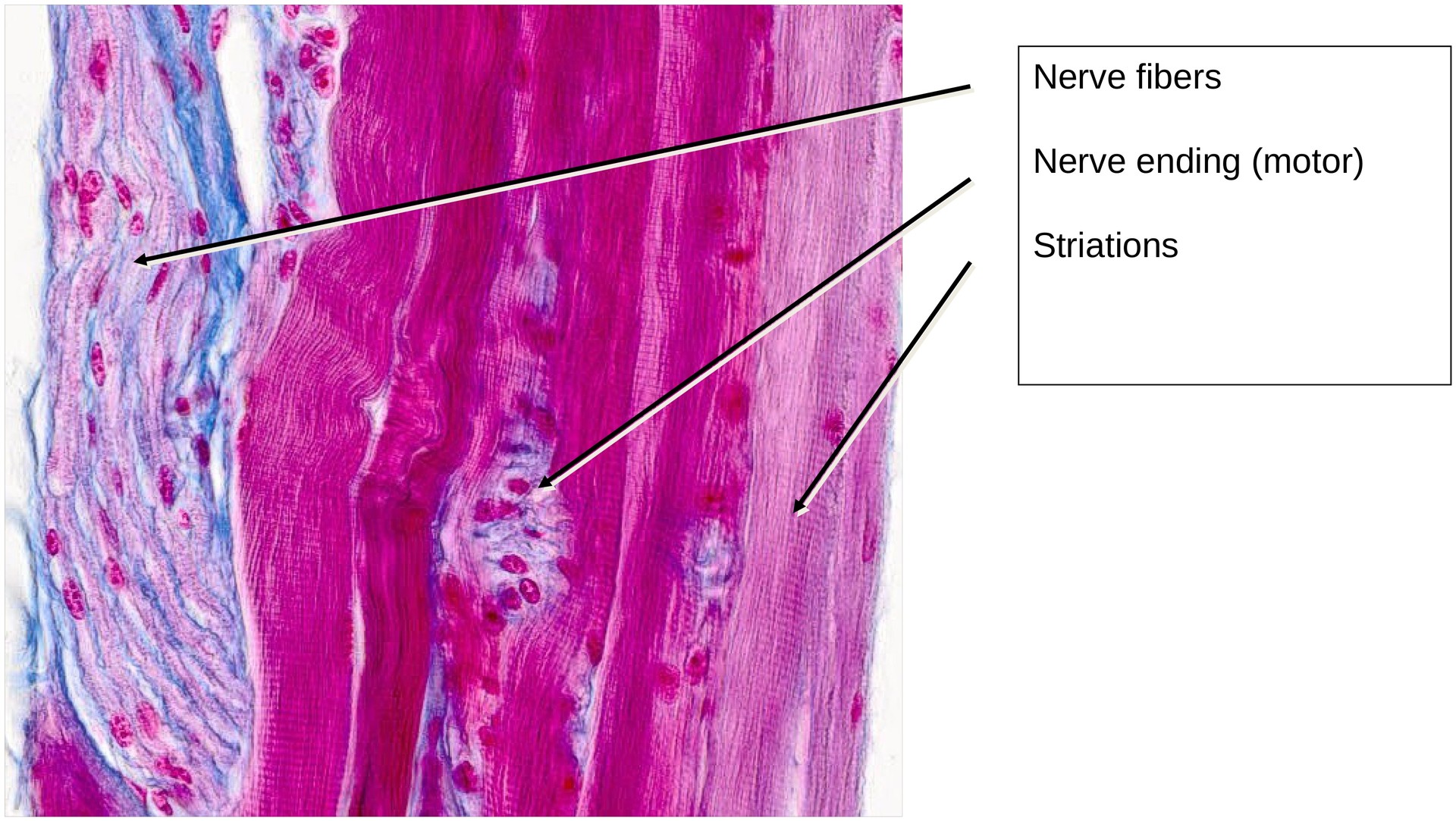
In longitudinally sectioned fibers, the cytoplasm shows longitudinal striations due to the parallel arrangement of myofibrils. These striations arise from the repetitive organization of sarcomeres within the myofibrils. The alternating A-bands (dark, anisotropic) and I-bands (light, isotropic) create the characteristic banding pattern.
Note: This appearance is caused by light refraction, not birefringence—the latter can only be observed using polarized light.
Myofibrils (High Magnification):
In precisely longitudinal sections, the A and I bands are easily recognizable. Depending on the state of muscle contraction, the H-zone and Z-line may also be visible.
Blood Supply and Innervation:
Within the perimysium—the connective tissue that bundles several muscle fibers into a fascicle—blood vessels and nerves are visible. These provide the muscle with its extensive vascular and neural supply.
Moiré Patterns:
In some areas, where muscle fibres are sectioned obliquely or tangentially, Moiré patterns may appear. These resemble striations but do not originate from sarcomeres. Rather, they result from optical interference—the superimposition of repetitive patterns, similar to the visual effect of a striped fabric filmed on a television screen.
Tasks:
• Identify the tissue as skeletal muscle based on its cross-striations and multinucleated syncytial fibers.
• Locate myofibrils (longitudinal striations within the muscle fibers).
• Find an area where the muscle fibres are not present through the entire section thickness — here the cross-striation is especially distinct.
• Identify the A and I bands and consider how striations arise from the repeating sequence of sarcomeric elements (Z–I–A–H–M–H–A–I–Z).
• Locate nerve fibers and blood vessels within the perimysium.
License
University of Basel
Downloads