NERVE TISSUE (GENERAL HISTOLOGY)
7.4
Spinal nerve (sciatic nerve)
Specimen Details:
Specimen Details:
Organ: Sciatic nerve
Origin: Dog
Staining: Fastblue/Kern Red
Method and Specimen Description:
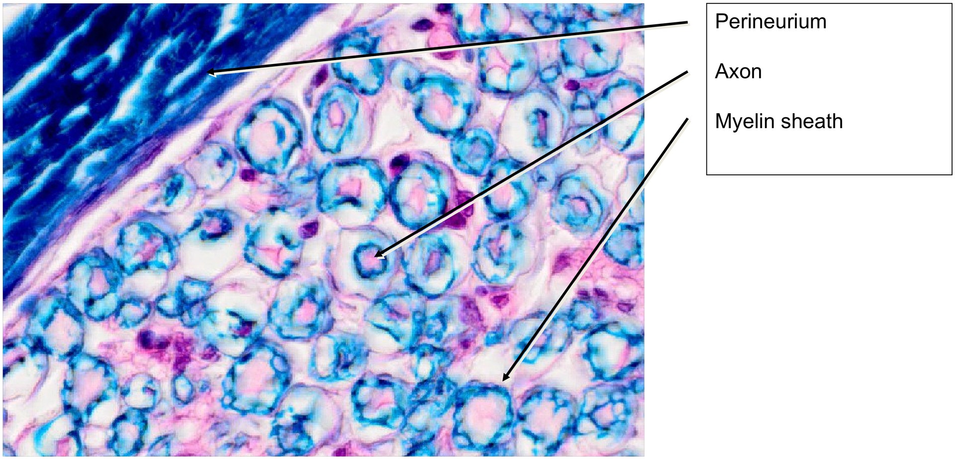
A normal histological specimen stained with Fast Blue and Kern Red. This staining combination allows for clear differentiation of the principal components of a peripheral nerve: axons appear pale pink, myelin sheaths light to dark blue, nuclei of Schwann cells and connective tissue red, and connective tissue fibers blue.
Objective of the Examination:
To gain knowledge of a peripheral nerve, its fasciculation, and its connective tissue coverings.
Special Features of the Specimen:
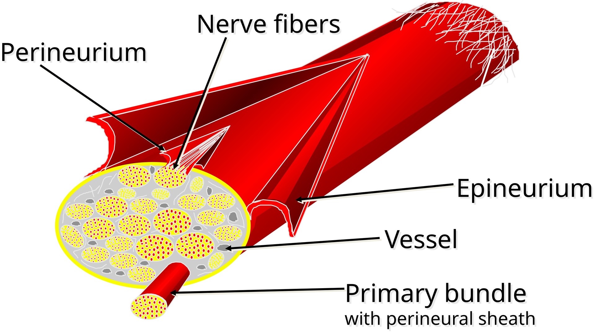
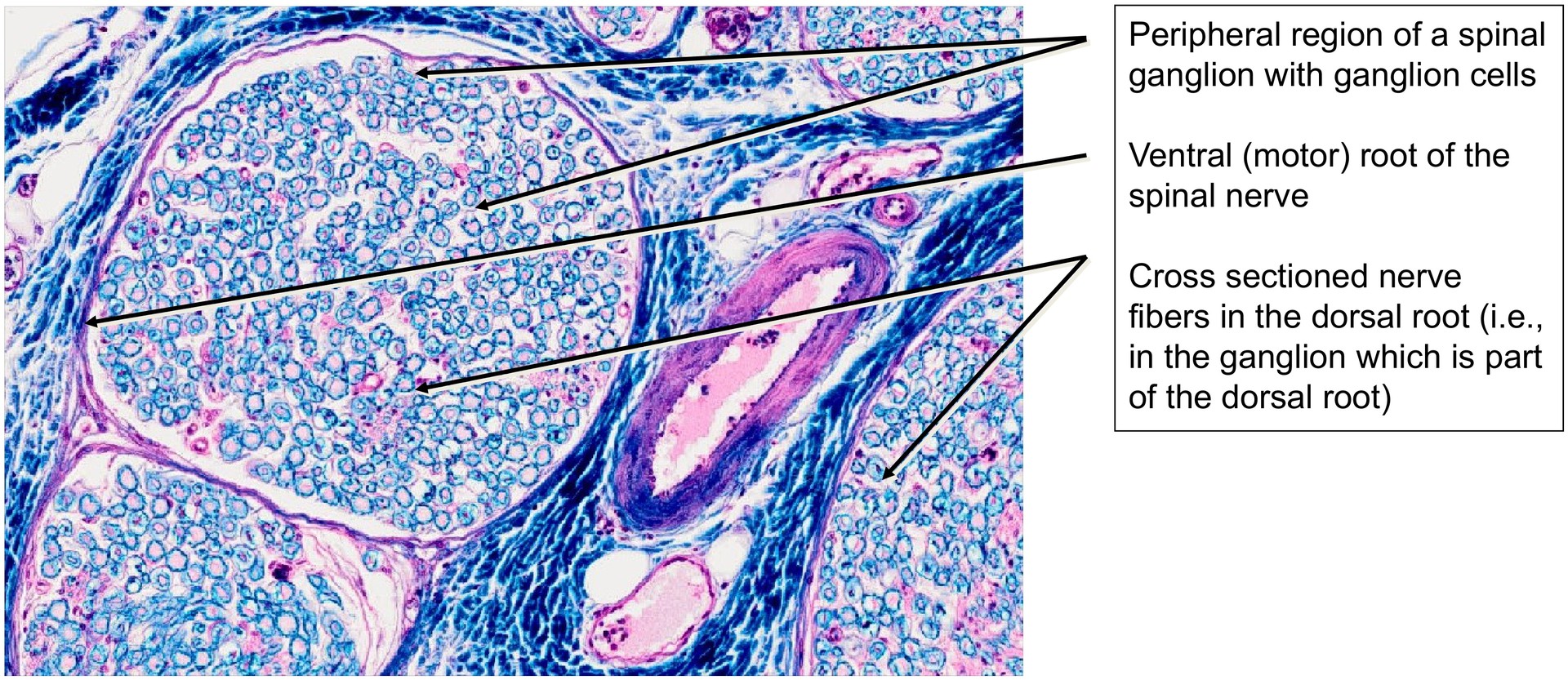
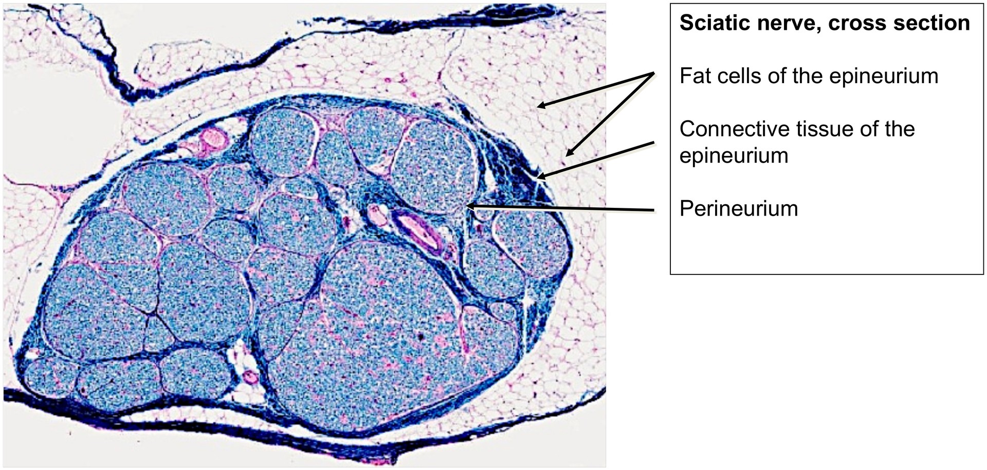
Even at low magnification, several nerve fiber bundles can be identified within the specimen. These bundles are embedded within the surrounding structures by the epineurium, a predominantly fatty, vascular connective tissue sheath.
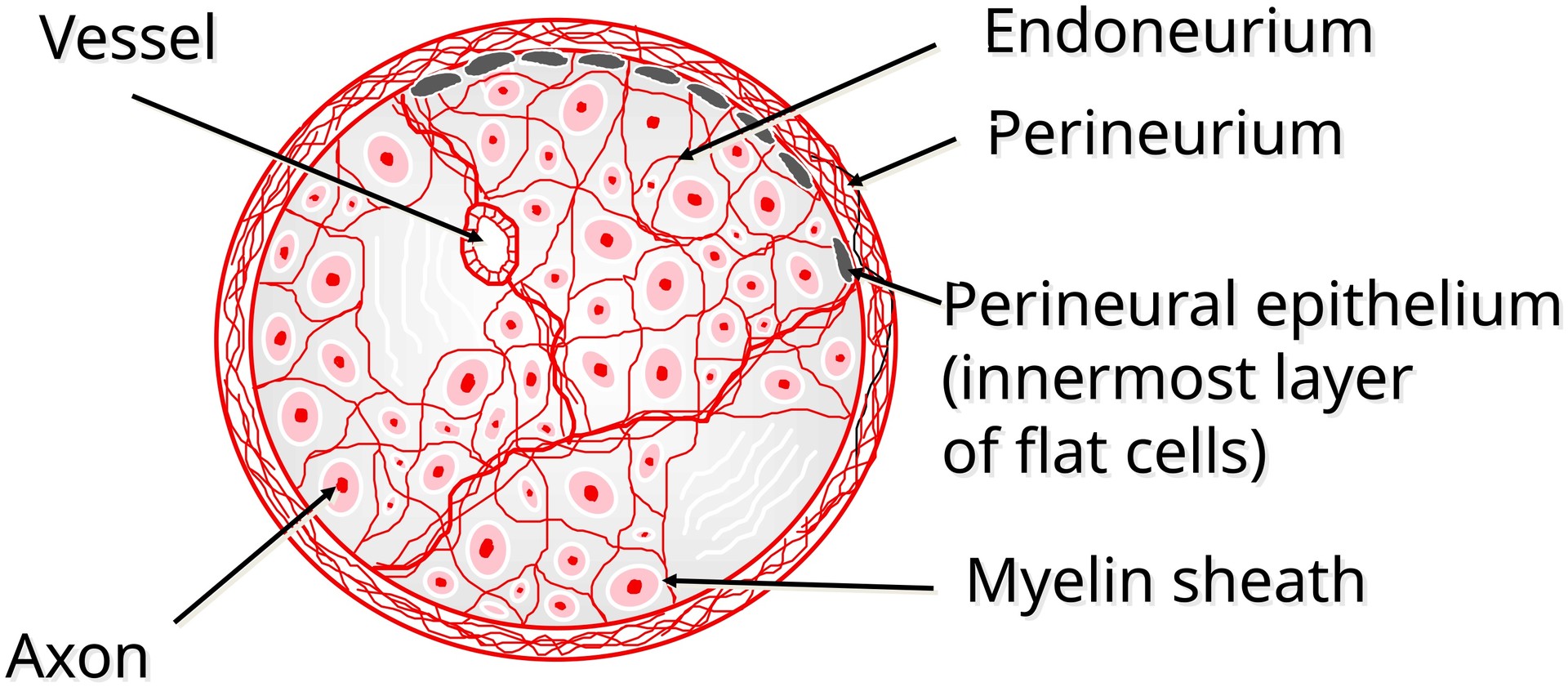
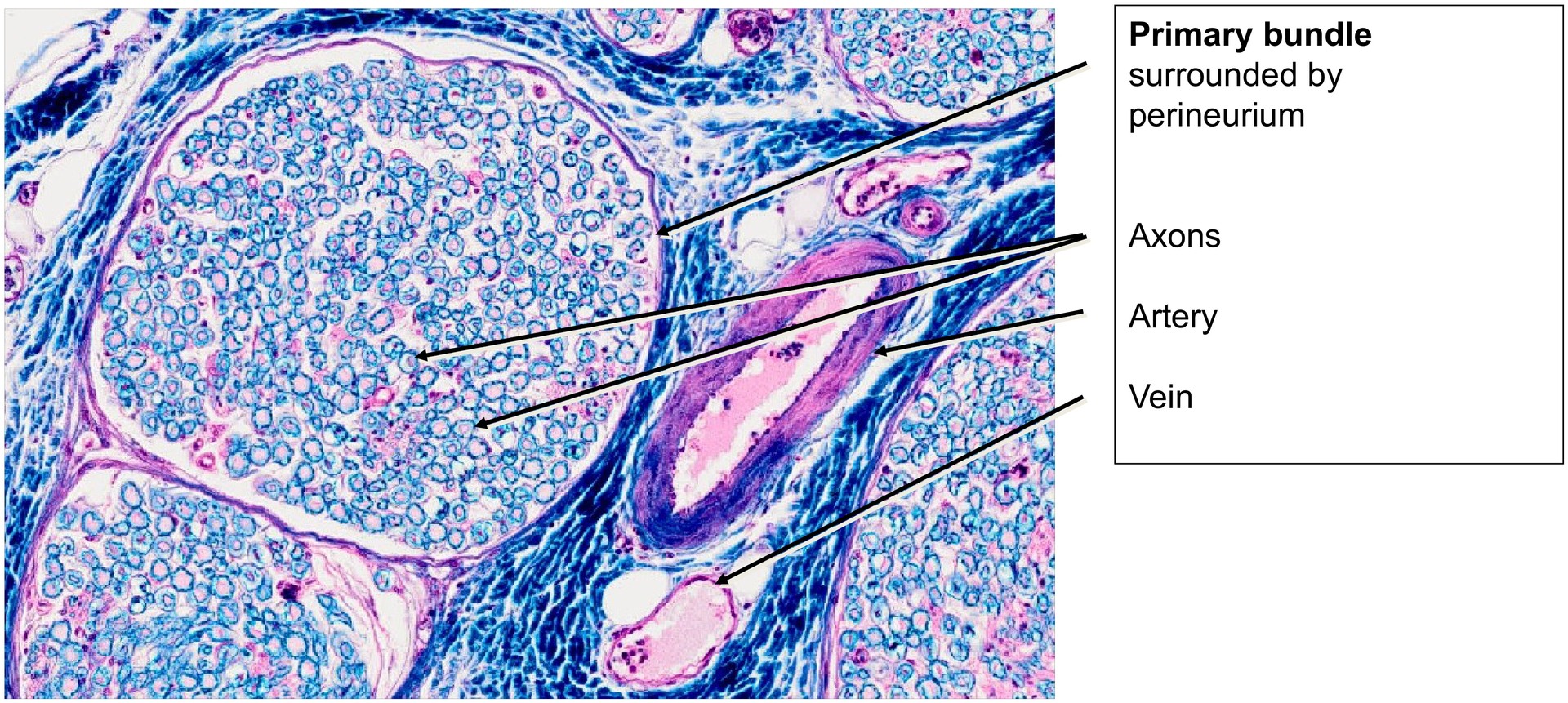
The entire nerve can be considered a secondary bundle, composed of several primary bundles. Each primary bundle is surrounded by perineurium, a connective tissue sheath whose innermost layer is formed by the perineural epithelium. The squamous perineural epithelial layer is not always completely preserved in histological sections.
Both sensory and motor fibers—afferent and efferent—run side by side within the primary bundles. Consequently, both heavily and lightly myelinated nerve fibers can be observed in large numbers in cross-section.
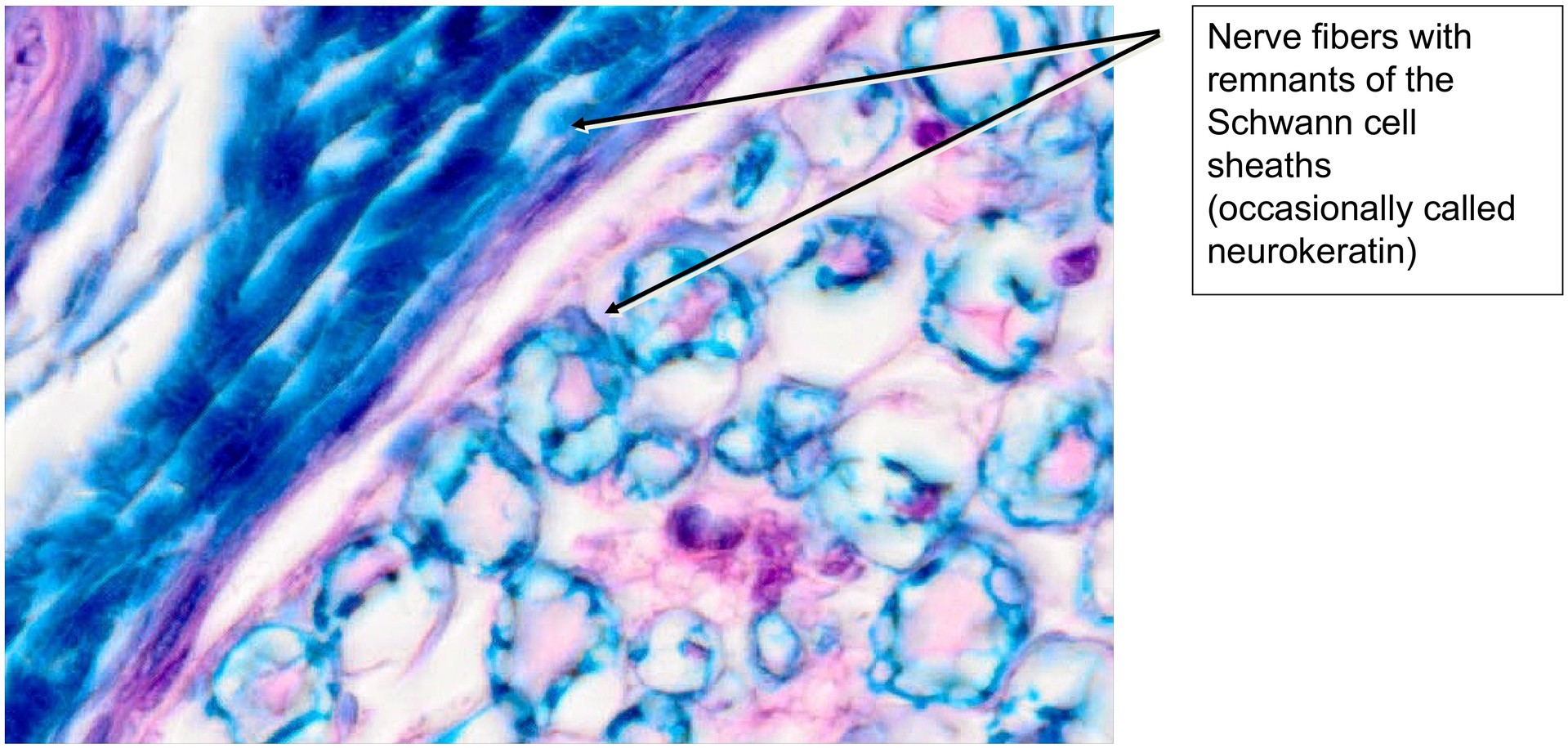
The residual material of the myelin sheaths remaining after fixation, dehydration, and embedding is sometimes referred to as neurokeratin. However, this term is a misnomer, as it has no relation to true keratin and contains no keratin components.
The nuclei of Schwann cells can be observed in many cross-sections of individual nerve fibers. Each nerve fiber is enveloped by fine reticular connective tissue fibers forming the endoneurium. Numerous blood vessels—including arteries, veins, and capillaries—are present within the epineurium, perineurium, and between the primary bundles.
Tasks:
Answer the following questions and identify the relevant structures:
-
Identify the epineurium. What does it enclose, and what is its composition?
-
What distinguishes a primary bundle from a secondary bundle?
-
Which structure surrounds a primary bundle?
-
Identify the myelin sheaths.
-
Describe the blood supply to the nerve and identify arteries and veins.
-
Identify the axons (neurites) and the fixation-related remnants of the myelin sheaths (neurokeratin).
License
University of Basel
Downloads