MALE REPRODUCTIVE ORGANS (ANATOMICAL MICROSCOPY)
11.1
Seminal vesicle 1
Specimen:
Specimen Details:
Organ: Seminal Vesicle
Origin: Human
Staining: Hematoxylin - Eosin (H&E)
Method and Specimen Description:
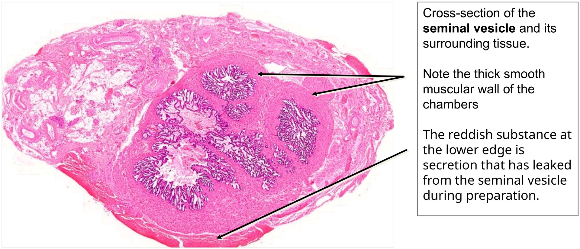
Normal histological section stained with the overview stain (H&E), showing a cross-section through the seminal vesicle.
Objective of the Examination:
To study the complex mucosal architecture of the seminal vesicle, including its numerous pouches, chambers, and niches, as well as the smooth muscle-rich wall of the organ.
Special Features of the Specimen:
This cross-section demonstrates the key features of the seminal vesicle.
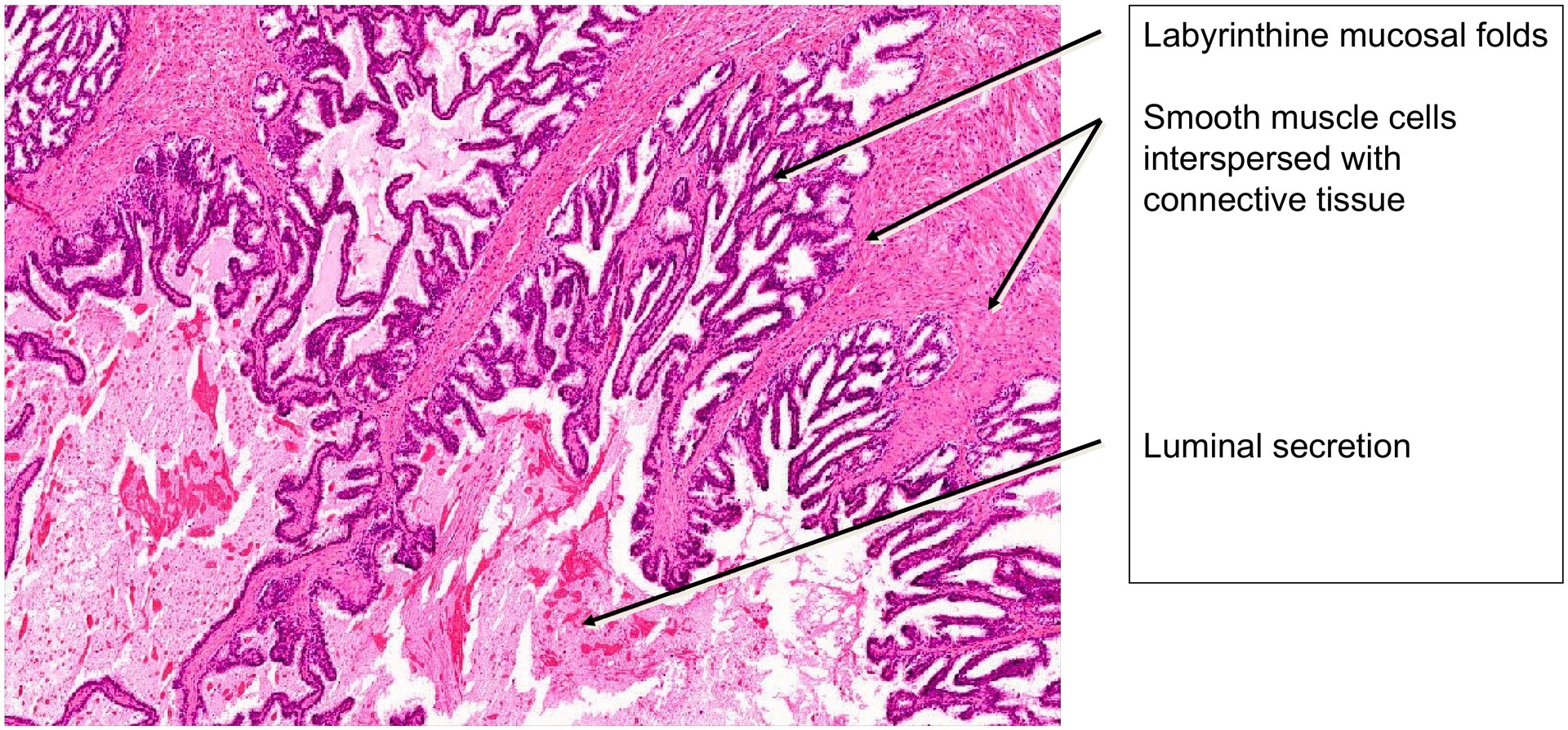
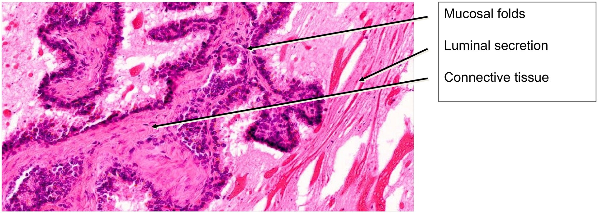
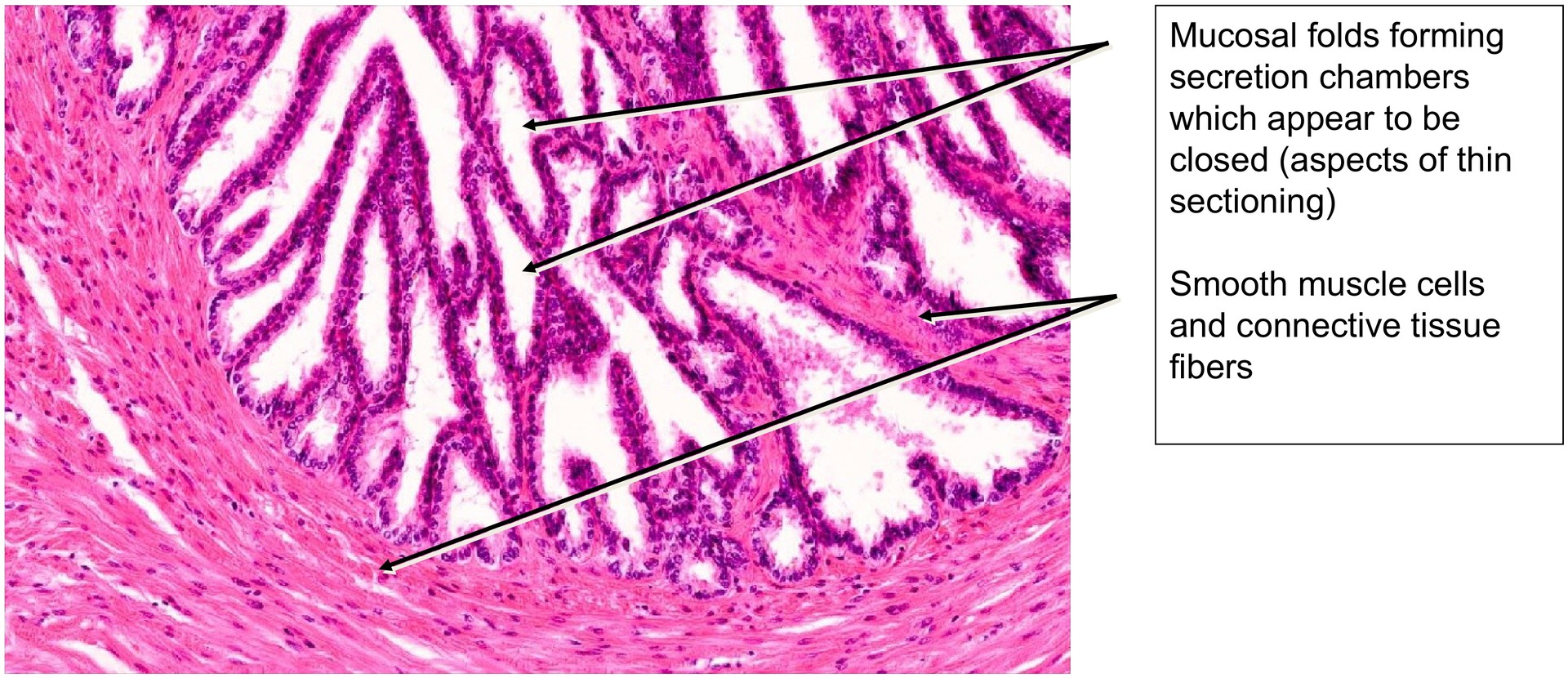
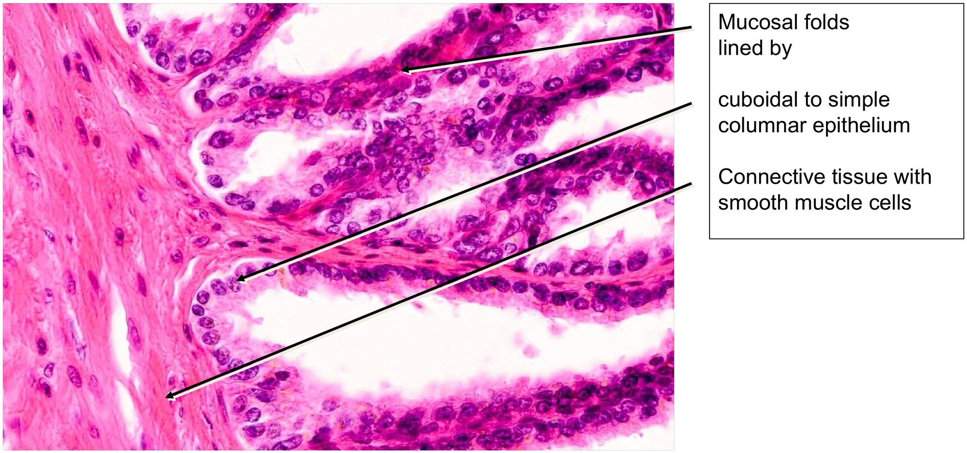
The lumen is lined by a mucous membrane that displays numerous folds, chambers, and recesses, producing the organ’s characteristic labyrinthine appearance. Surrounding the mucosa is connective tissue, which merges externally with a thick, smooth muscular wall. The surrounding connective tissue is highly vascularized, with numerous small and medium-sized vessels visible even under low magnification.
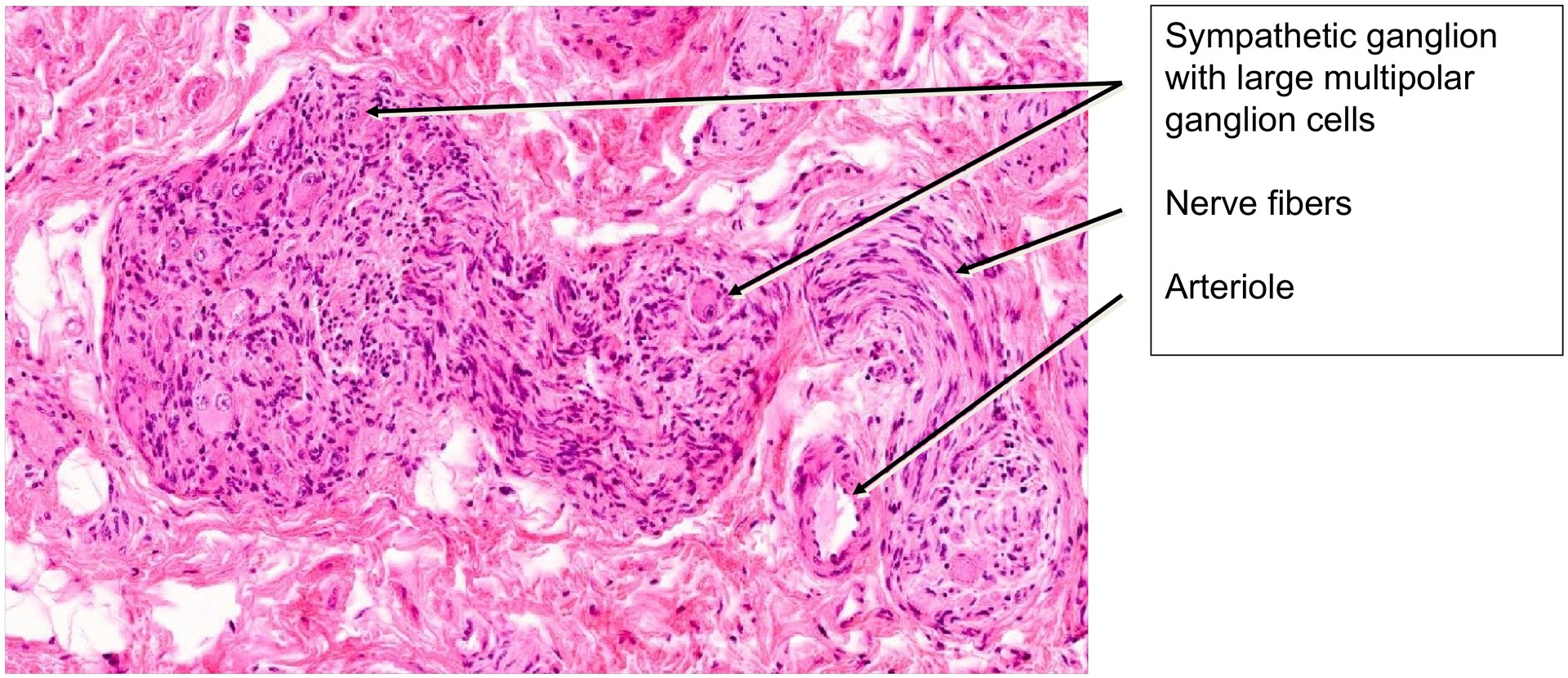
With higher magnification, several nerve fibers and sympathetic ganglia can be identified within the surrounding connective tissue, the latter containing characteristic large ganglion cells with prominent nuclei and nucleoli.
The epithelium varies in height and appearance. It is generally simple columnar to pseudostratified, although in this specimen, regions with cuboidal epithelium are also evident. The epithelial cells often contain secretory vacuoles, and their height can vary depending on the degree of organ activity.
The luminal secretion appears heterogeneous, which reflects artefactual separation during fixation. It contains both viscous, eosinophilic regions (staining bright red) and more fluid areas.
The wall of the seminal vesicle consists of smooth muscle bundles arranged in inner circular and outer longitudinal layers, interspersed with fibroblasts and collagen fibers. Some muscle cell strands extend into the larger mucosal folds, supporting their shape and mobility.
On the right side of the specimen, leaked secretion can be observed which was squeezed out during preparation.
Tasks:
• Using low-power magnification, familiarize yourself with the overall organization of the seminal vesicle and identify the characteristic mucosal folds.
• Examine the epithelium in different regions and assess the variation in cell height and layering (single- or two-layered).
• Trace connective tissue and smooth muscle bundles within the wall and follow their extension into the mucosal folds.
• Observe the heterogeneous appearance of the luminal secretion and consider its origin and functional relevance.
• Locate and follow mucosal pouches, tracing the continuity of the epithelium along their circumference.
• Search the surrounding connective tissue for nerve fibers and sympathetic ganglion cells.
• Identify arteries and veins among the numerous vascular profiles present.
License
University of Basel
Downloads