URINARY ORGANS (ANATOMICAL MICROSCOPY)
12.6
Ureter, dog
Specimen:
SPECIMEN DETAILS:
Organ: Ureter
Origin: Dog
Staining: Haematoxylin Eosin (H&E)
METHOD AND SPECIMEN DESCRIPTION:
Normal histological section of the ureter, stained with an overview stain (H&E).
OBJECTIVE OF THE EXAMINATION:
To gain knowledge of the histological structure of the ureter, with attention to its specific wall architecture and epithelial specialisations.
SPECIAL FEATURES OF THE SPECIMEN:
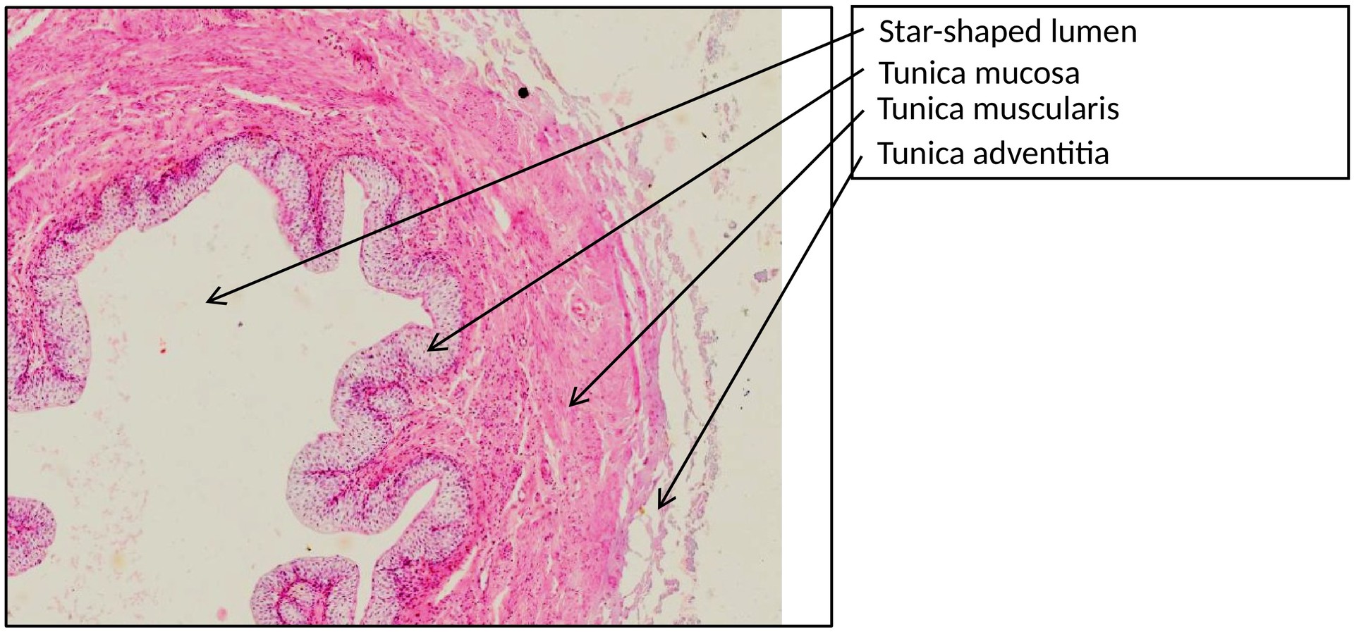
In general, the ureter is a muscular tube that, in the empty and contracted state, exhibits a star-shaped lumen due to the longitudinal folds of its mucosa (lined by urothelium).
The wall of the ureter is composed of three main layers, analogous to the other urinary outflow tracts:
- Tunica mucosa (mucosa)
- Tunica muscularis (muscular layer)
- Tunica adventitia (outer connective tissue covering)
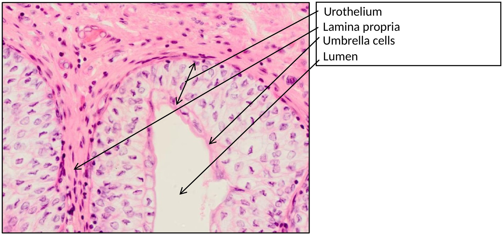
The mucosa consists of a stratified transitional epithelium (urothelium) and an underlying lamina propria.
- The urothelium is characterised by its superficial covering cells (umbrella cells), which are often multinucleated.
- These cells can enlarge their apical surface by incorporating subapical discoid vesicles into the plasma membrane, allowing the ureter to stretch.
- The membrane plaques, composed of uroplakins, increase both mechanical stability and chemical resistance to urine.
The lamina propria, part of the mucosa, consists of loose connective tissue rich in elastic fibers and blood vessels.
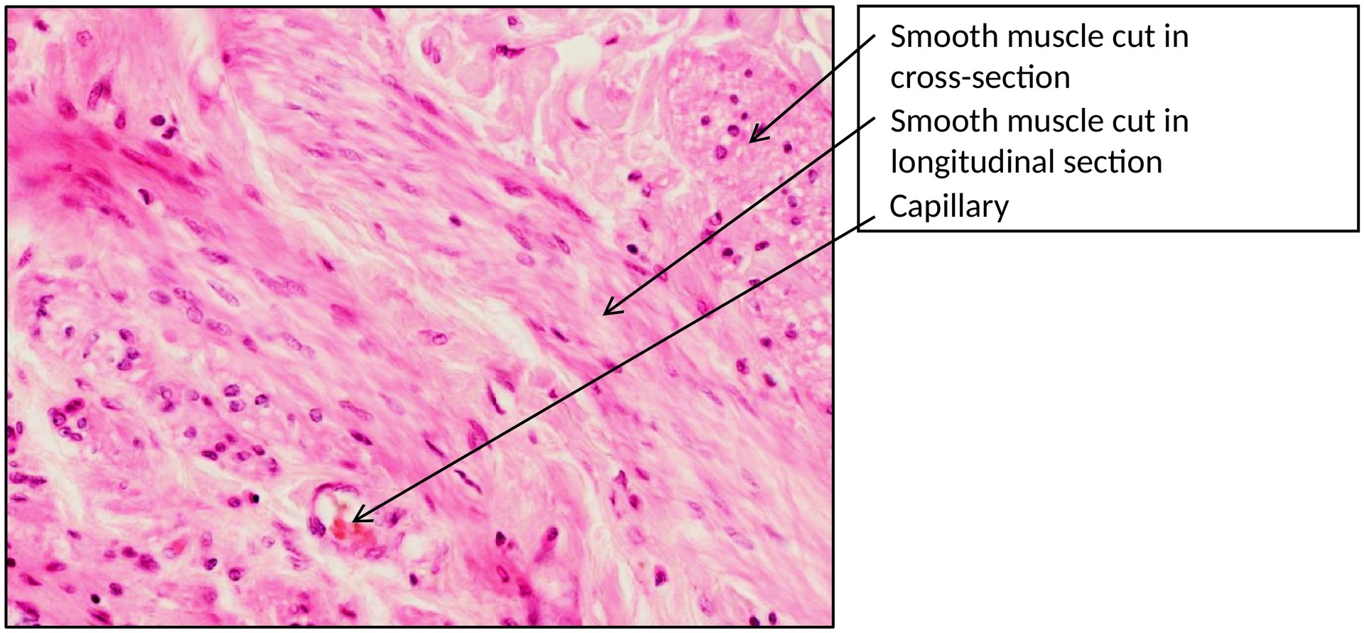
Beneath the mucosa lies the tunica muscularis, composed of smooth muscle strands arranged spirally and interspersed with connective tissue septa.
- In the proximal ureter, the muscularis consists of two layers (inner longitudinal and outer circular).
- In the distal ureter, a third outer longitudinal layer is added. This arrangement supports the peristaltic transport of urine towards the urinary bladder.
Externally, the tunica adventitia consists of connective tissue, containing blood vessels, nerves, and adipose tissue, which anchor the ureter to surrounding structures.
TASKS:
- Identify the tunica mucosa, tunica muscularis, and tunica adventitia.
- Examine the urothelium and recognise the superficial covering (umbrella) cells.
- Observe the longitudinal folds of the mucosa responsible for the star-shaped lumen in the contracted state.
- Differentiate between the muscle layers and assess the arrangement of smooth muscle fibres.
License
University of Basel