FEMALE REPRODUCTIVE ORGANS (ANATOMICAL MICROSCOPY)
10.8
Non-lactating breast tissue
Specimen:
SPECIMEN DETAILS:
Organ: Mammary gland (non-lactating)
Origin: Human
Staining: Haematoxylin Eosin (H&E)
METHOD AND SPECIMEN DESCRIPTION:
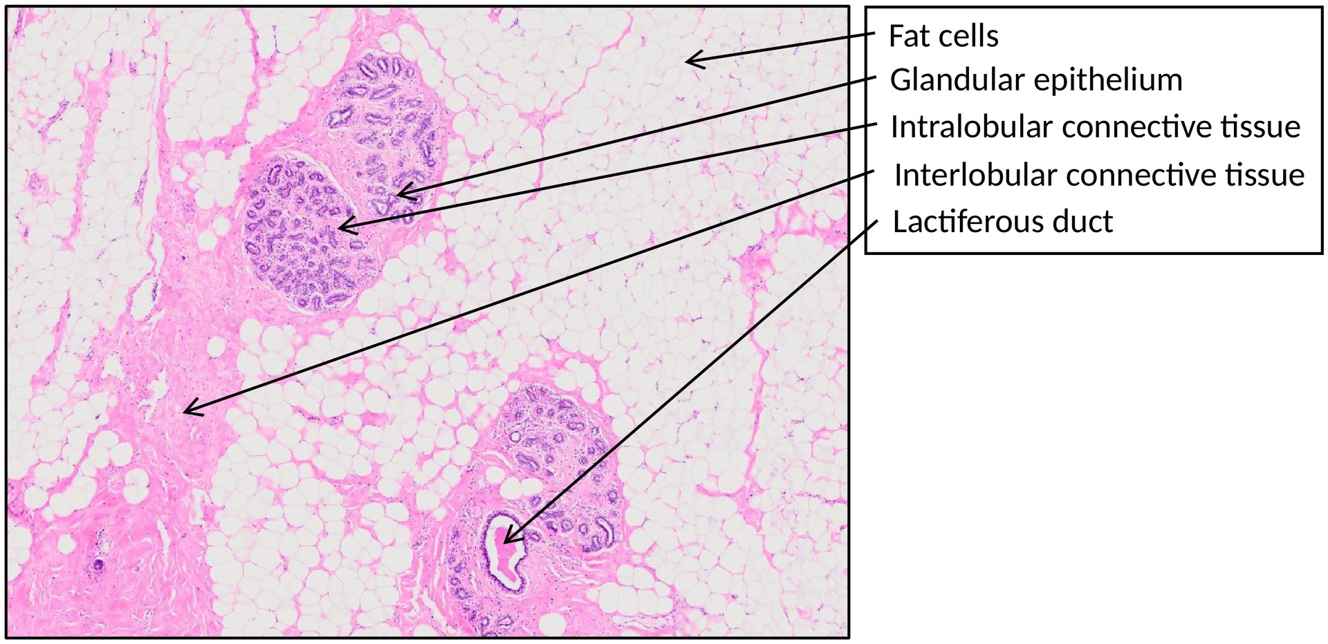
Routine histological section of a non-lactating human mammary gland. This specimen should be compared with the lactating mammary gland to appreciate structural and functional differences.
OBJECTIVE OF THE EXAMINATION:
To understand the histological structure of the non-lactating breast, which is composed mainly of fibrous connective tissue stroma with adipocytes, and only a small proportion of glandular epithelium.
Special Features of the Specimen:
The mammary gland is composed of 10–20 individual lobes, each drained by a main excretory duct that opens at the nipple.
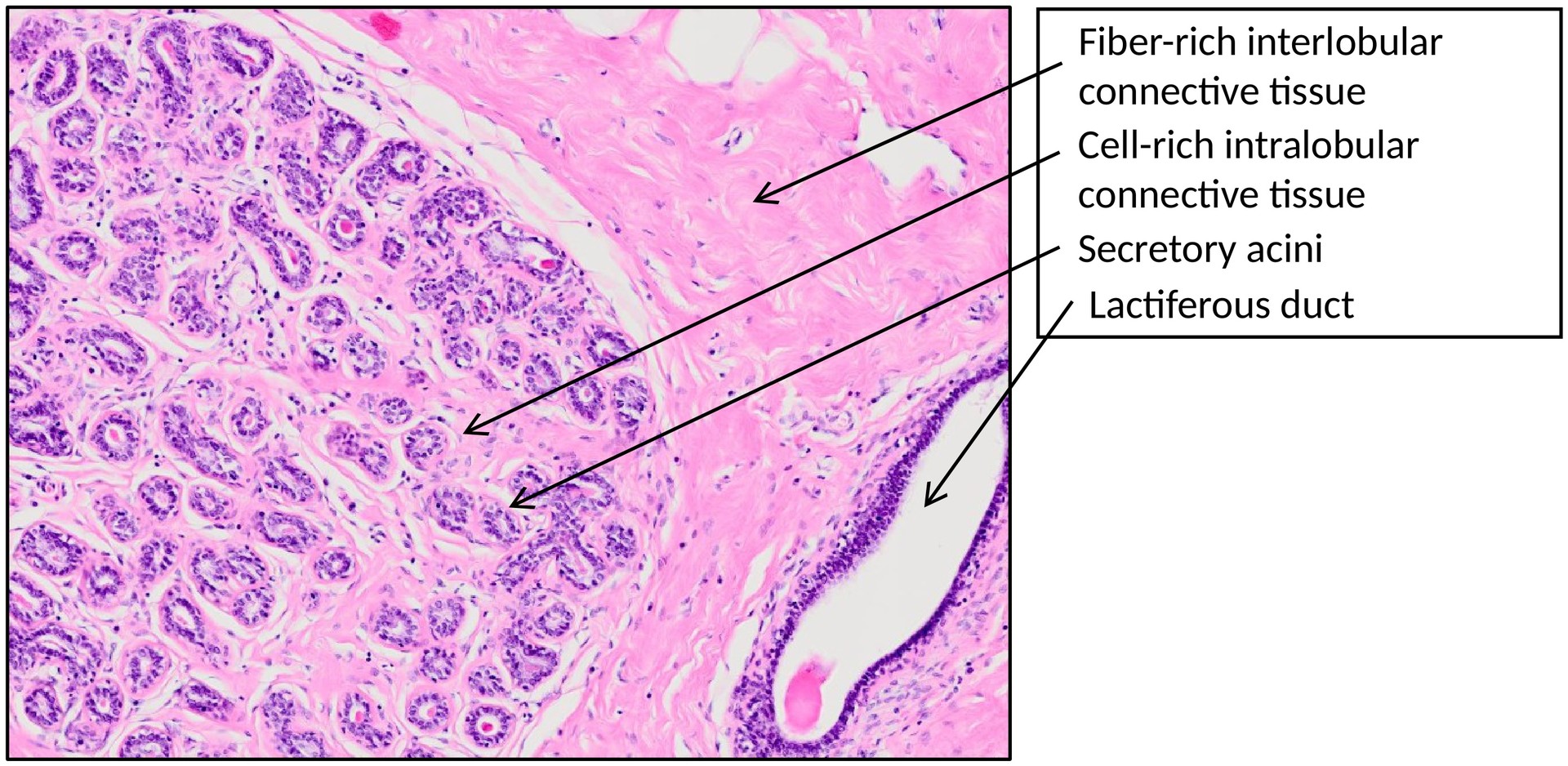
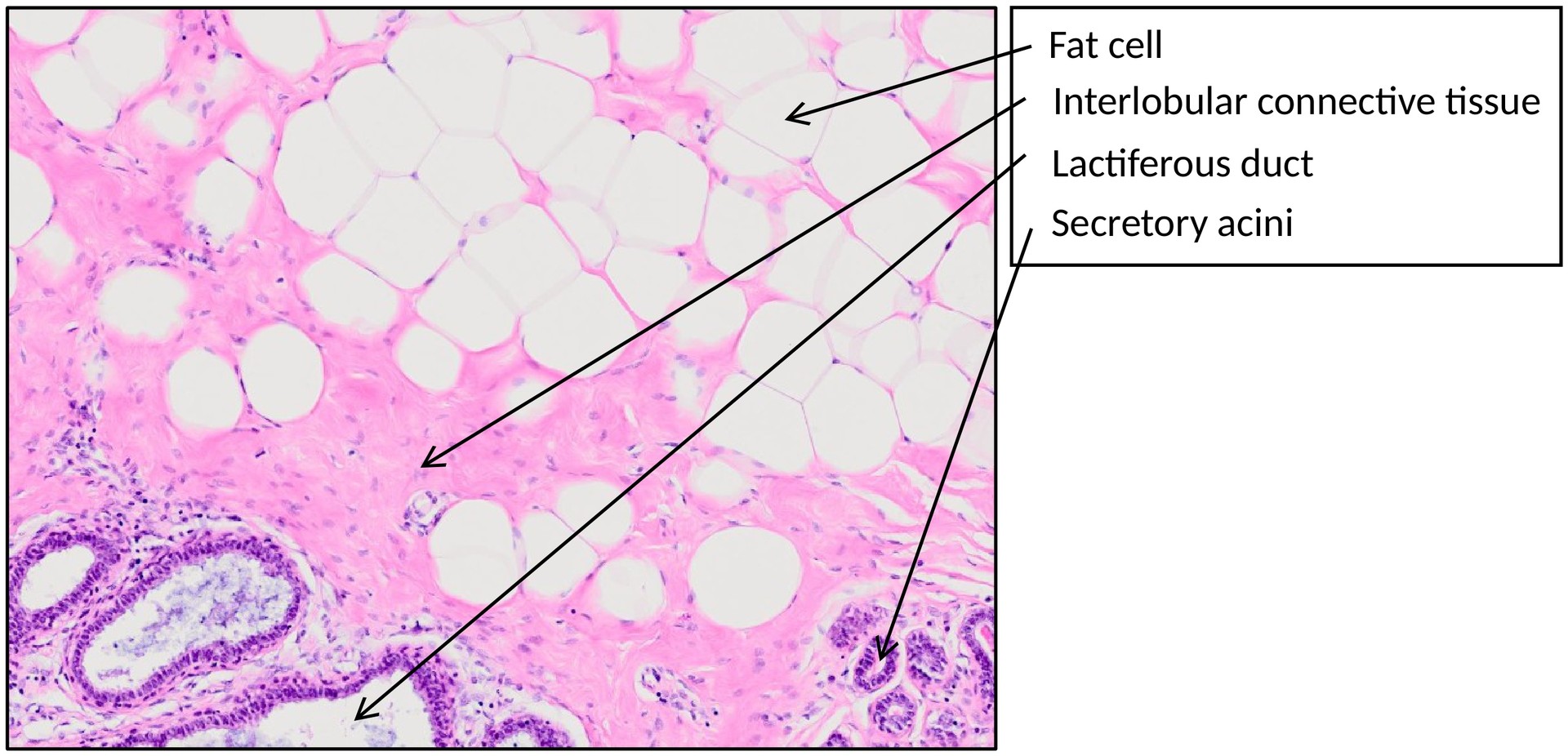
Each lobe consists of a branched ductal system, terminating in small rudimentary secretory end-pieces (acini), which are grouped into lobules. Each lobule is drained by a terminal duct. Together, the terminal duct and its lobule form the terminal ductulo-lobular unit (TDLU) — the key functional unit of the mammary gland.
Within each lobule, the intralobular connective tissue is loose and cell-rich, containing fibroblasts, immune cells, and stem cells that enable the cyclical and reproductive remodeling of the gland (e.g., during pregnancy and lactation).
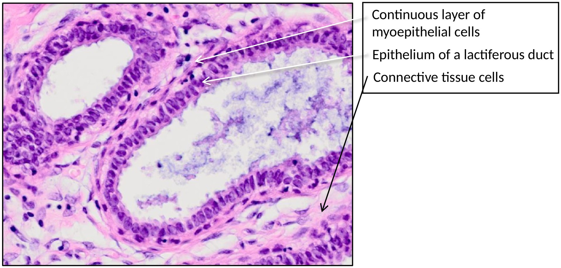
The epithelium of the acini and terminal ducts is cuboidal, supported by a discontinuous layer of myoepithelial cells that facilitate contraction during milk ejection.
Several terminal ducts converge into larger lactiferous ducts, which display bilayered epithelium (inner cuboidal/columnar and outer myoepithelial cells). These ducts eventually unite to form the main excretory ducts (not shown in this specimen), which open at the nipple.
In the non-lactating breast, only about 5–15% of the tissue consists of glandular elements. The remaining stroma is predominantly fibrous connective tissue, interspersed with adipose tissue, through which the ducts course.
The balance between fibrous and fatty stroma varies between individuals and with age, hormonal status, and physiological state.
TASKS:
-
Identify glandular epithelial islands embedded in loose, cell-rich intralobular connective tissue.
-
Distinguish between intralobular and interlobular connective tissue:
-
Intralobular: loose, vascular, and cell-rich.
-
Interlobular: dense, fibrous, with variable amounts of adipose tissue.
-
-
Observe the cuboidal glandular epithelium and underlying myoepithelial layer.
-
Trace the ductal system from small acini to larger ducts.
-
Compare the proportion of glandular tissue and stroma with that of a lactating breast specimen.
License
University of Basel