NERVOUS SYSTEM (ANATOMICAL MICROSCOPY)
16.2
Brain (Calcarine sulcus)
Specimen:
SPECIMEN DETAILS:
Organ: Brain
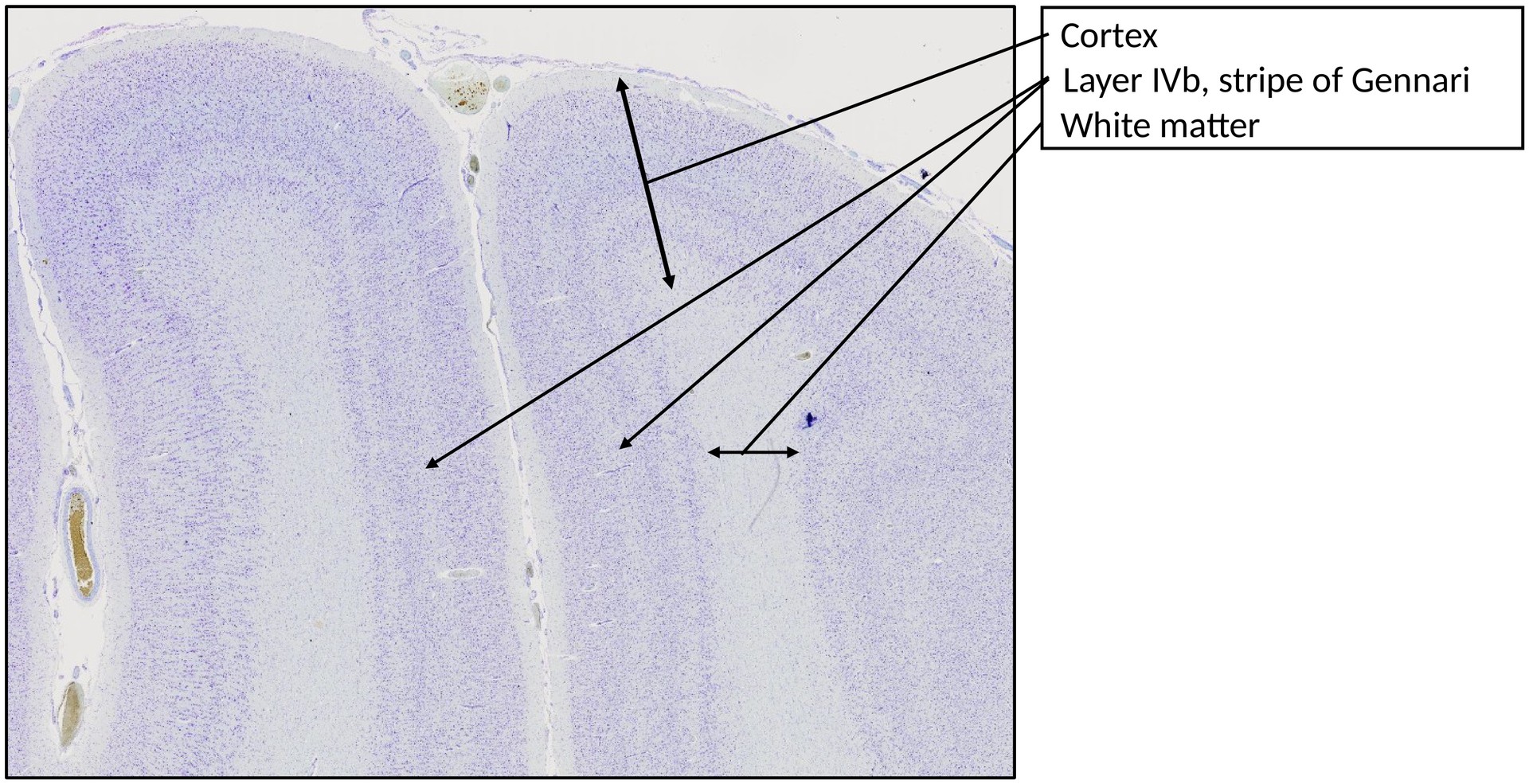
Origin: Human - Calcarine Sulcus (Primary Visual Cortex, V1)
Staining: Cresyl Violet (Nissl Staining)
METHOD AND SPECIMEN DESCRIPTION:
Cresyl violet is a basic dye that binds to acidic components such as RNA and DNA, staining the chromatin, nucleolus, and rough endoplasmic reticulum (Nissl substance) blue–violet. This staining method, commonly referred to as Nissl staining, provides a clear visualization of neuronal cell bodies and the laminar structure of the cortex.
OBJECTIVE OF THE EXAMINATION:
To study and understand the laminar organization of the cerebral cortex, particularly the distinct structure of the primary visual cortex (V1) located around the calcarine sulcus.
SPECIAL FEATURES OF THE SPECIMEN:
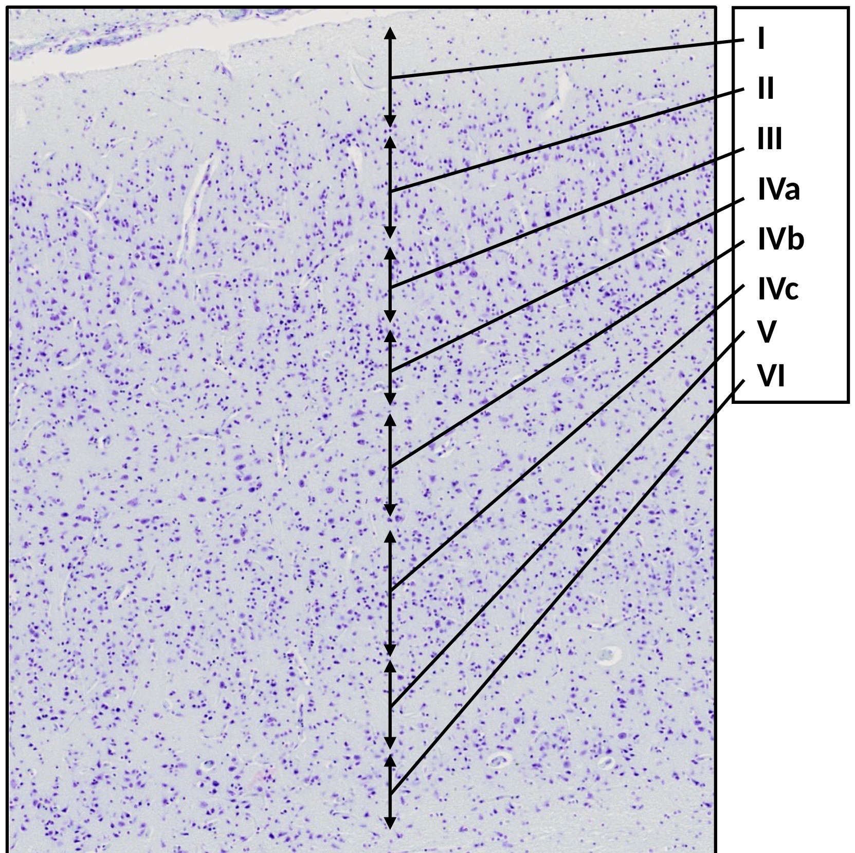
The visual cortex surrounding the calcarine sulcus is notable for its very broad and distinct layer IV, which can be subdivided into three sublayers: IVa, IVb, and IVc.
A prominent feature is the “stripe of Gennari”, located within layer IVb — a lightly cellular region rich in tangentially running myelinated fibers, which gives rise to a macroscopically visible white line in fresh brain tissue (hence the alternate name striate cortex).
Inhibitory neurons are difficult to distinguish with Nissl staining; therefore, orientation within the cortex relies mainly on the distribution and size of pyramidal cells.
Cortical Layer Structure (Isocortex):
| Layer | Name | Main Features |
|---|---|---|
| I | Molecular layer | Very few cells; mostly neuropil. |
| II | External granular layer | Thin; contains small pyramidal and non-pyramidal (granular) neurons. |
| III | External pyramidal layer | Thicker; larger pyramidal cells, many association fibres. |
| IV | Internal granular layer | Extremely thick in the visual cortex; subdivided into: IVa – thin layer, densely packed with small round cells IVb – sparsely cellular, contains large pyramidal cells, site of the Gennari stripe IVc – dense with round, non-pyramidal (spiny stellate) cells; termination site of fibers from the lateral geniculate body |
| V | Internal pyramidal (ganglionic) layer | Narrow; contains large pyramidal cells projecting to subcortical structures. |
| VI | Multiform layer | Broader; contains pyramidal and polymorphic cells; merges with the white matter. |
TASKS:
- List the laminae of the isocortex of the cerebral cortex.
- Explain what indicates that this specimen originates from the primary visual cortex (V1).
- Describe the Gennari stripe: what is it, and how does the macroscopic appearance of a thin white line arise?
- Identify the internal pyramidal layer (V) and sublamina IVc.
- Determine the approximate boundary between layer III and layer IVa.
License
University of Basel