NERVOUS SYSTEM (ANATOMICAL MICROSCOPY)
16.10
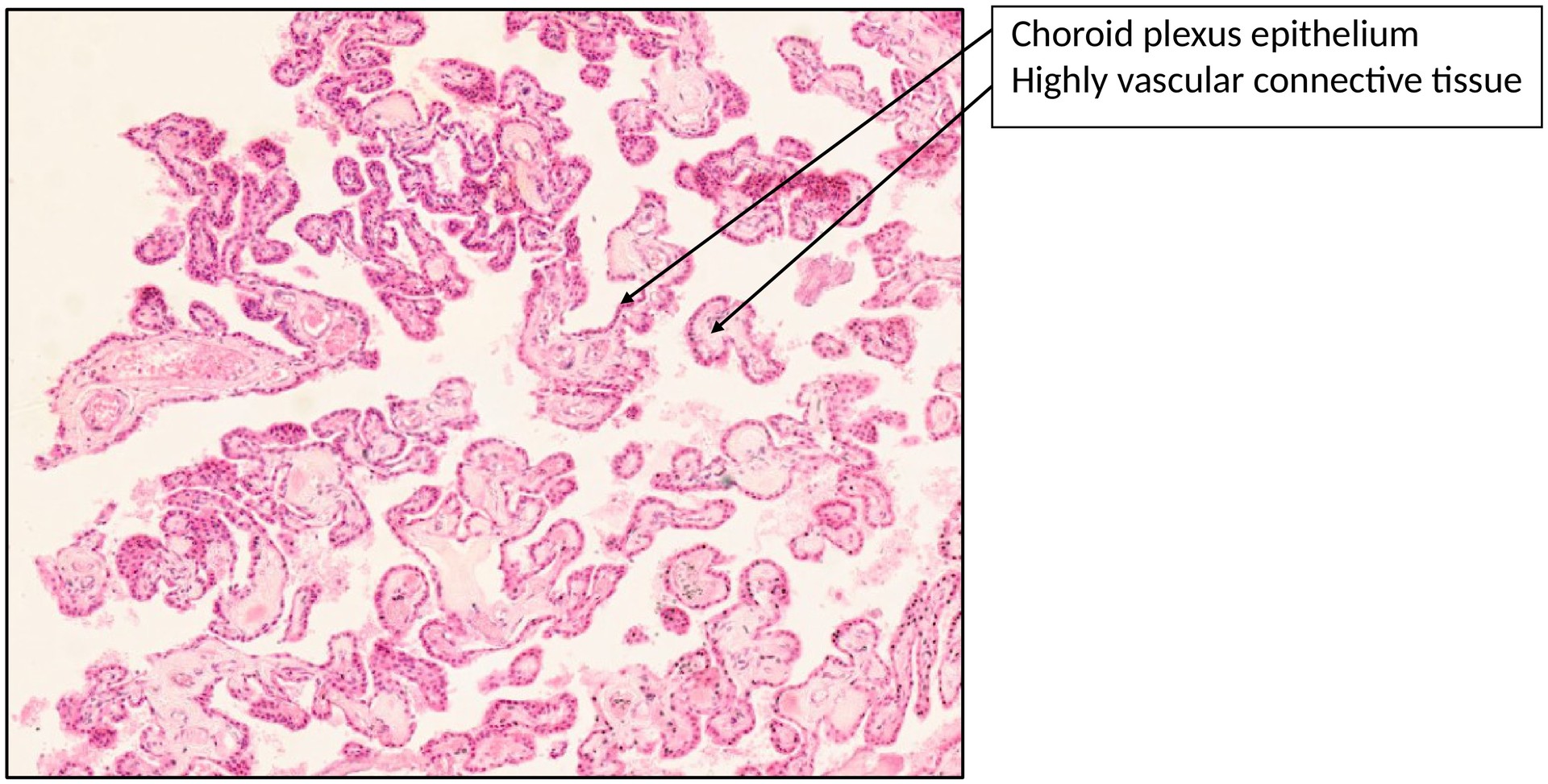
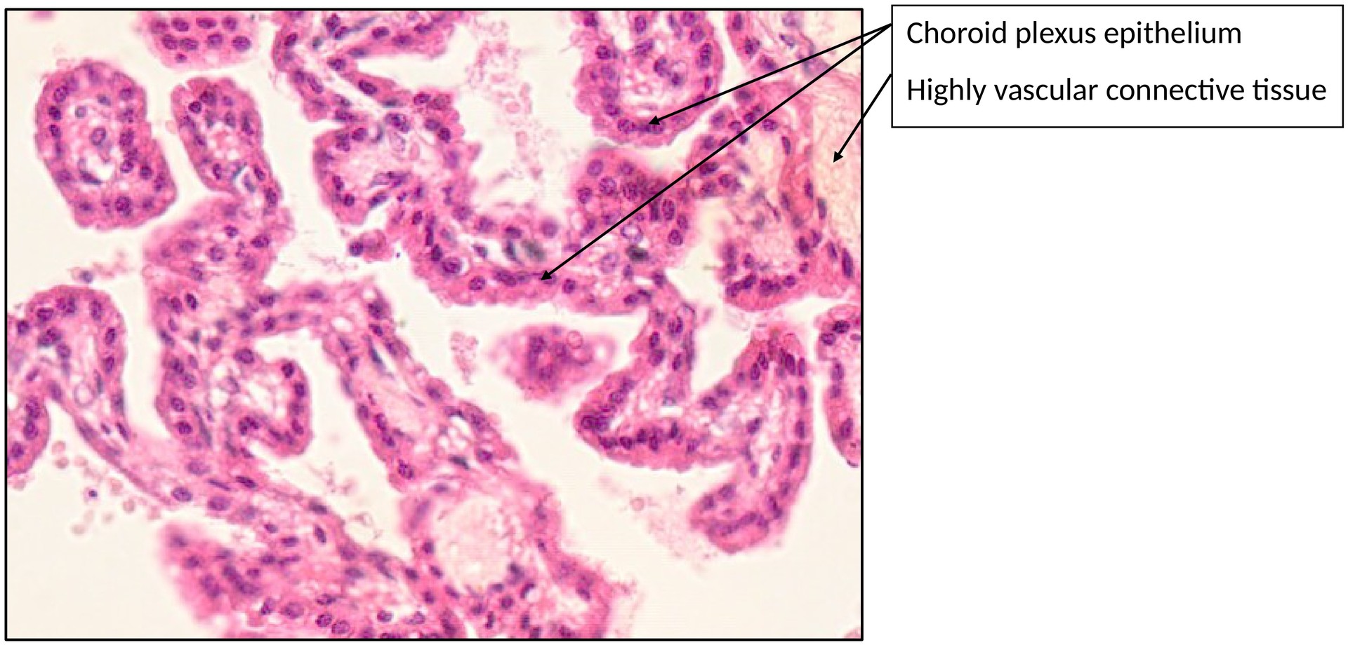
Choroid plexus
Specimen
SPECIMEN DETAILS:
Organ: Choroid plexus
Origin: Human
Staining: Hematoxylin and Eosin (H&E)
METHOD AND SPECIMEN DESCRIPTION:
Standard H&E-stained section of the human choroid plexus.
OBJECTIVE OF THE EXAMINATION:
To study the location, structure, and histological features of the choroid plexus.
SPECIFIC FEATURES OF THE SPECIMEN:
The choroid plexus is responsible for the production of cerebrospinal fluid (CSF) and is located within the ventricular system of the brain. It forms numerous folds and villi, increasing the surface area for secretion.
The surface is lined by a simple cuboidal epithelium (also referred to as the lamina epithelialis) consisting of modified ependymal cells. These epithelial cells possess numerous apical microvilli and kinocilia, and are joined by tight junctions, forming the blood–CSF barrier (analogous to the blood–brain barrier).
Beneath the epithelium lies the tela choroidea, a highly vascularised connective tissue layer containing fenestrated capillaries, small arterioles and venules, and dense collagenous fibers.
Differential diagnosis: At low magnification, the choroid plexus can resemble the fetal placenta, as both structures show a villous architecture. However, the choroid plexus is lined by a single layer of cuboidal epithelial cells, whereas the placenta is covered by syncytiotrophoblast and cytotrophoblast layers.
TASKS:
- Identify the plexus epithelium, consisting of a simple cuboidal layer of ependymal cells.
- Identify the tela choroidea, located beneath the epithelium, containing loose connective tissue, numerous fenestrated capillaries, arterioles, venules, and occasional larger vessels.
- Compare the choroid plexus with the placenta and describe the histological differences.
License
University of Basel