NERVE TISSUE (GENERAL HISTOLOGY)
7.1
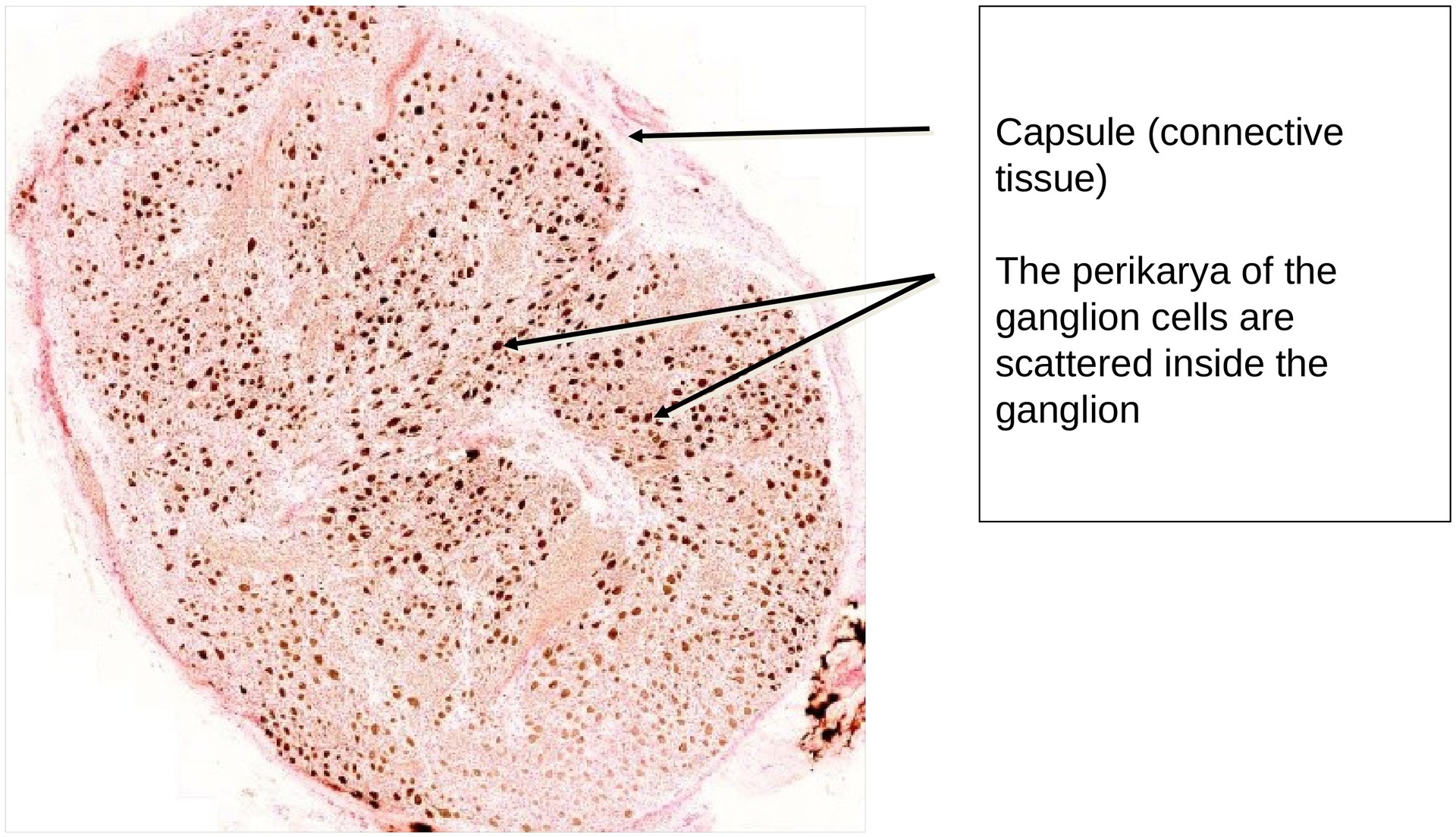
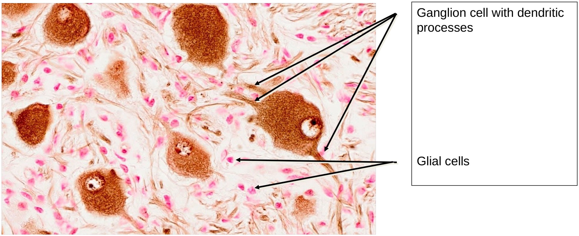
Sympathetic ganglion
Specimen Details:
Specimen Details:
Organ: Paravertebral ganglion
Origin: Bovine
Staining: Acid Phosphatase
Method and Specimen Description:
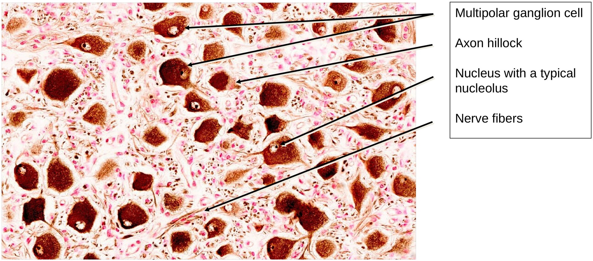
In the visualization of acid phosphatase, phosphate residues are precipitated with lead salts. This results in an intense staining of the perikarya and neurofibrils within dendrites and neurites.
Objective of the Examination:
To gain knowledge of the structure of a sympathetic ganglion, particularly the presence and arrangement of multipolar nerve cells.
Special Features of the Specimen:
Even at low magnification, the sympathetic ganglion cells are seen to be scattered throughout the ganglion. However, the ascending and descending processes of these multipolar neurons are sometimes grouped in smaller bundles.
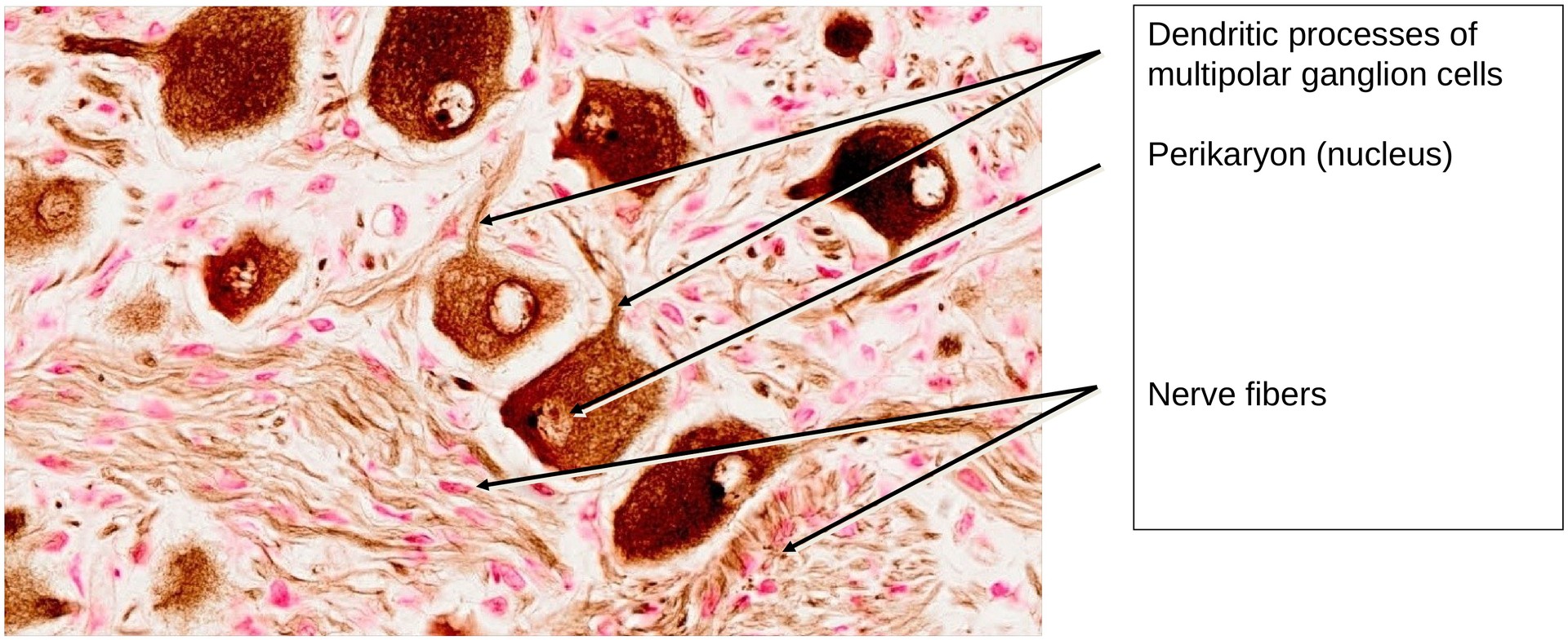
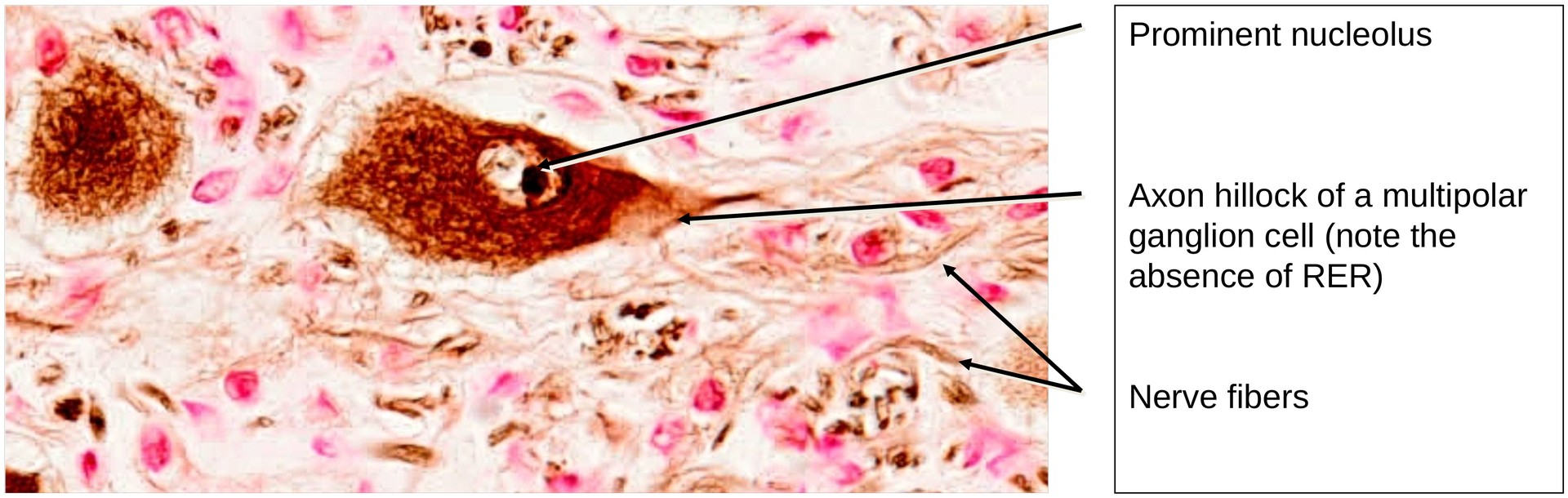
In contrast to a spinal ganglion, where a single process is often difficult to identify, the processes of the multipolar ganglion cells in this specimen are numerous and therefore frequently visible. With a favorable section orientation, the axon hillock of a neurite (axon) can be recognized by the parallel arrangement of neurofibrils and the absence of Nissl substance (rough endoplasmic reticulum).
The perikarya vary greatly in size. Surrounding each are satellite cells, identifiable by their prominent nuclei. A narrow space is often visible between the satellite cells and the perikaryon; this, however, is a preparation artefact.
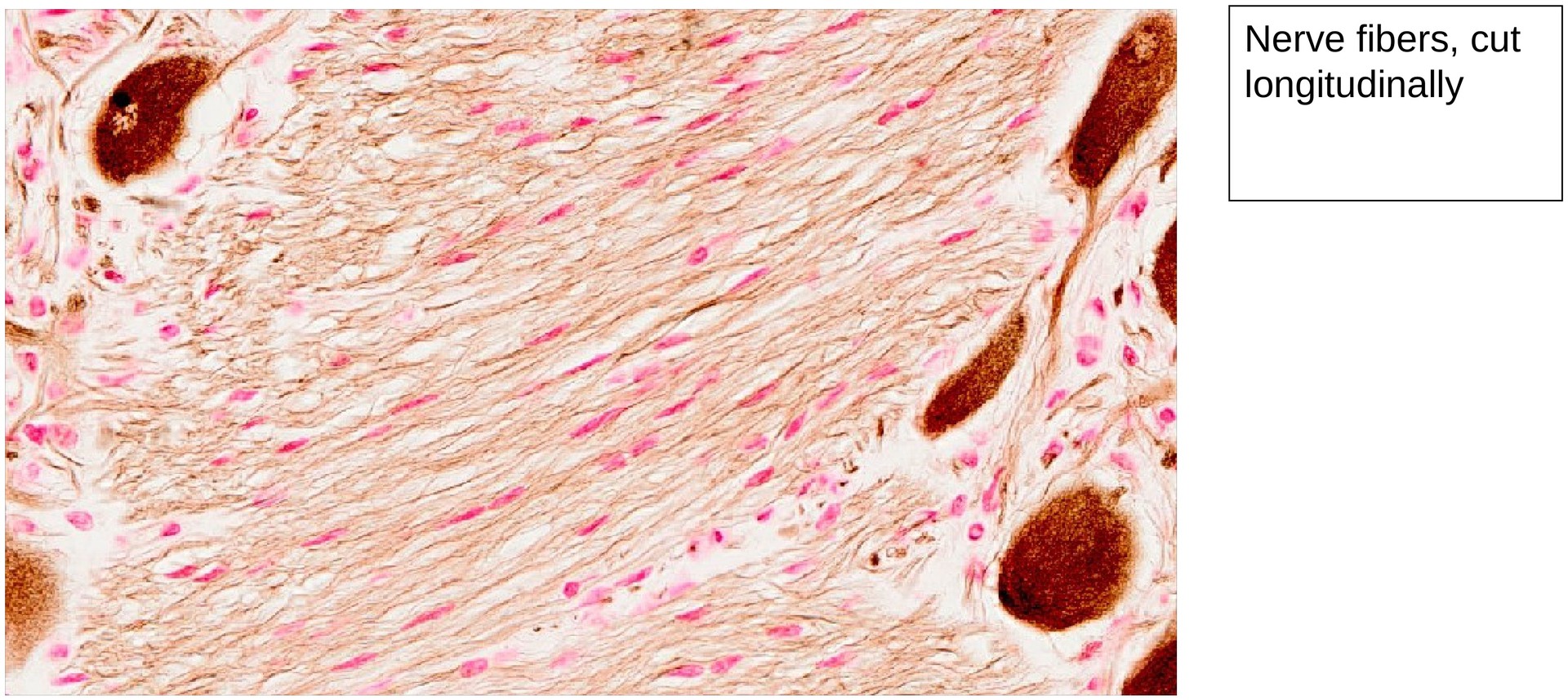
The nerve cell processes are seen in multiple orientations — longitudinal, transverse, and tangential — depending on the plane of section. Between the perikarya and the nerve fibers, numerous small nuclei are visible; these belong to glial cells and vascular cells supplying the ganglion.
Tasks:
• Locate different multipolar ganglion cells and note their distribution and the variation in perikaryon size.
• Identify regions containing nerve fibers and determine their orientation (transverse, longitudinal, or tangential).
• Search for an axon hillock where a neurite (axon) emerges from the perikaryon (rare in this specimen).
• Identify and observe blood vessels within the ganglion.
License
University of Basel
Downloads