SENSORY ORGANS (ANATOMICAL MICROSCOPY)
17.1
Eye
Preparation:
Preparation Details:
Organ: Eye
Origin: Pig
Staining: Hematoxylin - Chromotrope
Method and Specimen Description:
Standard histological section of the pig’s eye stained with hematoxylin–chromotrope, a combination particularly well suited for demonstrating the different coats of the eyeball and the retinal layers.
Objective of the Examination:
To study the structure of the eye, including the outer, middle, and inner coats, and to understand their differences in the anterior and posterior portions of the eyeball. The examination also aims to demonstrate the blind spot and its significance.
Special Features of the Specimen:
Many key details are already clearly visible at low magnification, including the lens, iris, sclera, cornea, optic nerve (region of the blind spot), the anterior and posterior chambers, and the ciliary body. It is recommended to examine the anterior and posterior halves of the eye separately.
Anterior third of the eye
The eye is best considered as comprising an anterior third and a posterior two-thirds, rather than being divided precisely at the midline.
In the anterior portion, the three coats of the eyeball are represented as follows:
- Outer coat: Cornea
- Middle coat: Iris stroma
- Inner coat: Pigment epithelium behind the iris and the ciliary body
At the front, the anterior chamber is enclosed by the cornea, which has a greater curvature than the sclera and contributes significantly to light refraction.
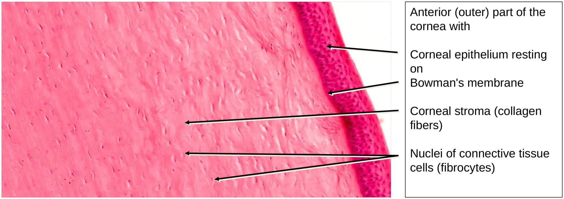
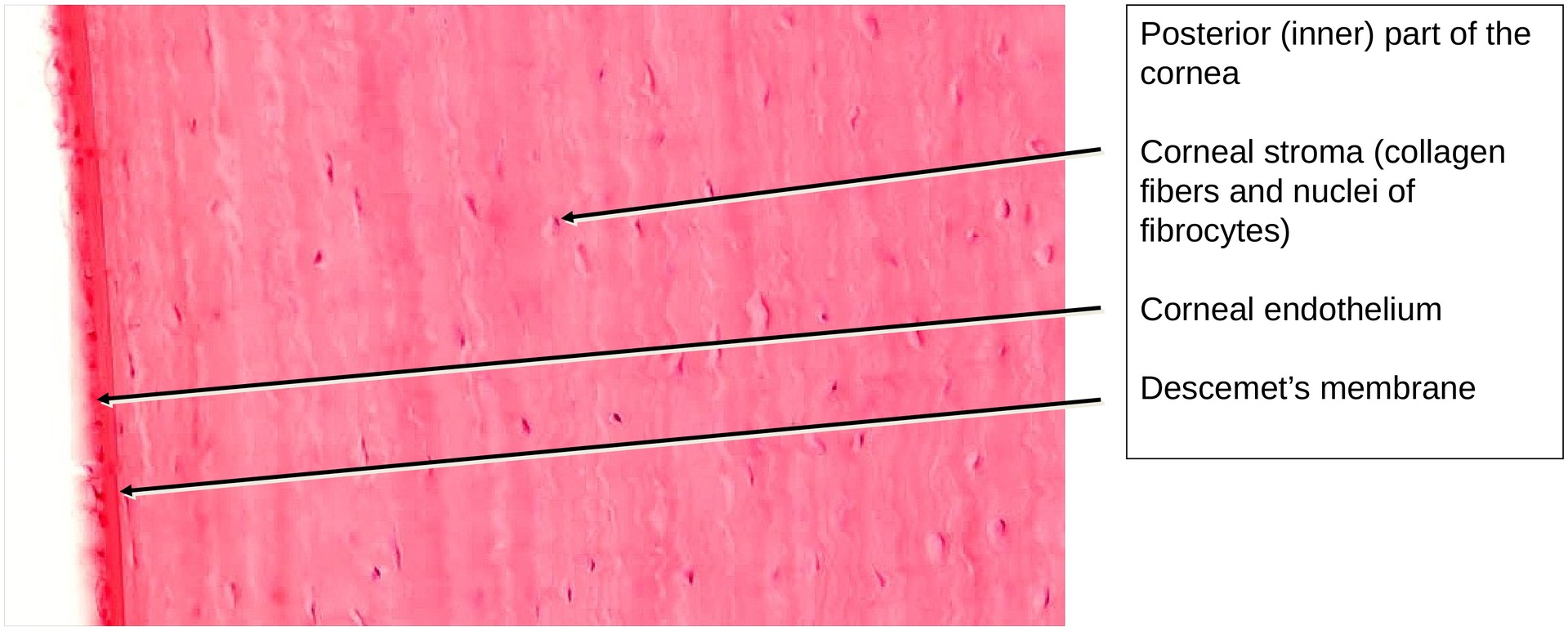
The cornea is composed of three main layers:
- The outer corneal epithelium, resting on Bowman’s membrane
- The avascular stroma, consisting predominantly of regularly arranged collagen fibers with few cells (visible only by their nuclei)
- The inner corneal endothelium, resting on Descemet’s membrane
Both Bowman’s and Descemet’s membranes play important roles in regulating corneal hydration.
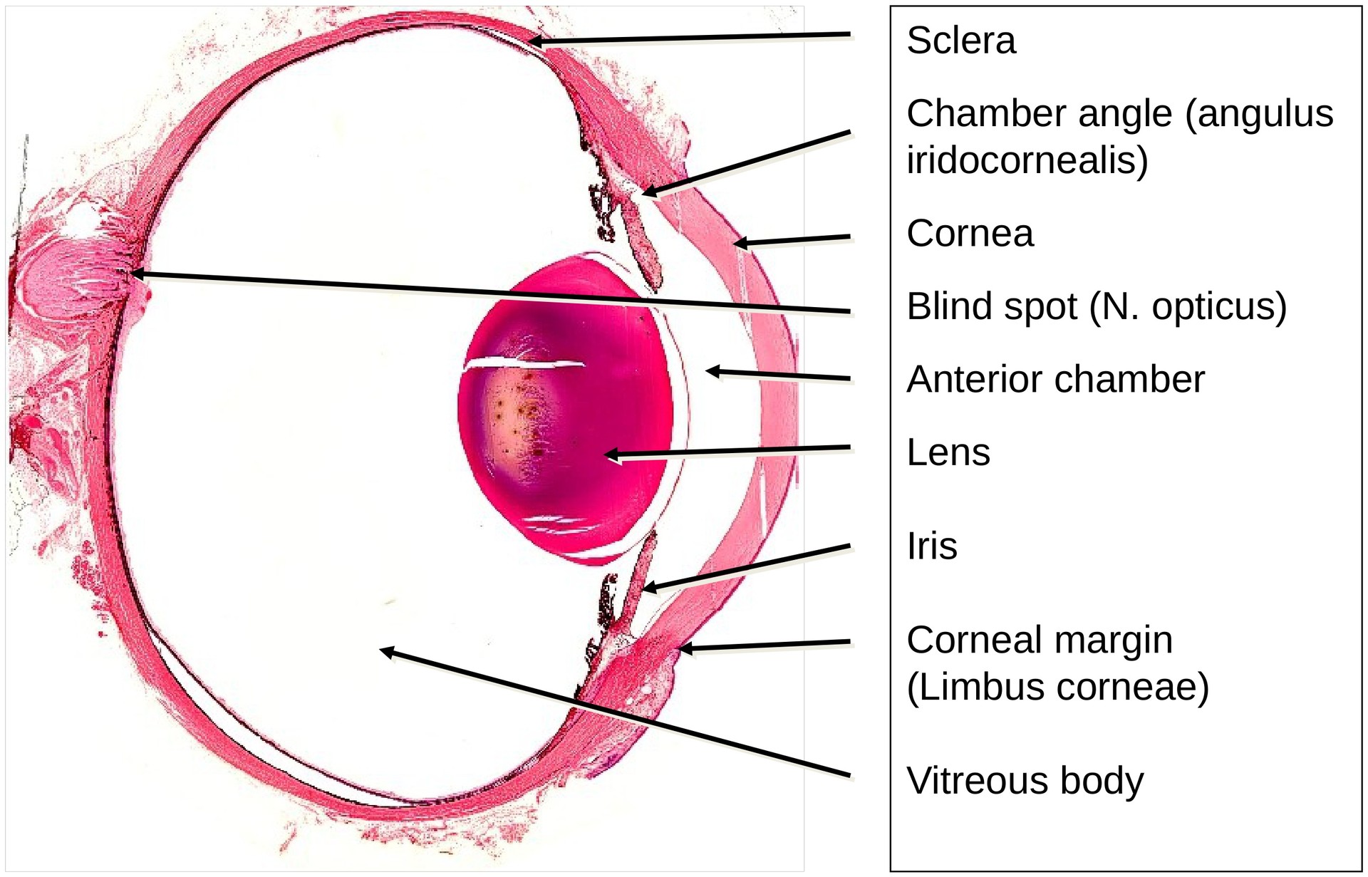
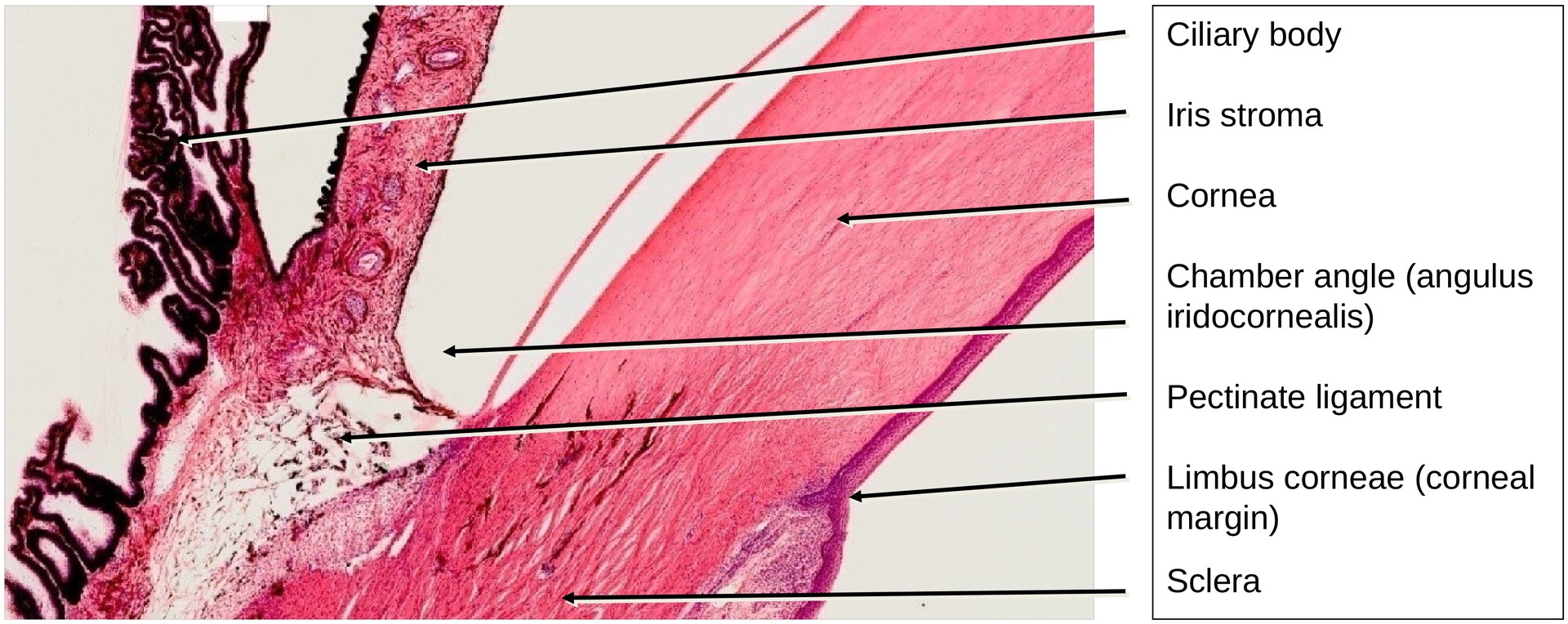
At the limbus corneae (corneal margin), the cornea transitions into the sclera, forming the junction between the transparent, refractive part of the eye and the opaque, fibrous tunic.
The anterior chamber lies behind the corneal endothelium, while the posterior chamber is bounded posteriorly by the ciliary body and separated from it by the iris. Aqueous humor circulates between these two chambers, produced by the ciliary processes of the ciliary body.
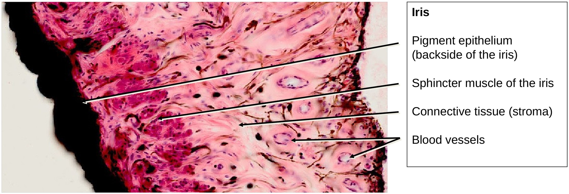
Both the posterior surface of the iris and the ciliary body are covered by pigmented epithelium. The iris forms the pupil, whose diameter adjusts to light intensity via smooth muscle fibers in the iris stroma:
- The sphincter pupillae muscle (parasympathetically innervated) lies near the free edge of the iris and is readily visible in this preparation.
- The dilator pupillae muscle (sympathetically innervated) lies in close contact with the pigment epithelium and may be less distinct.
The iris constitutes part of the middle coat. Its stroma lacks an anterior epithelial covering and instead terminates in a loose arrangement of fibroblasts and fibers. The color of the iris depends on several factors, including pigment cells in the stroma, the density and arrangement of stromal fibers, vascularization, and the transparency of the pigment epithelium.
A comb-like network of fibers (the pectinate ligament) in the eye’s iridocorneal angle alleviates the humor drainage from the anterior eye chamber into the Schlemm canal. The pectinate ligament is covered by an endothelium. At the base of the iris the connective tissue transitions into the ciliary body, which with its smooth muscle fibers serves two functions: Accommodation and production of the humor. Accommodation is achieved through the contraction of the ciliary muscle (M. ciliaris). Its contraction relaxes the lens of the eye, thus leading to a more convex shape, which in turn increases the refractive index of the lens.
The ora serrata is the serrated junction between the choroid (choroidea) and the ciliary body. This junction marks the transition from the simple, non-photosensitive area of the ciliary body to the complex, multi-layered, photosensitive region of the retina.
Posterior two Thirds of the Eye:
In the posterior two thirds of the eye it is composed of three coats or tunics:
- The outer or fibrous coat: sclera (mostly collagen fibers)
- The middle vascular coat: choroid (blood vessels)
- The inner coat: retina
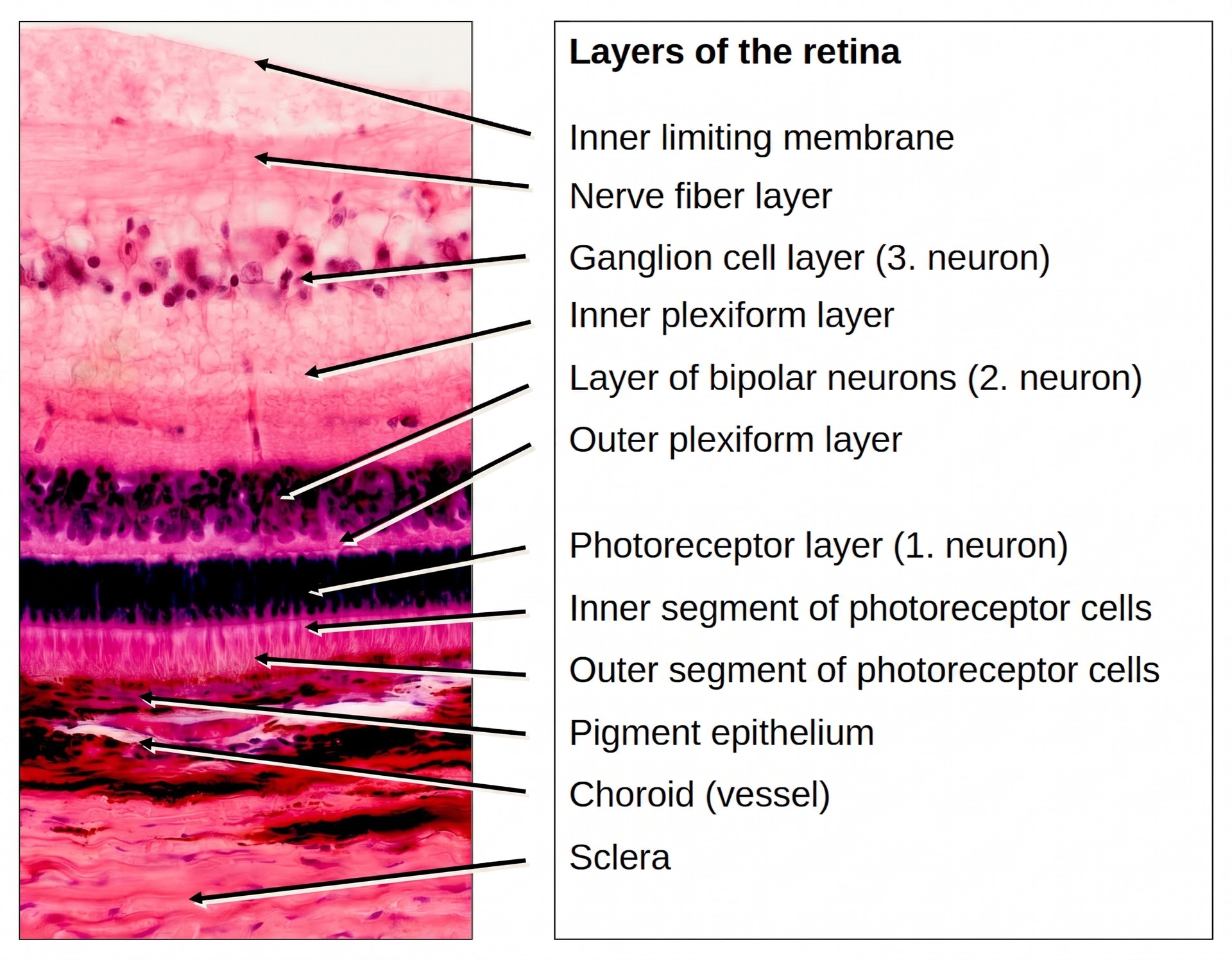
The inner coat, or retina, is the light-sensitive component of the eye. Histologically, the retina is classically divided into ten layers, though not all can be clearly distinguished in this preparation.
The three principal neuronal layers are identifiable and essential for understanding retinal organization:
- Photoreceptor layer – containing rods and cones (outer and inner segments can be differentiated: the outer segments appear light and slender; the inner segments, darker and broader)
- Inner granular (bipolar cell) layer
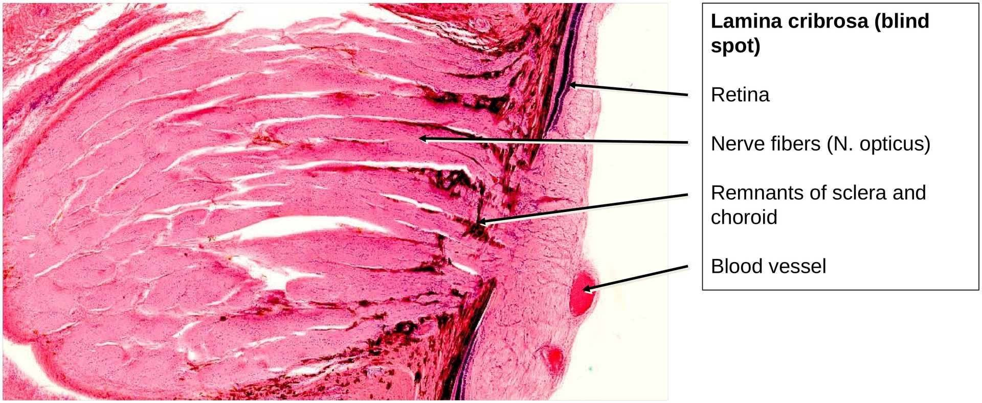
- Ganglion cell layer – whose axons converge, pass through the sclera at the lamina cribrosa, and form the optic nerve
Between these lie the outer and inner plexiform layers, representing synaptic zones between neuronal cell types.
At the lamina cribrosa (a perforated region of the sclera, where the nerve fibers forming the optic nerve exit the eye), here the retinal layers are reduced or absent, creating the blind spot.
The vitreous body (corpus vitreum) occupies the largest portion of the eyeball. It appears almost unstained because it contains very few cellular or fibrous structures. It fills the space between the lens and the retina.
Tasks:
Identify the following structures in the anterior half of the eye, and answer the related questions based on the preparation:
- Cornea: Identify the corneal epithelium, Bowman’s membrane, corneal stroma, corneal endothelium, and Descemet’s membrane.
- Anterior chamber: What are its boundaries?
- Posterior chamber: What structures delimit it?
- Locate the chamber angle — which structures define it?
- What is Schlemm’s canal?
- Where are the Fontana spaces located?
- How is the iris connected to the anterior chamber?
- Which muscle constricts the pupil, and how is it innervated?
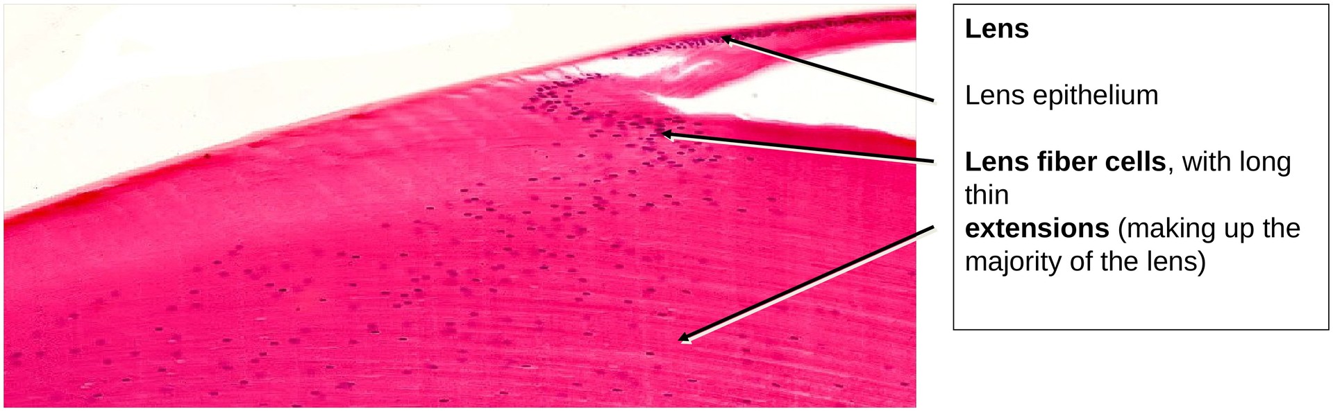
- What is meant by the term lens epithelium?
- Which structures make up the lens?
- How is the iris stroma organized? Are pigment cells present in it?
License
University of Basel
Downloads