FEMALE REPRODUCTIVE ORGANS (ANATOMICAL MICROSCOPY)
10.7
Fallopian tube (uterine tube)
Specimen Details:
Specimen Details:
Organ: Fallopian tube
Origin: Human
Staining: Hematoxylin - Eosin (H&E)
Method and Specimen Description:
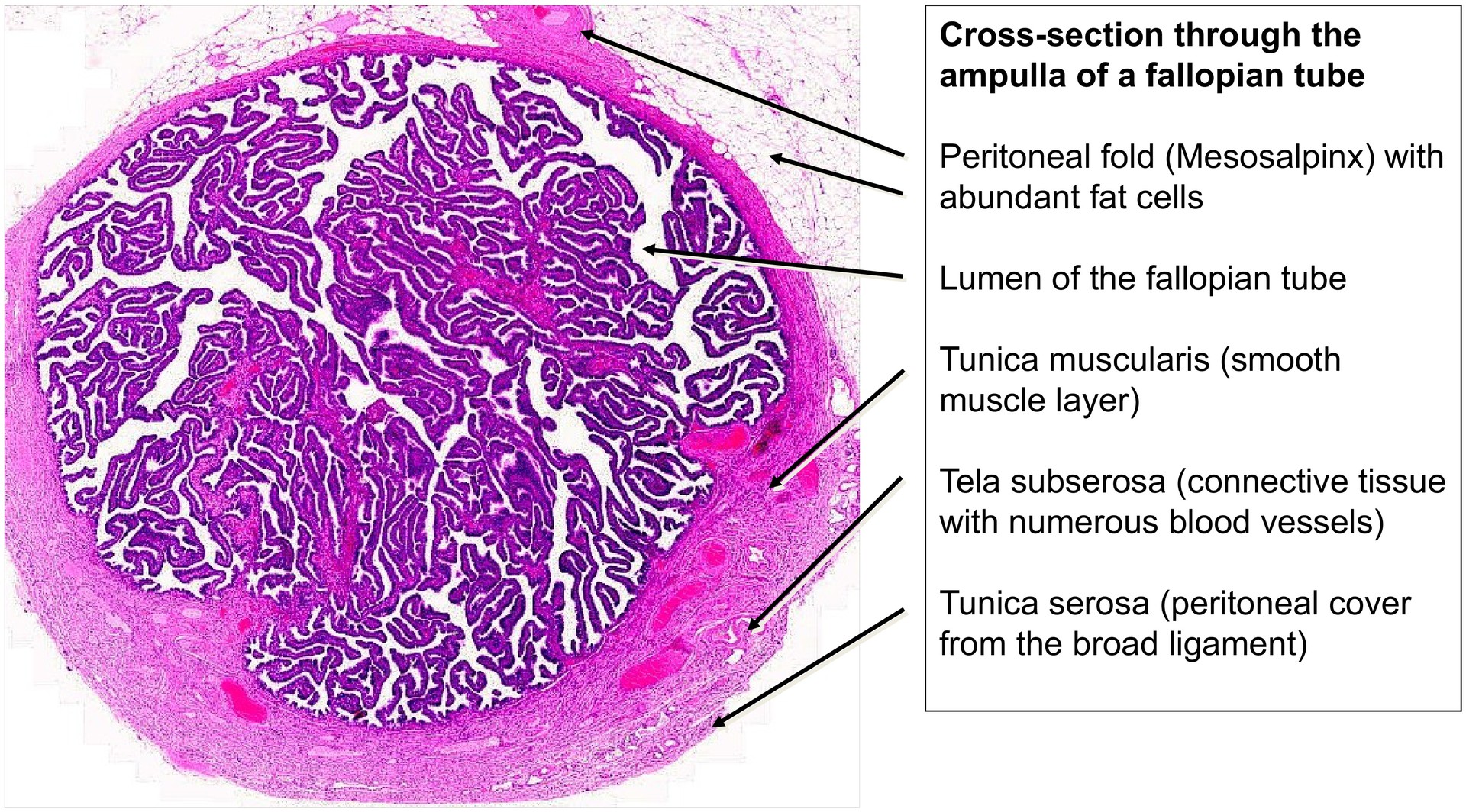
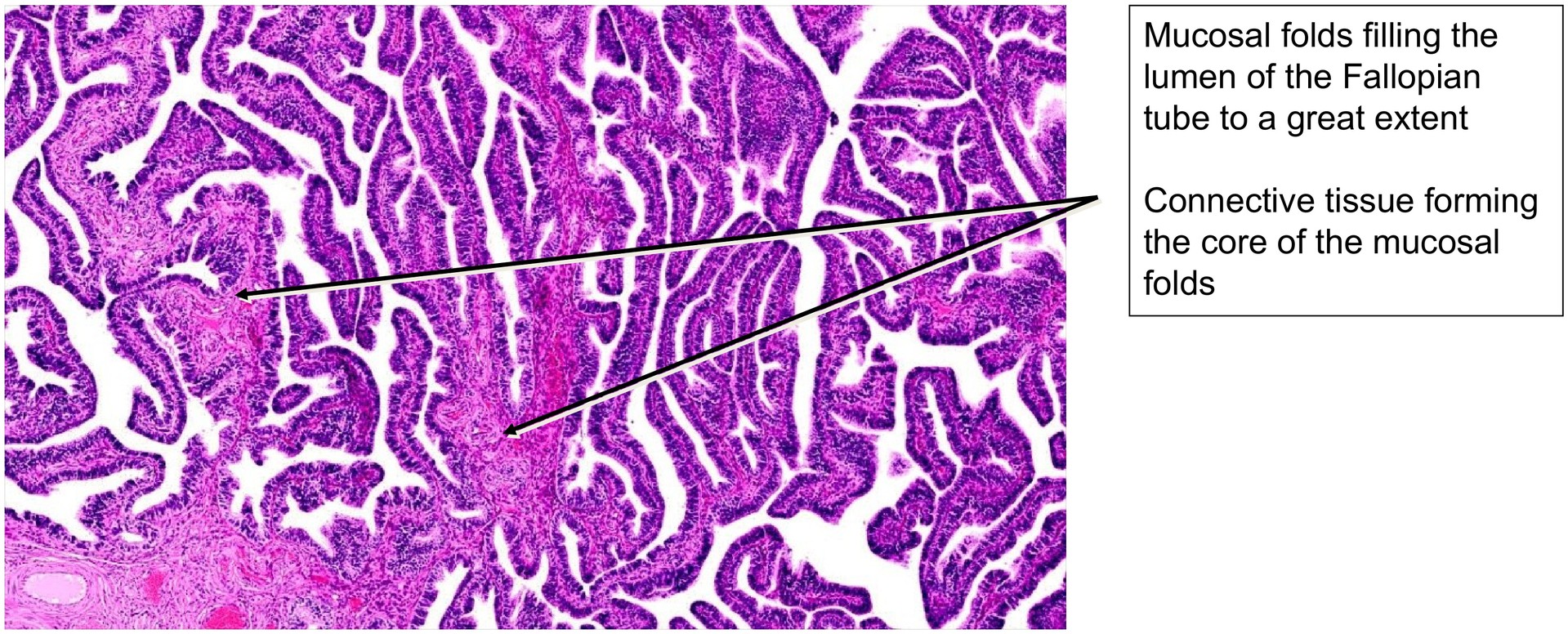
Routine histological section of the fallopian tube, stained with the general overview stain H&E. The specimen includes both the infundibulum and ampulla.
Objective of the Examination:
To study the histological organization of the fallopian tube, with particular focus on the mucosal folds and the ampulla, the region most significant for fertilization and tubal transport.
Special Features of the Specimen:
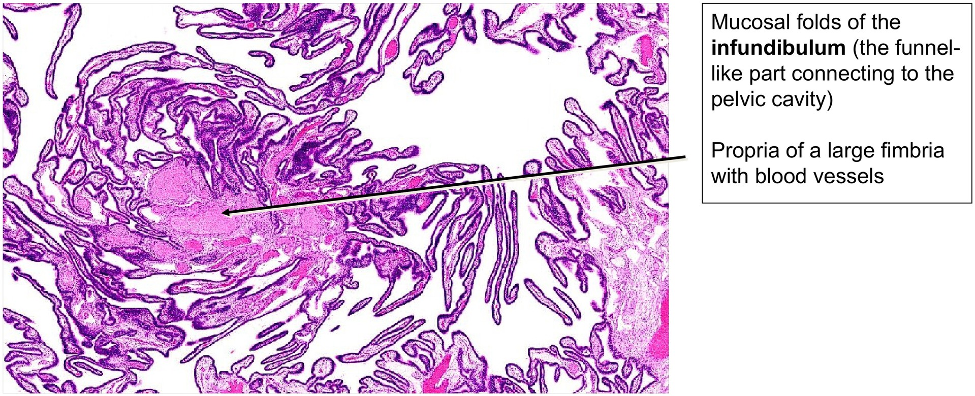
Following ovulation, the oocyte is captured by the infundibulum and passes into the ampulla, where fertilization may occur. The fertilized egg remains in the fallopian tube for about 4–6 days before entering the uterine cavity for implantation. During this time, the tubal environment supports the early stages of development through secretions that nourish and protect the embryo.
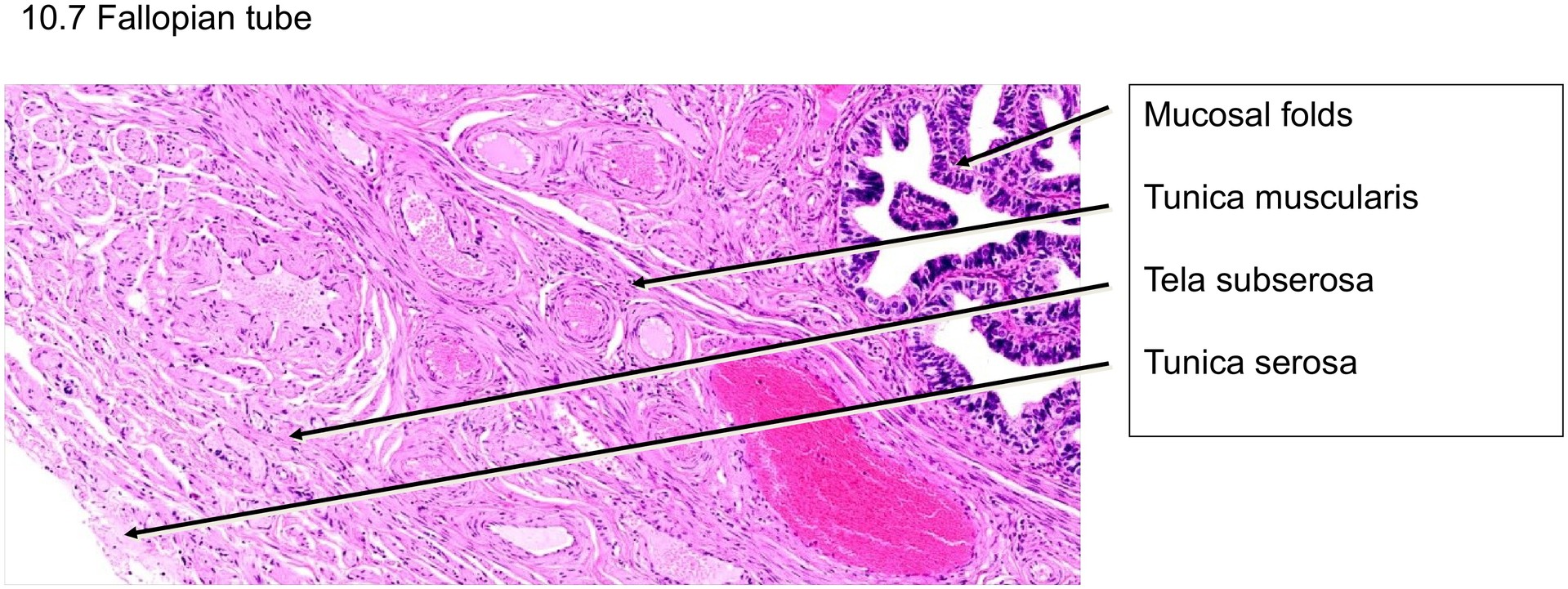
The wall of the fallopian tube displays four distinct layers:
-
Tunica mucosa – the mucosa with its thin lamina propria; the mucosa forms extensive primary, secondary, and sometimes tertiary folds.
-
Tunica muscularis – the muscular layer, which consists of interwoven circular and longitudinal smooth muscle fibers, responsible for peristaltic movement and transport.
-
Tela subserosa – a loose connective tissue layer containing blood vessels and small muscle bundles.
-
Tunica serosa – the outermost peritoneal covering, a continuation of the broad ligament.
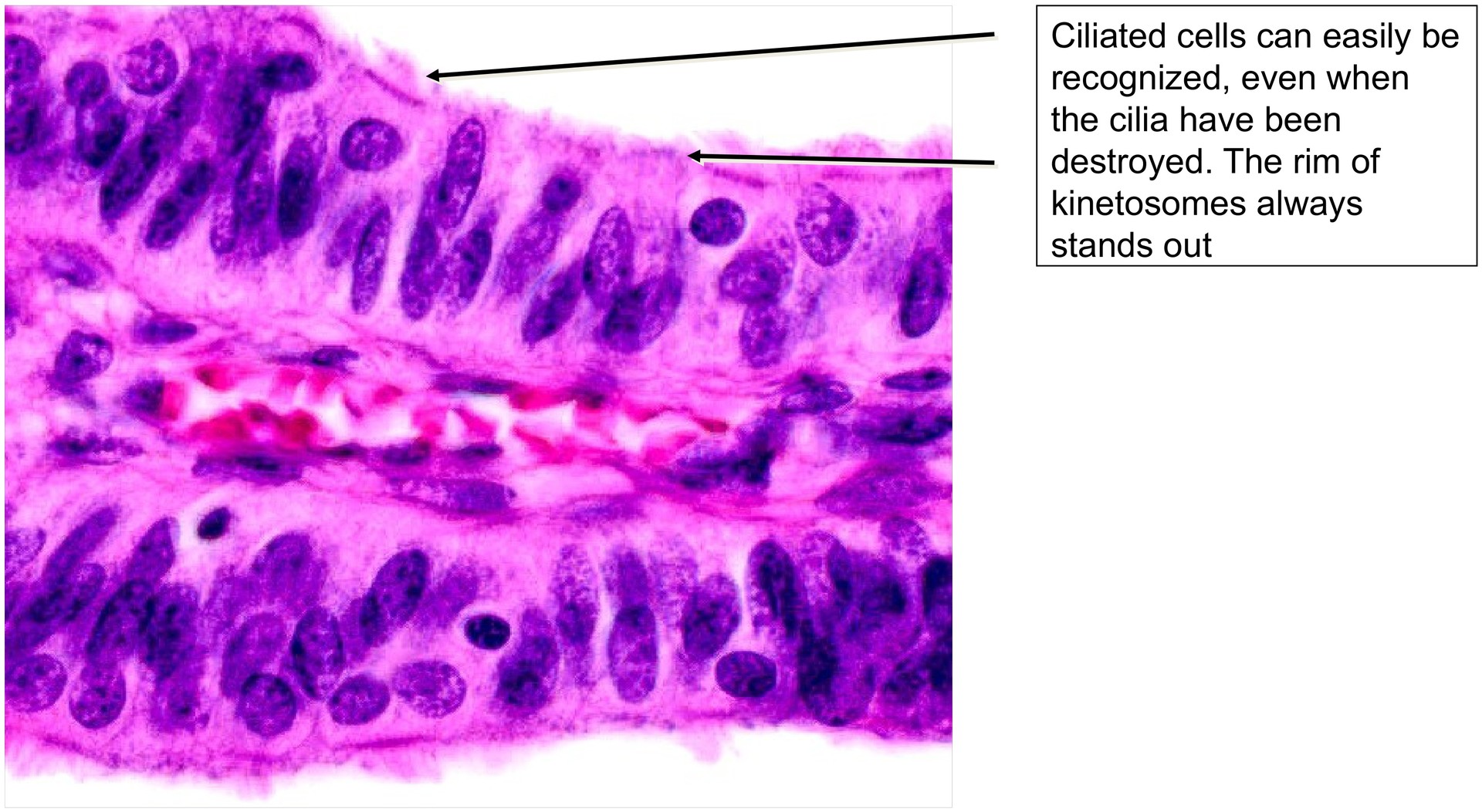
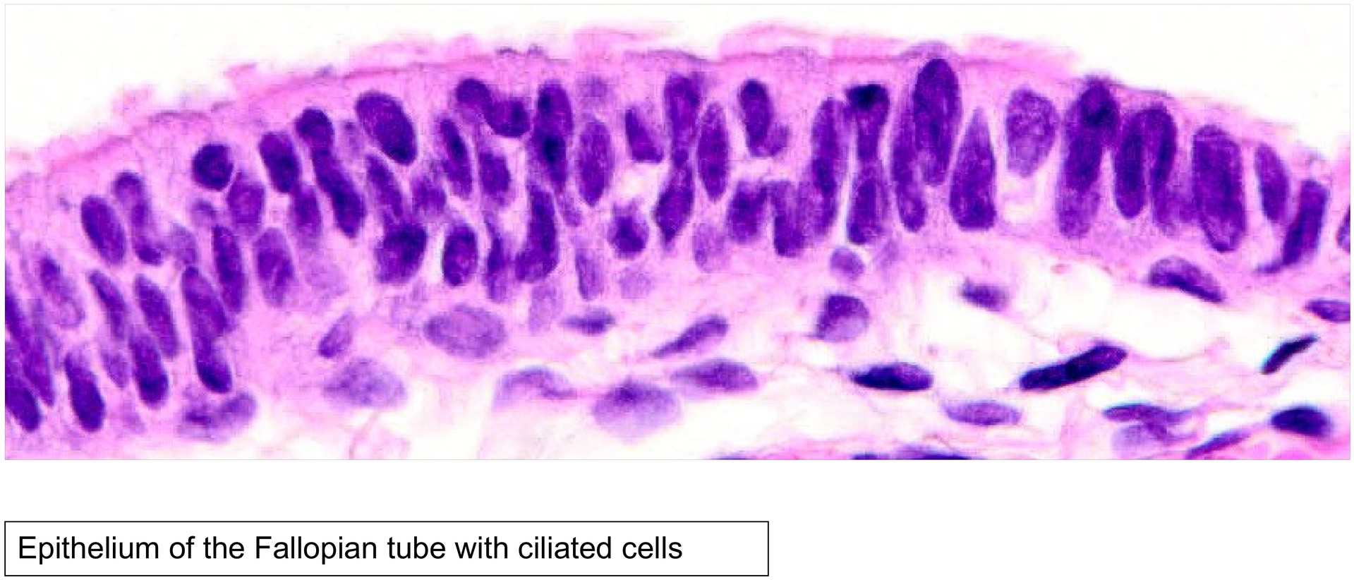
The epithelium of the mucosa is composed of two main cell types:
-
Ciliated cells, with rounded to oval nuclei and cilia that aid ovum transport.
-
Secretory (non-ciliated) cells, with elongated nuclei, which produce nutritive fluid for the oocyte and early embryo.
These cell types can be distinguished by nuclear shape even where the cilia are not clearly visible.
The so-called peg cells, sometimes described in older literature, are now recognized as a fixation artefact. In optimally fixed specimens, such as fresh intraoperative material, peg cells are typically absent.
The tunica muscularis comprises the tubal-specific smooth muscle, arranged in interwoven circular and longitudinal layers. Additional muscle bundles may be present within the tela subserosa, following the blood vessels, and beneath the serosa. These contribute to the motility of the tube, important during ovum pick-up and transport.
In some sections, particularly through the ampulla, the mesosalpinx is visible — a peritoneal fold derived from the broad ligament (mesometrium). It contains blood vessels and, in this specimen, abundant adipose tissue.
Tasks:
• various regions in overview magnification: cross-section through the ampulla, and tangential section through the ampulla and infundibulum.
• Determine how the cross-section through the ampulla can be recognized.
• Identify the macroscopic regions of the fallopian tube and describe how they differ.
• Differentiate secretory cells from ciliated cells at high magnification. What features allow this distinction?
• Trace a mucosal fold through its branching pattern and identify primary, secondary, and tertiary subdivisions.
• Identify the tela subserosa and tunica serosa. How can they be recognized histologically?
• Locate blood vessels within the tela subserosa and distinguish arteries from veins.
License
University of Basel
Downloads