MALE REPRODUCTIVE ORGANS (ANATOMICAL MICROSCOPY)
11.5
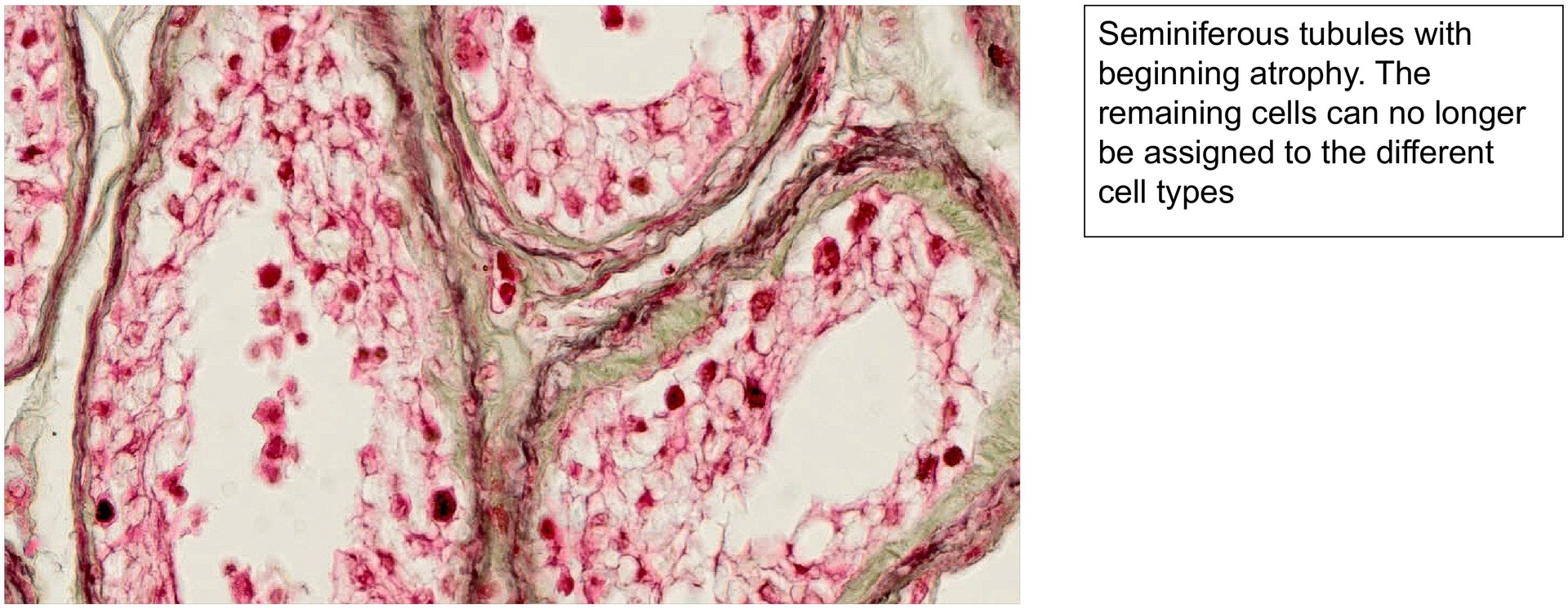
Testis, elderly
Specimen Details:
Specimen Details:
Organ: Testis
Origin: Human (Elderly)
Staining: RGAN
Method and Specimen Description:
Normal histological section stained with RGAN, a method used to highlight elastic fibres, which are significantly increased in the elderly testis.
Objective of the Examination:
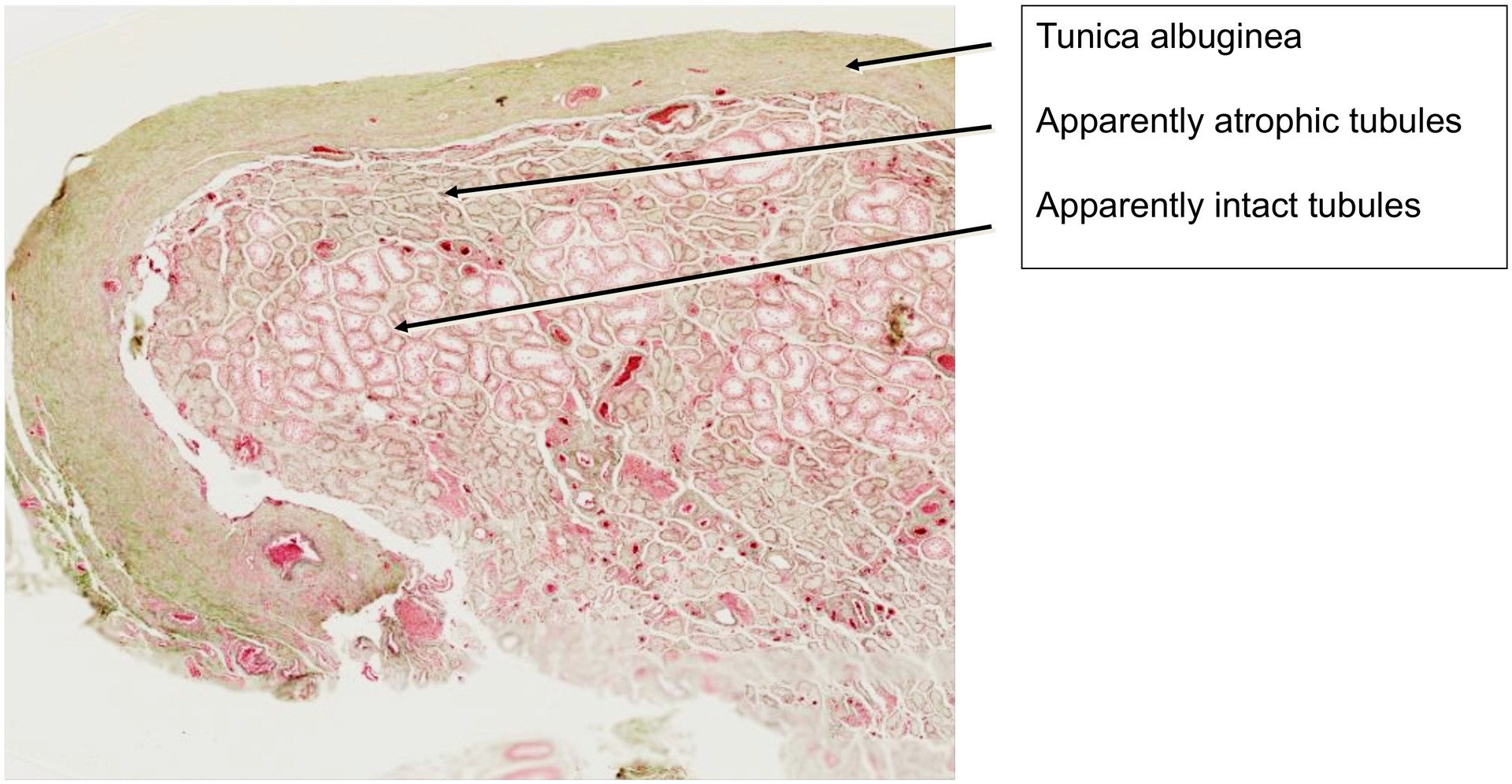
To study the age-related structural changes of the testis, in which many seminiferous tubules become atrophic and non-functional as a result of hyaline degeneration.
Special Features of the Specimen:
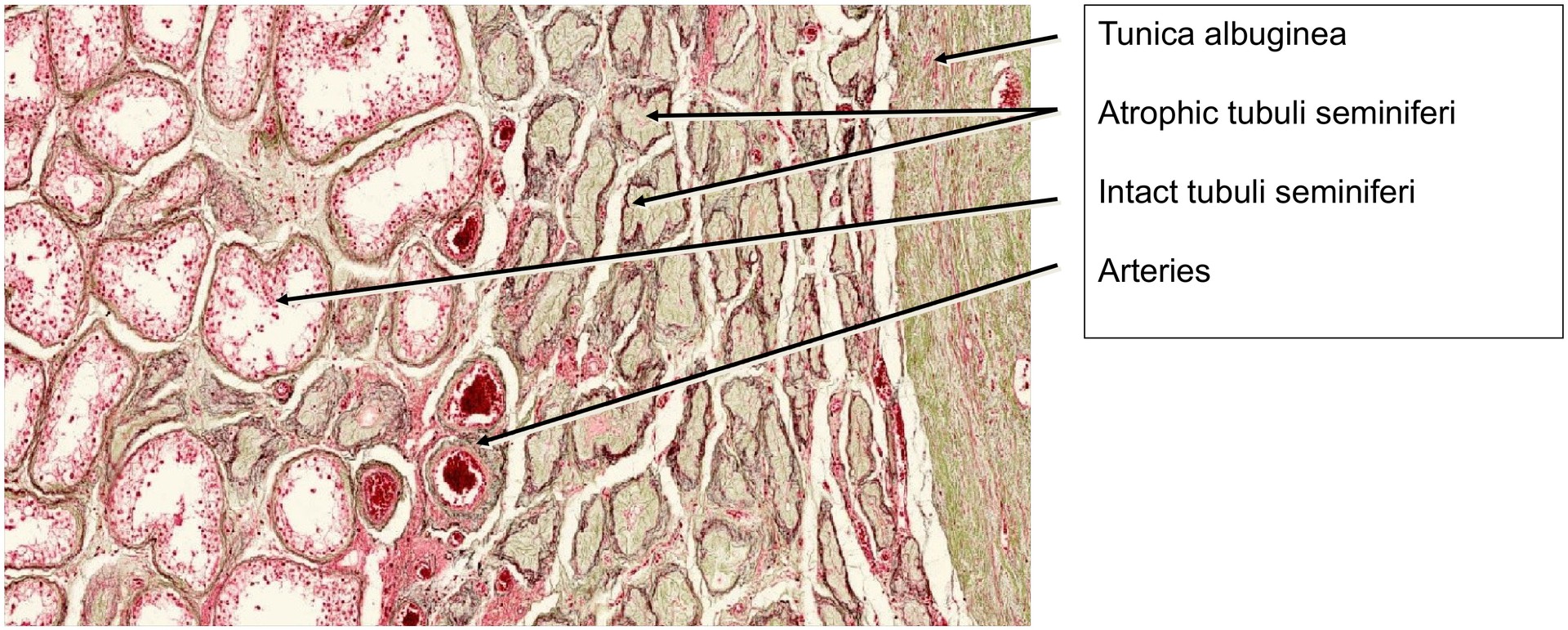
At low magnification, the dense tunica albuginea is immediately recognizable. The section shows two distinct types of seminiferous tubules:
-
Relatively intact tubules, which retain a discernible lumen and remnants of the seminiferous epithelium, and
-
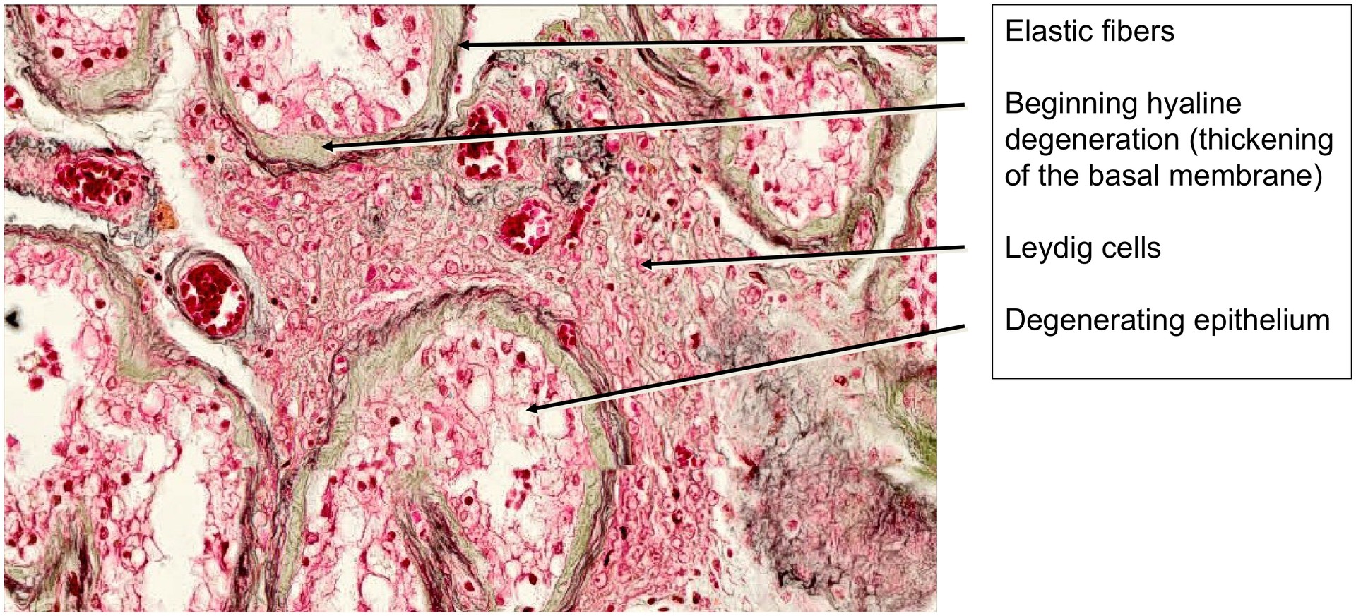
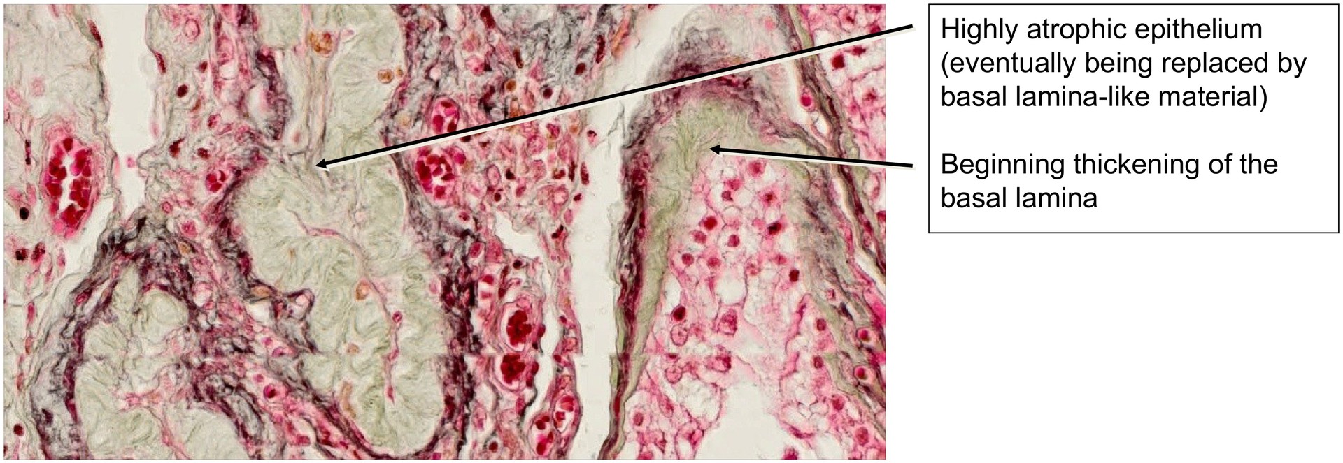
Atrophic tubules, where the epithelial lining has undergone hyaline degeneration, leading to loss of the lumen and replacement by homogeneous, basement membrane-like material.
In the remaining active tubules, the seminiferous epithelium is markedly thinner than in the adult testis, and the cell types of spermatogenesis are poorly defined. The interstitial tissue shows a reduction in Leydig cells, resulting in decreased testosterone production.
A characteristic feature of the senescent testis is the accumulation of elastic fibers, particularly in the tunica albuginea and the interstitial connective tissue, which can be clearly visualized due to the RGAN staining.
Tasks:
• Identify the intact and atrophied seminiferous tubules and compare their structure.
• Compare the elderly testis with the adult testis and describe the histological differences.
• Observe the abundance and distribution of elastic fibres in the specimen.
• Search for remaining stages of spermatogenesis within the seminiferous epithelium, if present.
License
University of Basel
Downloads