LYMPHATIC ORGANS (ANATOMICAL MICROSCOPY)
15.6
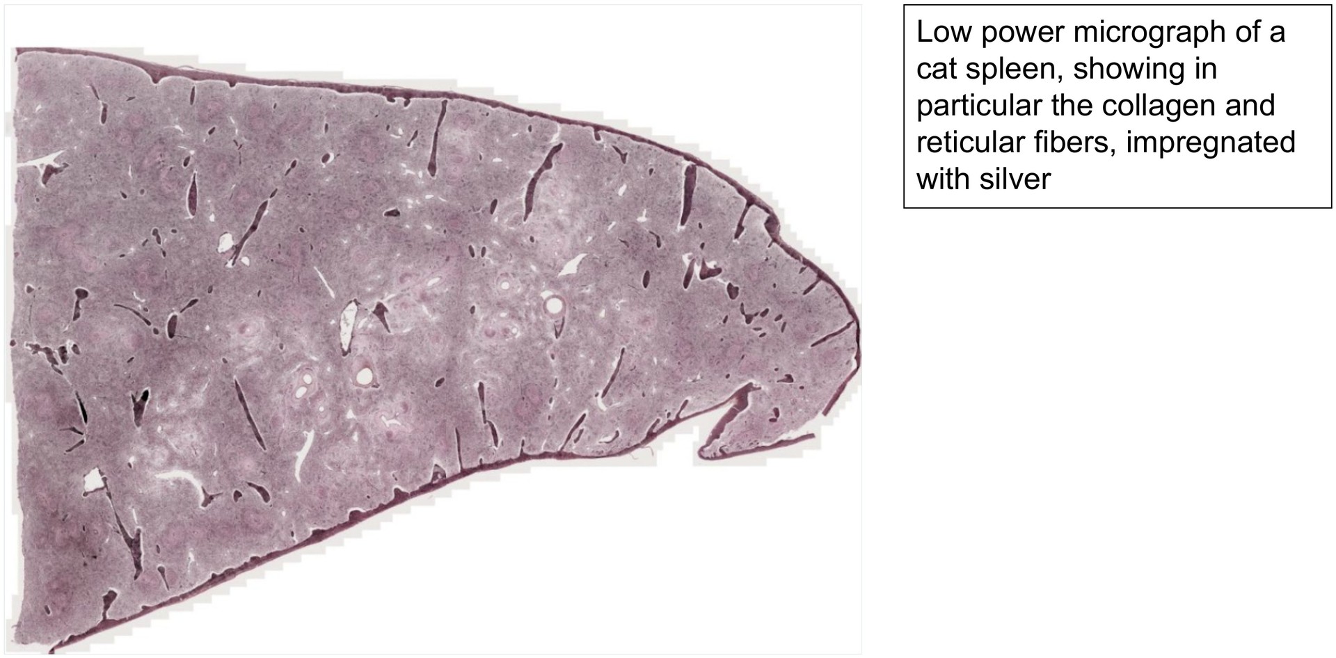
Spleen, cat 2
Specimen Details:
Specimen Details:
Organ: Spleen
Origin: Cat
Staining: Gomöri Silver Impregnation
Method and Specimen Description:
The Gomöri silver impregnation technique primarily highlights collagen fibers, including reticular fibers (collagen type III). In the spleen, this staining is particularly useful for distinguishing the different components of the vascular system, which vary in their collagenous sheaths.
Objective of the Examination:
To study the vascular system of the spleen and its relationship to the red and white pulp.
It is recommended to examine this preparation after the following specimens:
- Spleen, Cat 1 (Goldner stain)
- Spleen, Human (H&E stain)
Specific Features of the Specimen:
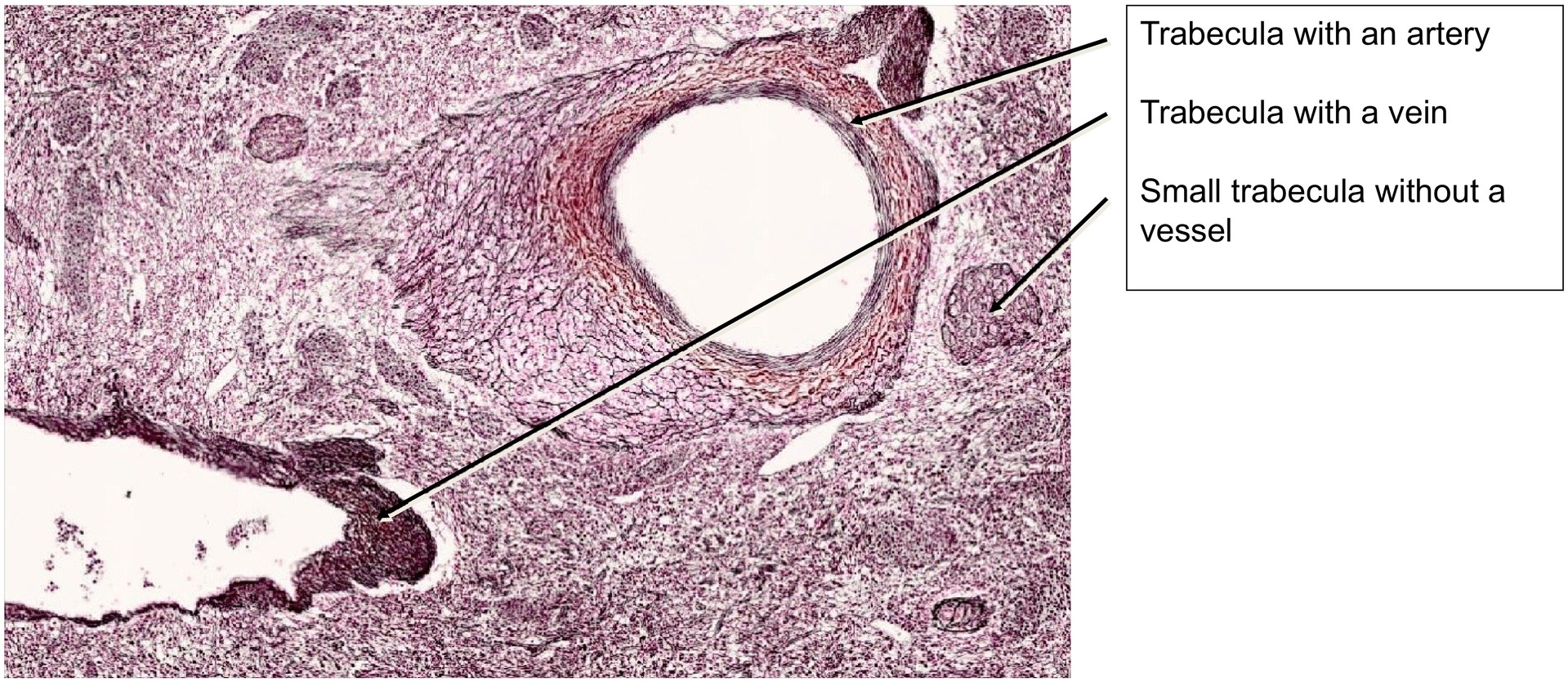
At low magnification, the trabeculae with their vessels extending from the hilum into the splenic parenchyma are easily recognized. Trabeculae containing arterial branches appear less darkly impregnated (less black) than those with venous branches, reflecting the differing density of their collagen fibers.
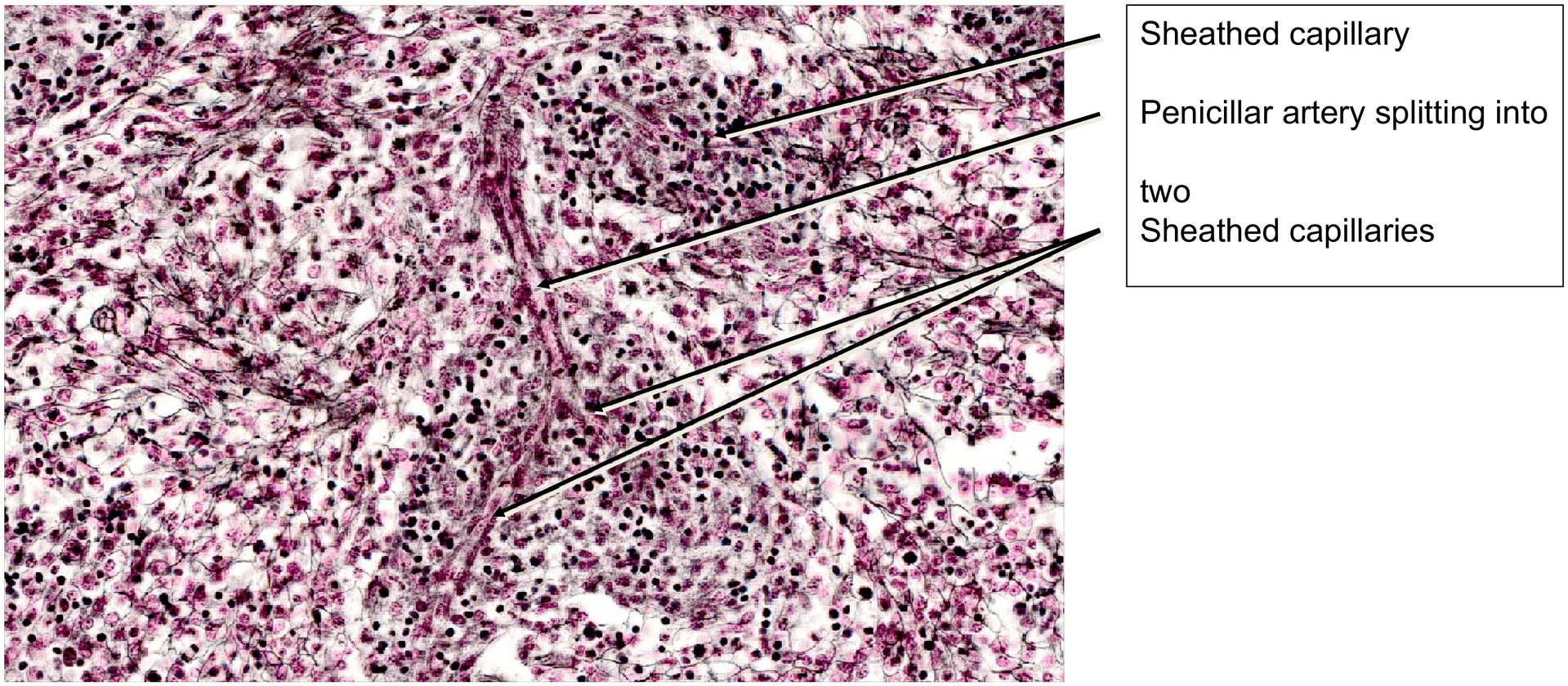
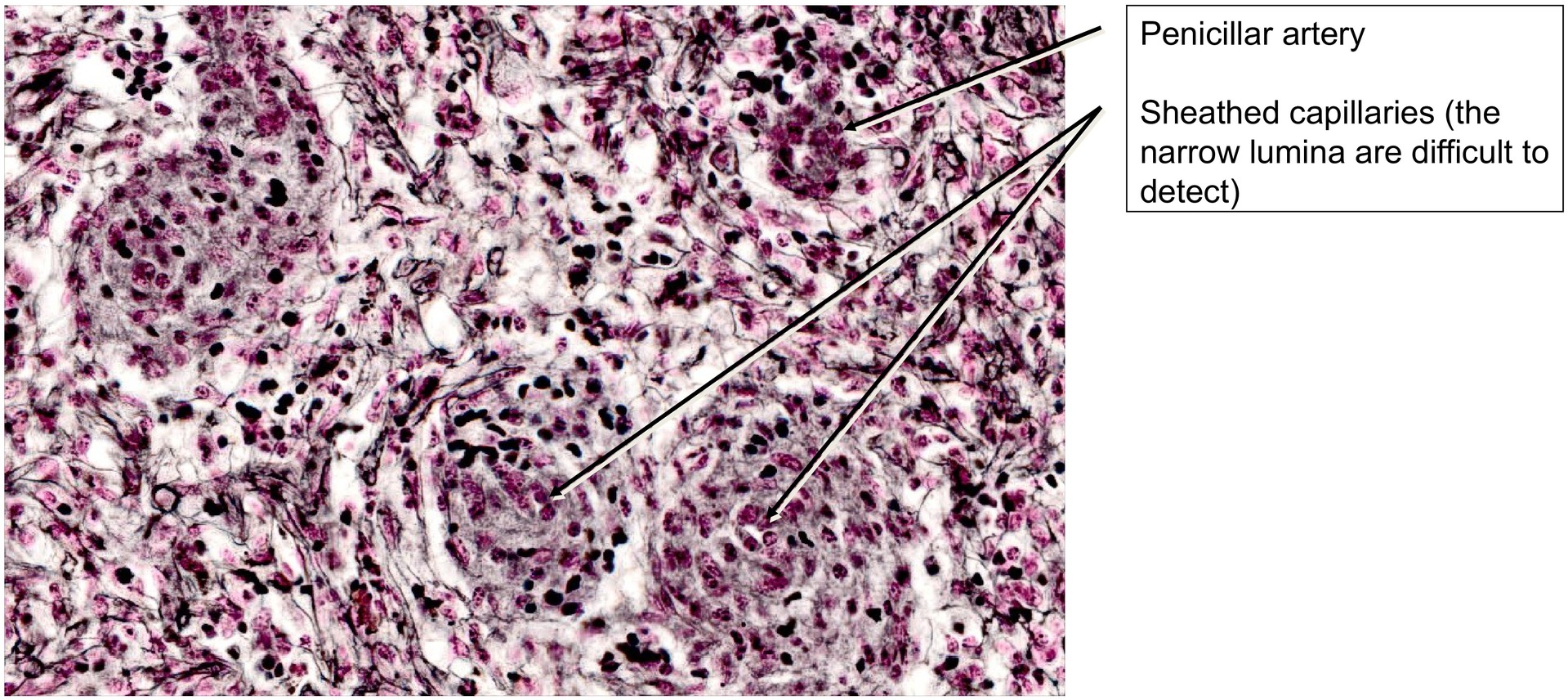
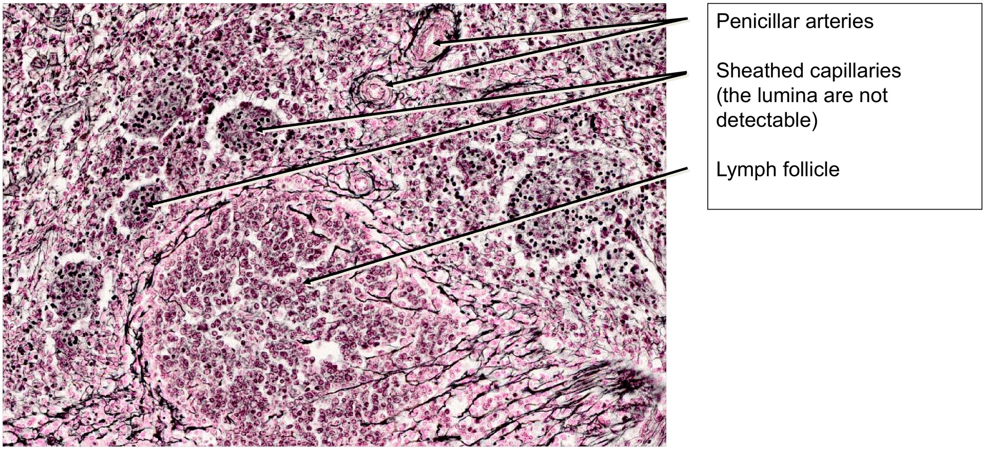
At medium magnification, cell clusters in the sheaths of the capillaries are conspicuous, sharply demarcating these regions from the surrounding tissue. Lumina are usually not visible in most sheathed capillaries, as they are very narrow and may not be included depending on the section plane. In contrast, the penicillar arteries show a clearly visible lumen but are less frequently encountered when scanning the specimen.
Because the collagen fibers are clearly displayed by the silver impregnation, the various regions of the spleen can be differentiated by the distribution and density of collagen fibers (collagen type III).
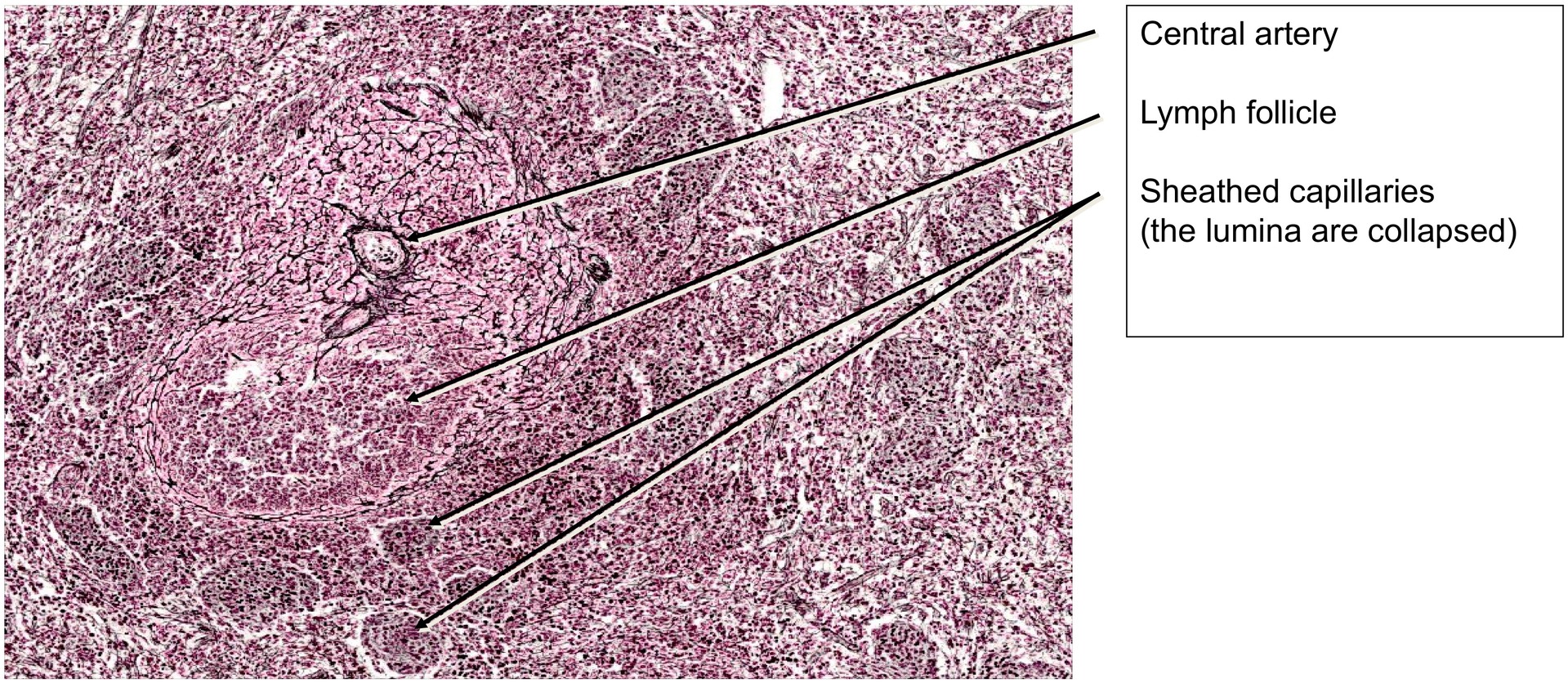
- Lymph follicles contain relatively few collagen fibers.
- Central arteries, usually located nearby, have more numerous but loosely arranged collagen fibers.
- Sheathed capillaries are notable for their cell clusters and possess collagen fibers only in their outer zones.
- The trabeculae contain the densest and most prominent collagen fibers.
- Penicillar arteries are surrounded by a fine collagen fiber network, unlike the capsular capillaries.
Tasks:
- At low magnification, assess the relative amount of connective tissue, such as trabeculae and collagen fibers, in comparison with the overall parenchymal volume of the spleen.
- Locate the sheathed capillaries and observe their structural characteristics.
- Identify penicillar arteries, which are frequently found in proximity to the capsular capillaries.
- Search for lymph follicles and determine the position of the central artery within different sectional planes.
- Distinguish between arteries and veins within the trabeculae based on their wall structure and fiber density.
License
University of Basel
Downloads