DIGESTIVE ORGANS: LIVER, GALLBLADDER, PANCREAS (ANATOMICAL MICROSCOPY)
20.1
Gallbladder
Specimen:
Specimen Details:
Organ: Gallbladder Origin: Cat Staining: Haematoxylin - Eosin (H&E)
Method and Specimen Description:
Normal histological specimen stained with H&E (a general overview stain).
Objective of the Examination:
To study the structure of the gallbladder, with emphasis on its wall organization, epithelial features, and the characteristic mucosal bridges formed by the folding of the mucosa.
Specific Features of the Specimen:
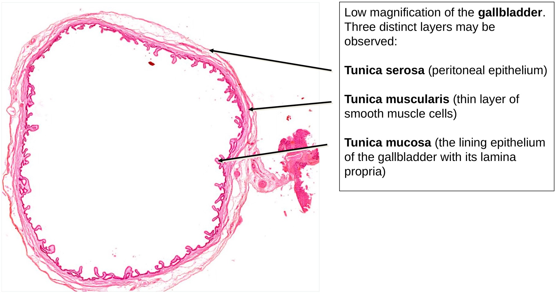
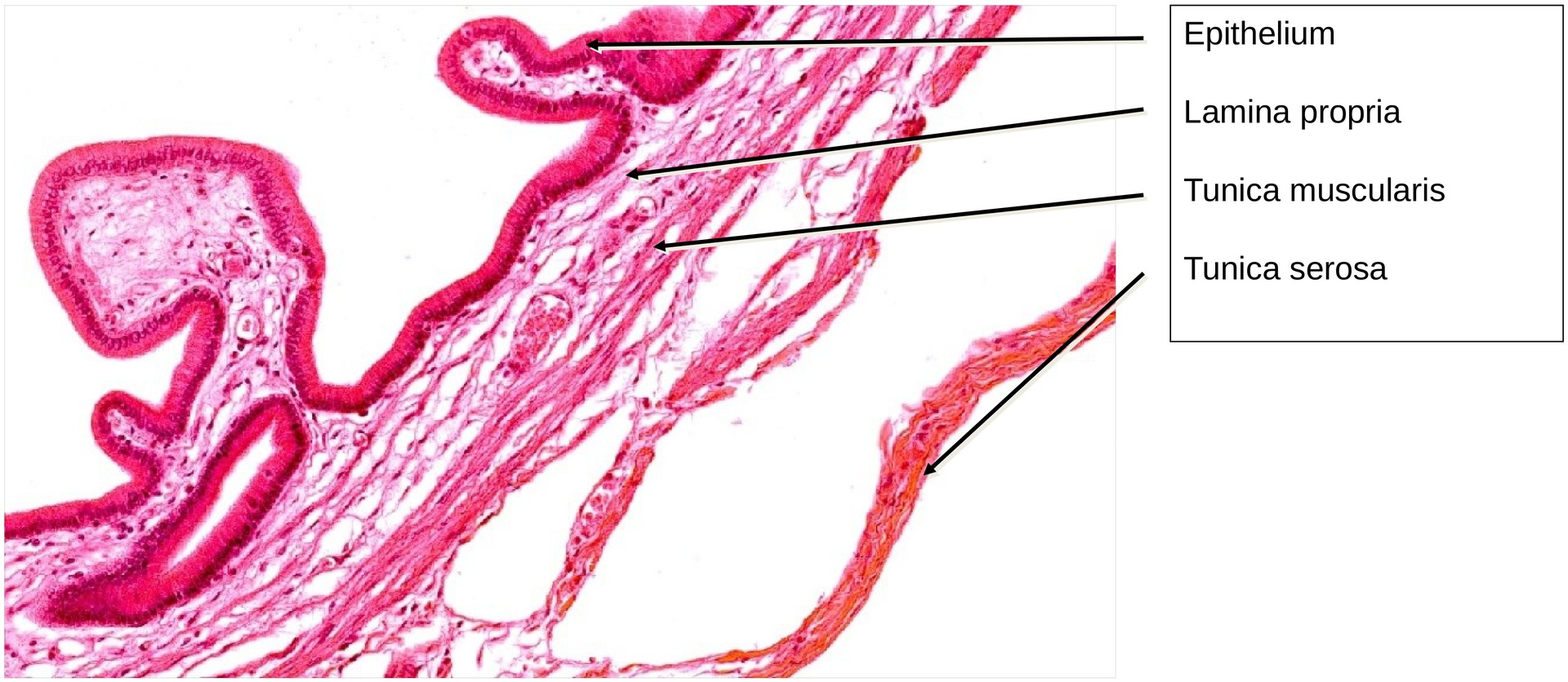
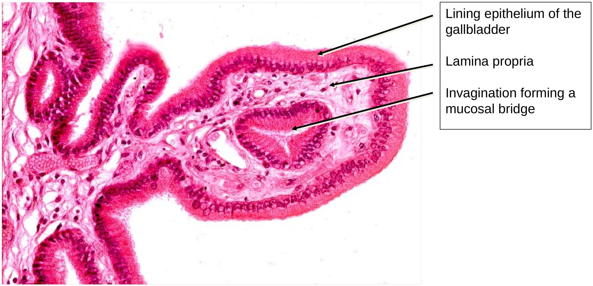
The wall of the gallbladder consists of three layers, from the outside inwards: 1. Tunica serosa – a peritoneal epithelium (mesothelium) with a subepithelial layer of dense connective tissue. 2. Tunica muscularis – a thin, irregular layer of smooth muscle cells, arranged in interlacing bundles without a clear directional pattern. 3. Tunica mucosa – composed of a simple columnar epithelium and its lamina propria (connective tissue core).
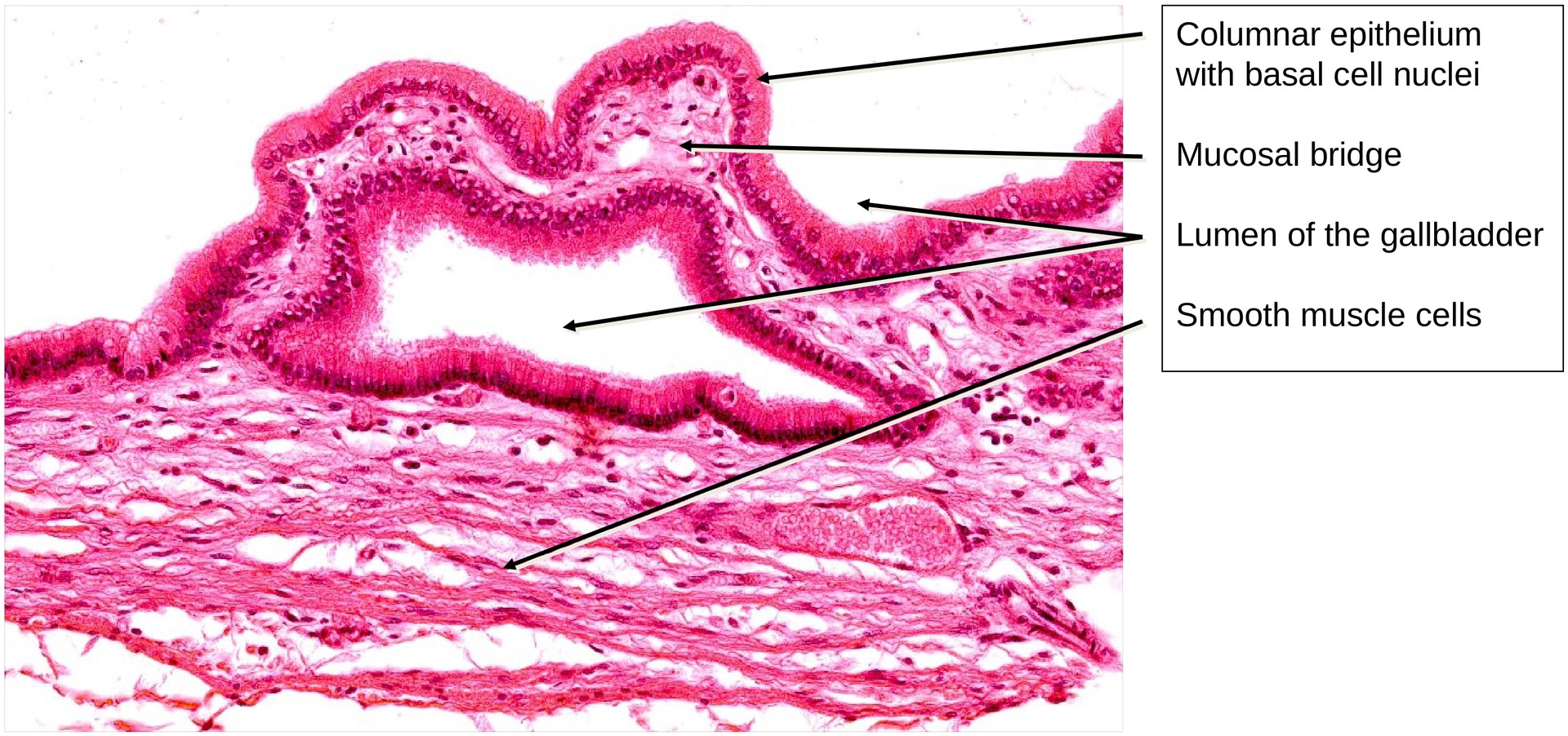
The mucosa shows a highly folded surface, forming mucosal bridges that often appear as closed loops in section. However, the lumina of these folds always communicate with the main cavity of the gallbladder — no completely isolated chambers are present.
The epithelial cells are tall columnar cells with numerous apical microvilli, which increase the absorptive surface area. Their main function is the concentration of bile through water resorption.
The cells also contain secretory granules, which produce a protective mucus layer that shields the epithelium from the potentially irritating bile contents.
The tunica muscularis is thin, and its smooth muscle bundles are interwoven, forming a network rather than distinct circular or longitudinal layers.
The tunica serosa forms the outermost covering and consists of mesothelium and underlying connective tissue, which merges seamlessly with the surrounding peritoneum.
Tasks:
- Examine the entire specimen and observe that its wall structure is consistent throughout, as expected for a bladder-like hollow organ.
- Identify the three layers of the gallbladder wall: tunica serosa, tunica muscularis, and tunica mucosa.
- Describe the epithelium — note the columnar cells, microvilli, and secretory granules.
- Explain the concept of mucosal bridges and verify that their lumina remain in continuity with the main cavity.
- Observe the tunica muscularis, focusing on the interlacing pattern of its smooth muscle bundles.
- Characterise the tunica serosa, noting the mesothelial covering and its connection to the peritoneum.
License
University of Basel
Downloads