FEMALE REPRODUCTIVE ORGANS (ANATOMICAL MICROSCOPY)
10.2
Chorion
Specimen Details:
Specimen Details:
Organ: Chorion
Origin: Human
Staining: Azan
Method and Specimen Description:
Routine histological section from an early stage of pregnancy, stained with Azan, which colors epithelial cells and erythrocytes red, and connective tissue fibers blue.
Objective of the Examination:
To study the formation of early chorionic villi, with particular attention to the syncytiotrophoblast and cytotrophoblast layers.
Special Features of the Specimen:
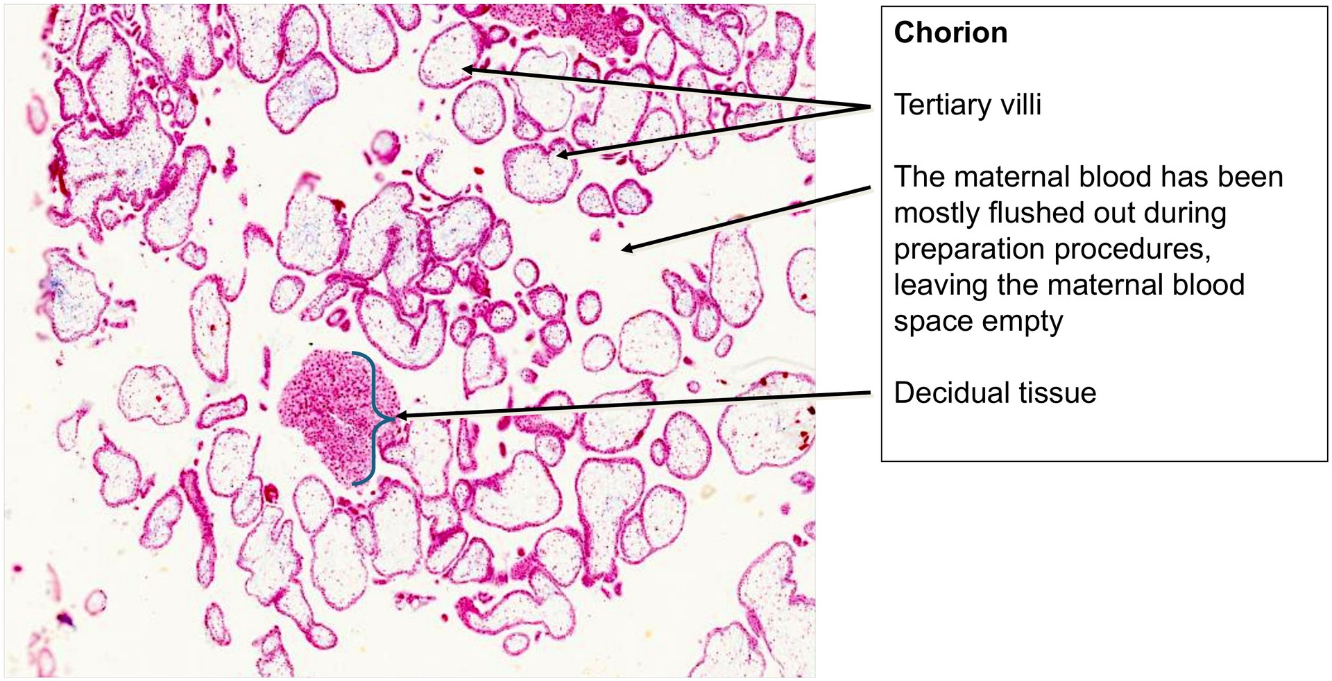
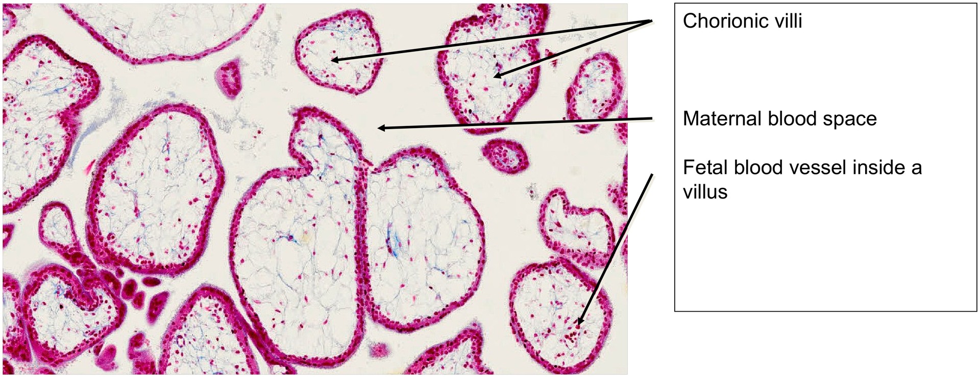
This specimen represents the chorion during early pregnancy, at a stage when chorionic villi are already formed. The villi are surrounded by maternal blood, which has mostly been washed out during specimen preparation.
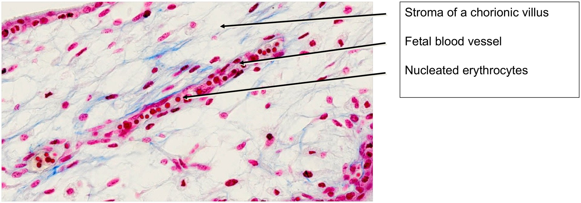
Within the chorionic villi, embryonic blood vessels are already present, identifying them as tertiary villi.
For orientation:
-
Primary villi – solid projections of trophoblast composed only of epithelial cells.
-
Secondary villi – invasion of extraembryonic mesenchyme into the villous core.
-
Tertiary villi – formation of embryonic blood vessels within the mesenchyme.
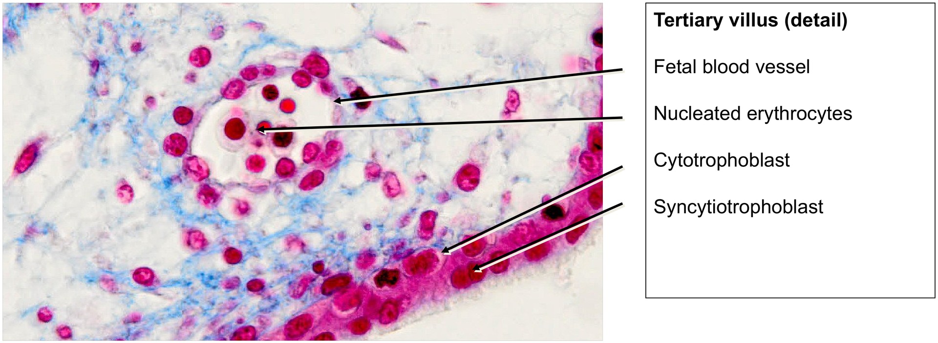
The blood within tertiary villi contains nucleated embryonic erythrocytes, characteristic of early development.
The villi exhibit the typical two-layered trophoblastic structure:
-
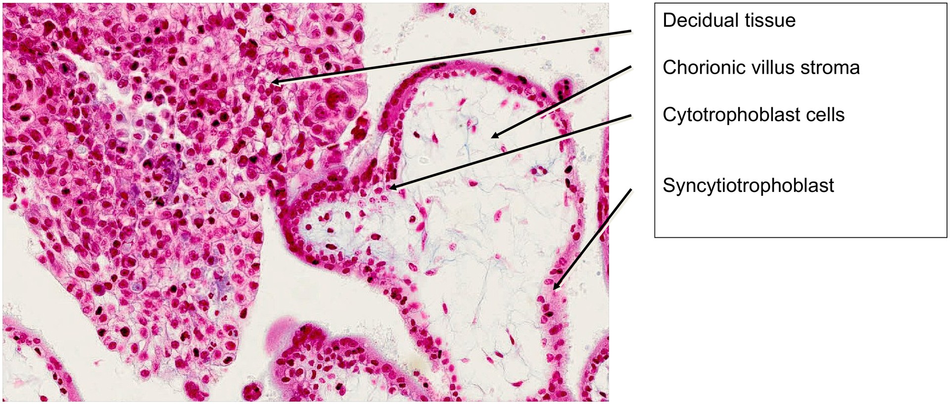
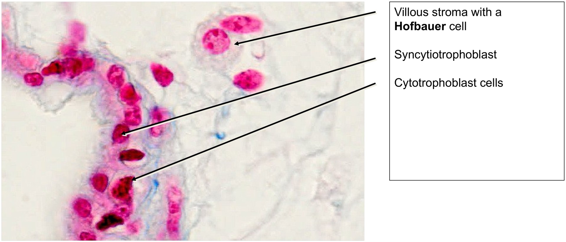
Outer layer: the syncytiotrophoblast, a multinucleated cell layer formed by the fusion of trophoblastic cells.
-
Inner layer: a single layer of cytotrophoblast cells with distinct cell boundaries and nuclei, visible in most regions.
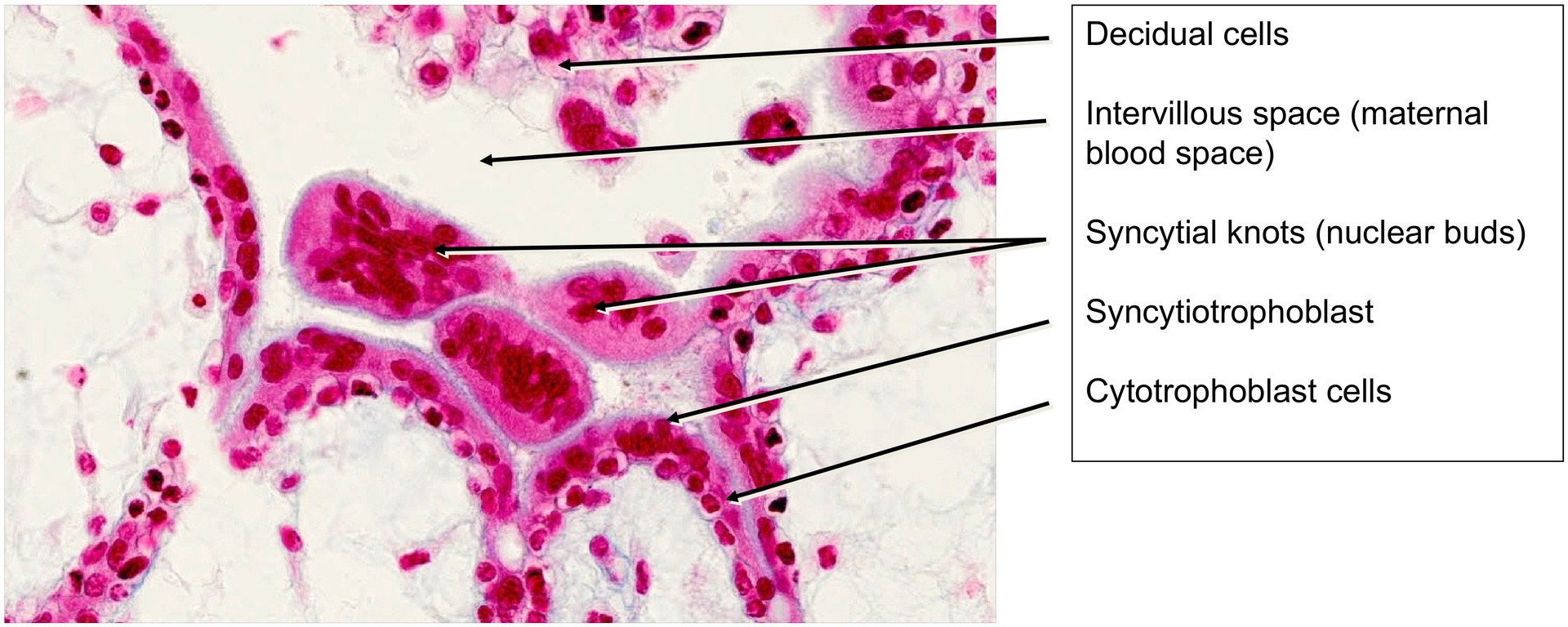
Larger tissue masses within the specimen correspond to decidual tissue derived from the maternal endometrium. Within these regions, remnants of degenerating endometrial glands can still be identified.
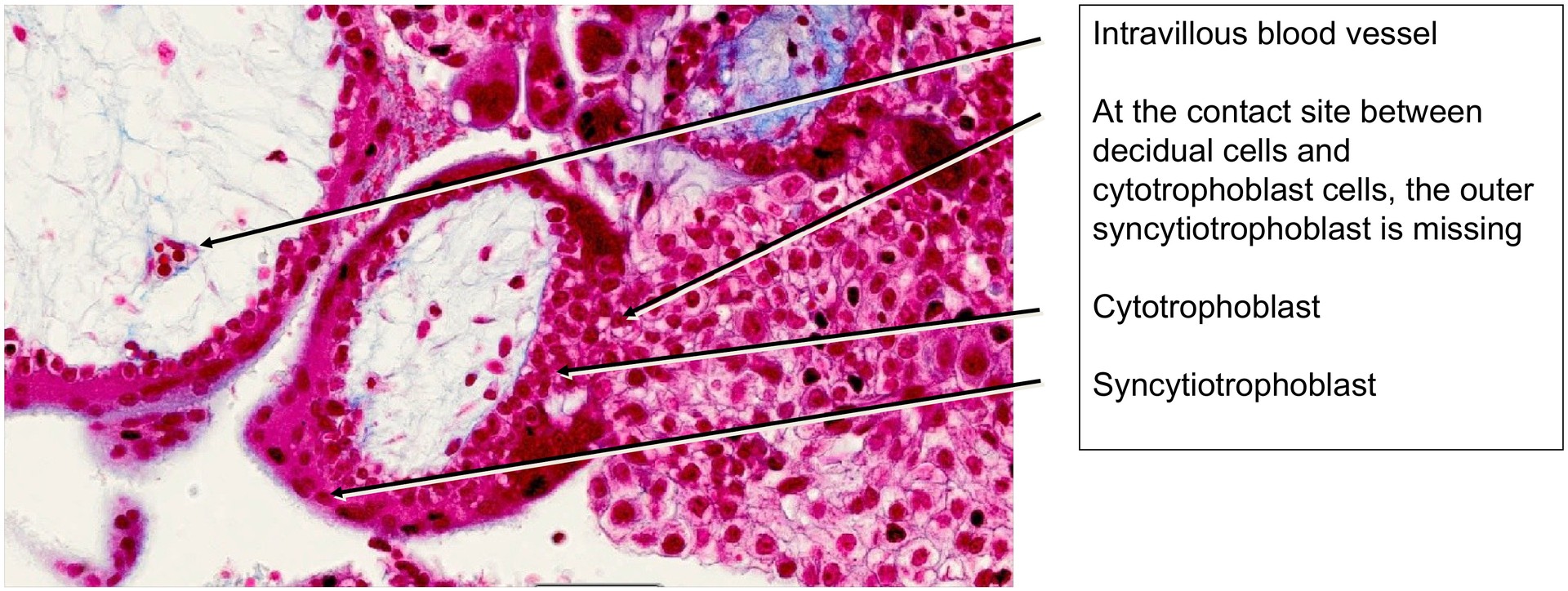
At sites where chorionic villi contact decidual cells, there is partial breakdown of the syncytiotrophoblast, allowing direct contact between the cytotrophoblast (or villous mesenchyme) and the maternal decidual cells. These junctions represent anchoring villi, which attach the developing fetal tissue to the maternal endometrium. Such contact zones are sites of important materno–fetal immunological interactions, essential for maintaining pregnancy.
In some areas, accumulations of syncytial nuclei and cytoplasmic fragments can be seen protruding from the villi. These structures are known as nuclear buds or syncytial knots.
Within the villous stroma, larger cells containing lysosomes and phagocytic inclusions are visible. These are Hofbauer cells, specialized fetal macrophages with functions including phagocytosis and immunoregulation.
Tasks:
-
Examine the chorionic villi and determine whether they are secondary or tertiary villi. What features help you make this distinction?
-
Identify blood vessels within the villi and note the predominant cell type present in their lumina.
-
Locate sites of anchoring villi formation. Which villous layer is disrupted at these attachment points?
-
Differentiate between syncytiotrophoblast and cytotrophoblast cells based on structure and staining.
-
Identify nuclear buds (syncytial knots) within or between the villi.
-
Locate decidual cells and identify any residual endometrial glandular tubules within the maternal tissue.
License
University of Basel
Downloads