CARDIOVASCULAR ORGANS (ANATOMICAL MICROSCOPY)
14.9
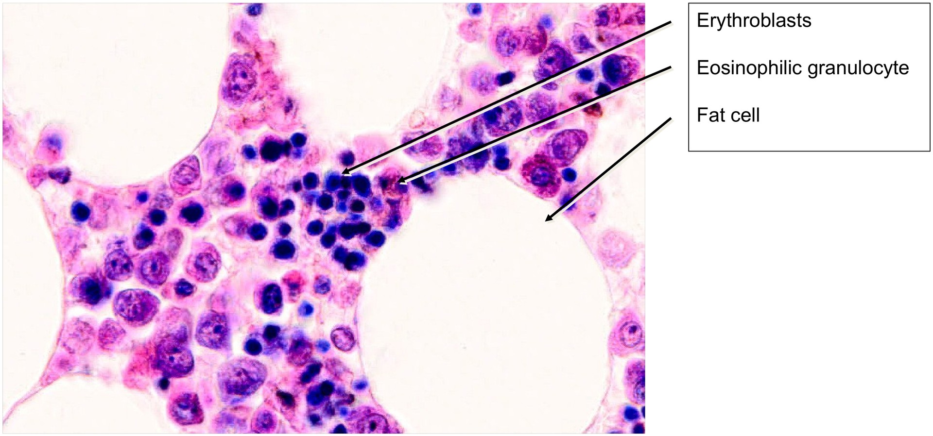
Bone marrow
Specimen:
Specimen Details:
Organ: Bone marrow
Origin: Human
Staining: May–Grünwald–Giemsa (MGG)
Method and Specimen Description:
This preparation consists of a section of red bone marrow that was fixed, embedded, and stained with May–Grünwald–Giemsa, allowing clear differentiation between the various hematopoietic cell types.
Objective of the Investigation:
To study the microscopic structure of red (hematopoietically active) bone marrow, to identify the precursor stages of blood cell development, and to recognize megakaryocytes, the progenitor cells of platelets.
Special Features of the Specimen:
Hematopoietic bone marrow is composed of a reticular connective tissue framework, within which the blood-forming cells are embedded. The reticular cells themselves are difficult to distinguish from the surrounding hematopoietic cells.
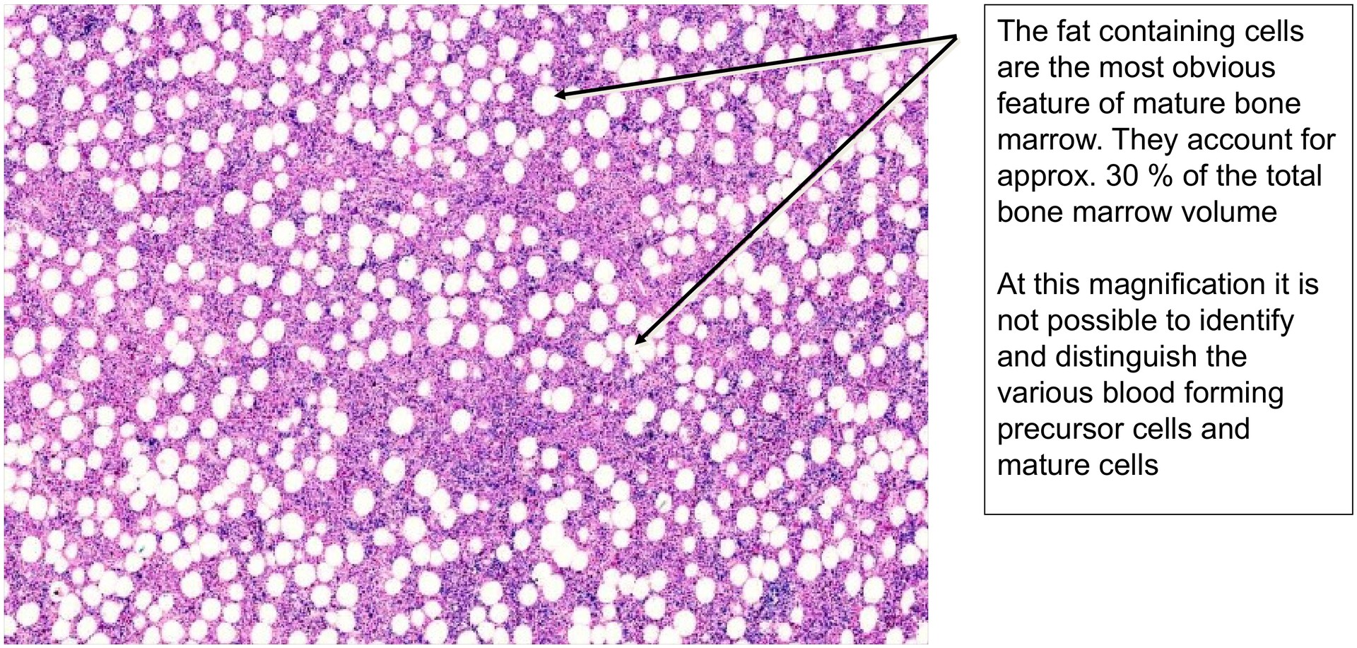
Many of the stromal areas contain fat-laden cells, which appear as empty spaces after preparation and account for approximately 30% of the bone marrow volume. This proportion decreases during enhanced hematopoietic activity (e.g. after blood loss).
During hematopoiesis, precursor cells undergo a sequence of maturation stages, resulting in the coexistence of mature and immature cells within the same field of view. The following structures can typically be identified:
- Fat cells – large, round, clear spaces (lipid content lost during processing).
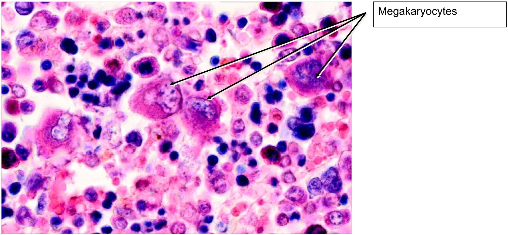
- Megakaryocytes – very large cells with lobulated nuclei; precursors of platelets.
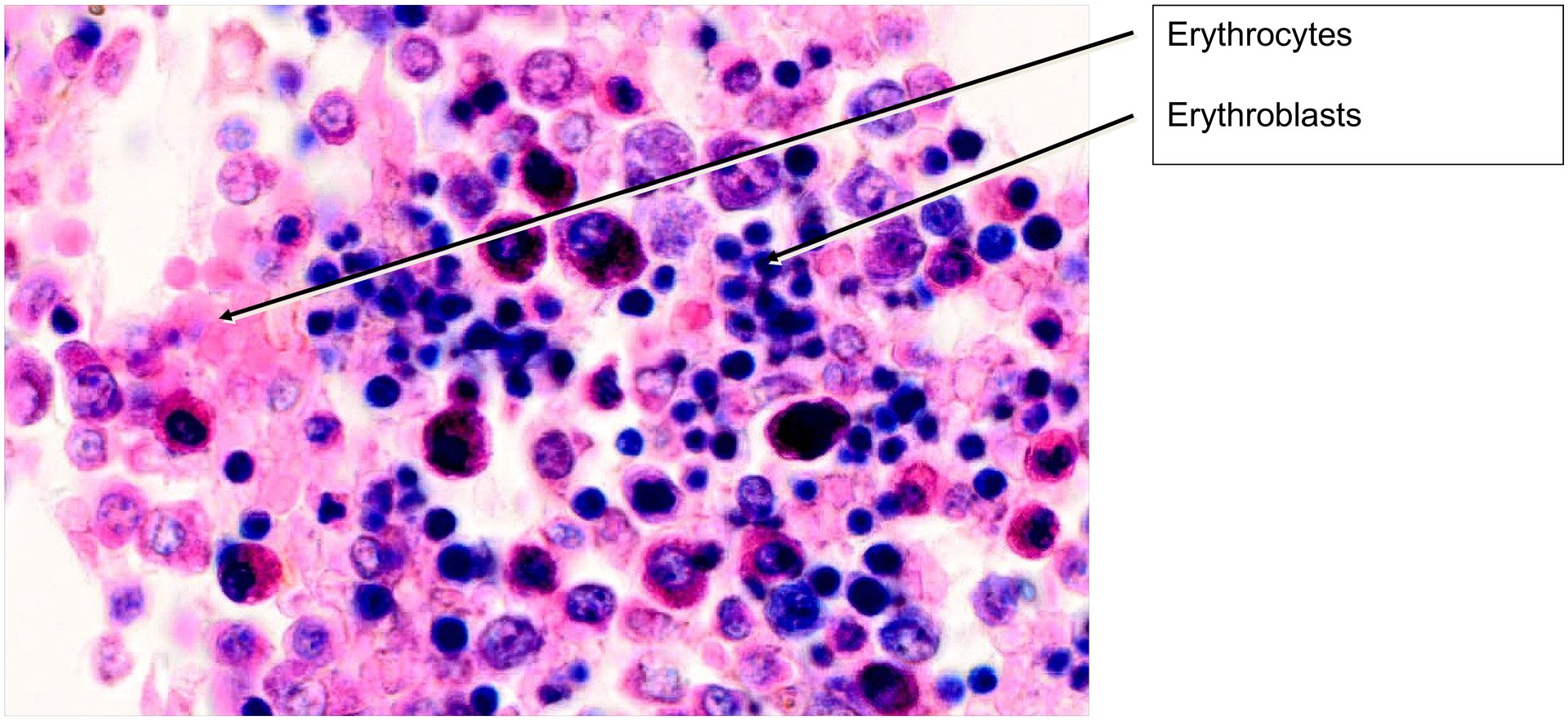
- Erythroblasts and erythrocytes – various maturation stages (E1–E5), not morphologically distinct in this specimen.
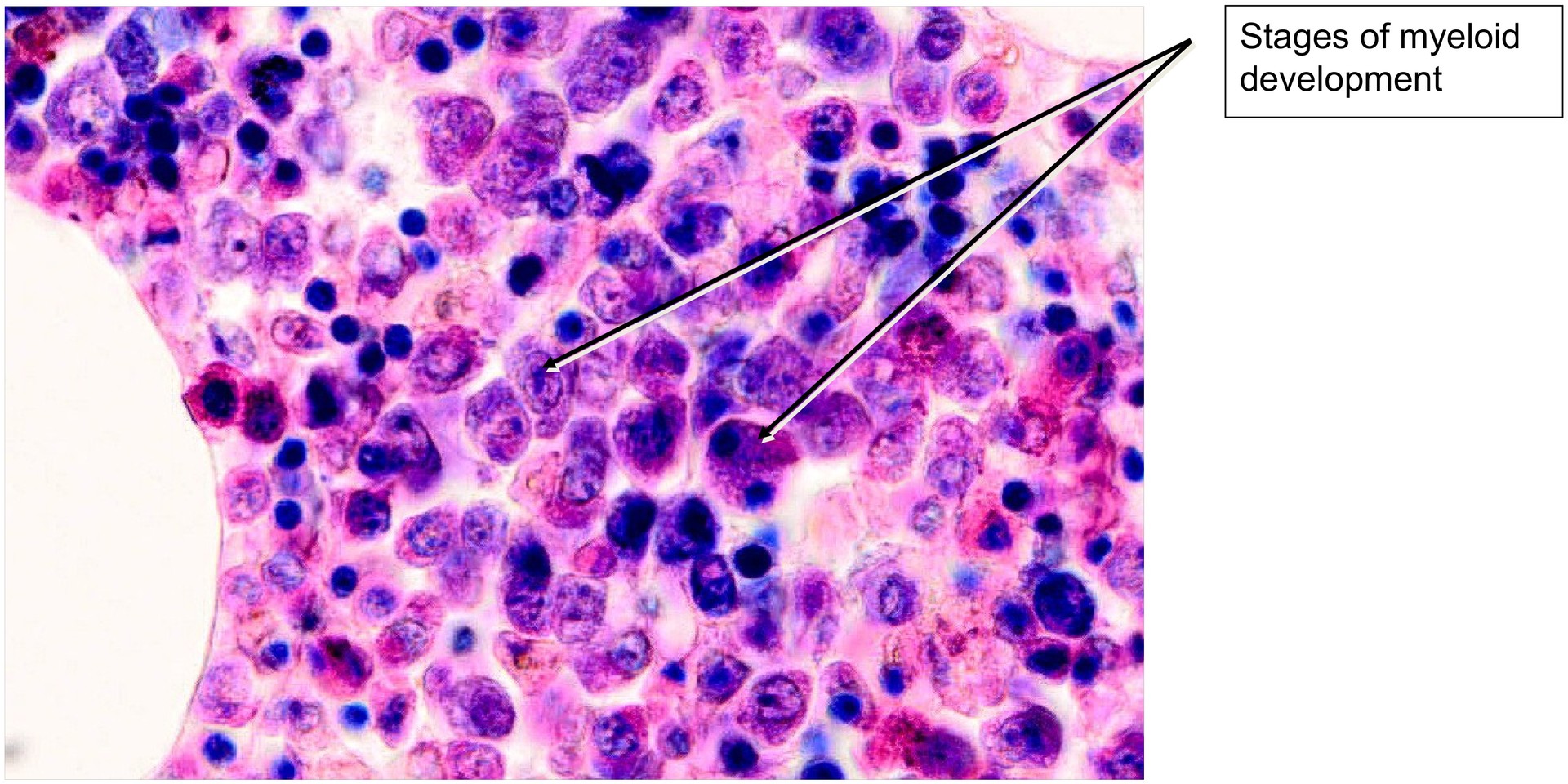
- Myelocytes and granulocytes – different stages of granulocytic development; mature granulocytes can be distinguished as eosinophilic and basophilic forms.
Further identification of the myeloid developmental stages and lymphocytes requires advanced microscopic experience and higher magnification.
Tasks:
- Begin at low magnification and observe the overall structure, noting the large proportion of lipid-containing cells.
- Locate megakaryocytes, the largest and most striking elements of the bone marrow.
- Identify mature erythrocytes and note their distribution.
- Observe clusters of erythroblasts, which appear as compact groups (colony-forming units).
- Search for mature granulocytes and distinguish between eosinophilic and basophilic types.
License
University of Basel
Downloads