CONNECTIVE TISSUE (GENERAL HISTOLOGY)
2.5
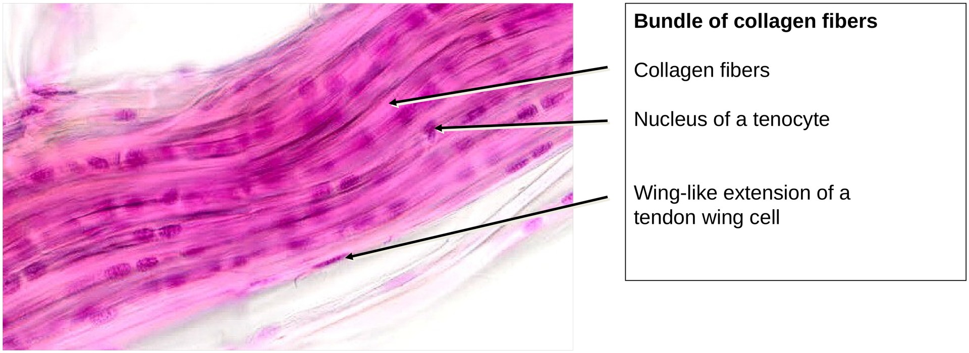
Tendon, longitudinal aspect (collagen fibers)
Specimen Details:
Specimen Details:
Organ: Tendon
Origin: Rat
Staining: Hematoxylin - Chromotrope
Method and Specimen Description:
Bundles of tendon fibers were isolated from the rat tail tendon and carefully teased apart with a fine needle. This preparation therefore represents an unsectioned whole-mount specimen, rather than a microtome section.
Objective of the Examination:
• To study the organization of dense regular connective tissue using the tendon as a model.
• To recognize that the extracellular matrix (ECM) dominates the tissue composition, while tendon cells (tenocytes or tendinocytes) occupy minimal space between collagen fibrils and fibers.
• To appreciate the functional significance of the collagen fiber arrangement in relation to mechanical stress and elasticity.
Special Features of the Specimen:
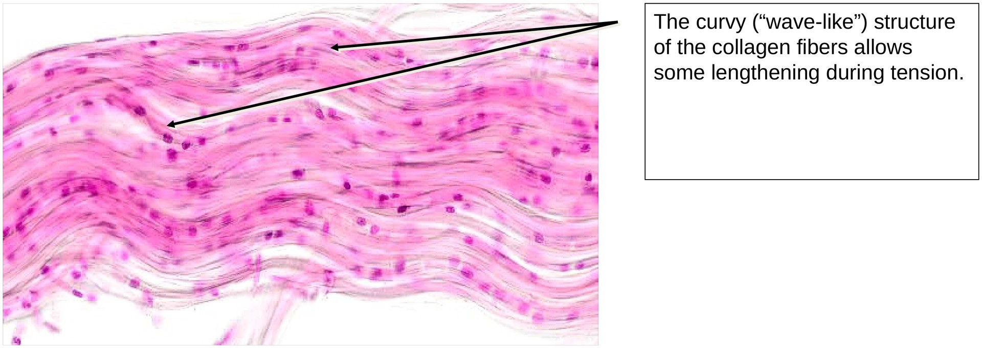
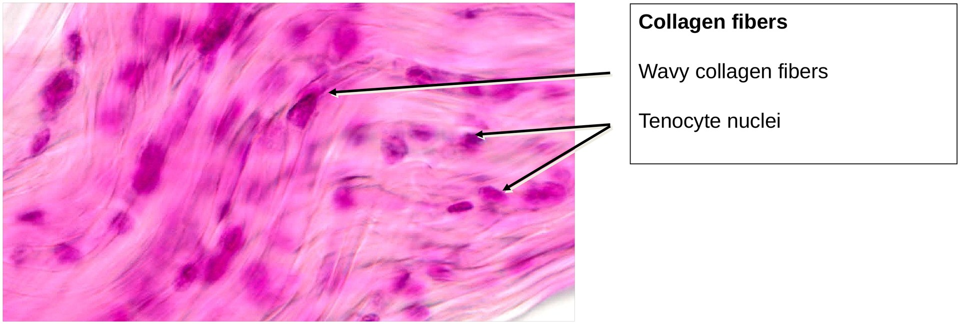
In accordance with the longitudinal mechanical stress to which tendons are subjected, the collagen fibers are arranged in parallel bundles along the longitudinal axis. Despite their alignment, individual fibers display a characteristic wavy pattern, which plays a crucial role during the initial phase of muscle contraction. This wavy arrangement allows the tendon to stretch slightly under tension, providing a spring-like response that cushions the onset of muscle activity.
The tenocytes (also known as tendinocytes) are elongated fibroblast-like cells arranged in rows resembling a string of pearls. Because of the dominance of collagen, there is limited intercellular space, and the tenocytes must extend lateral processes between adjacent collagen fiber bundles. These processes give the cells a wing-like appearance, which is why they are also termed tendon wing cells.
Under the microscope, the appearance of tenocytes depends on the focal plane during scanning:
-
When the cell processes are in focus → the cells appear narrow and elongated.
-
When the nuclear region is in focus → the cells appear wider and more distinct.
Tasks:
• At high magnification, identify the tenocytes, noting their arrangement in longitudinal rows (“string-of-pearls” pattern).
• Distinguish between thicker and thinner bundles of collagen fibers.
• Observe the natural waviness (curling) of the collagen fibers — it is a real structural feature, not an artefact.
• Estimate the relative volume ratio of cells to extracellular matrix within the tendon, recognizing that the ECM predominates.
License
University of Basel
Downloads