CONNECTIVE TISSUE (GENERAL HISTOLOGY)
2.1
Elastic fibers (nuchal ligament)
Preparation:
Preparation Details:
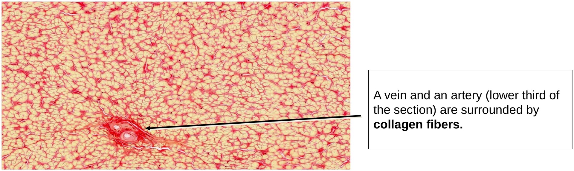
Organ: Nuchal ligament
Origin: Cow
Staining: Van Gieson
Method and Preparation Description:
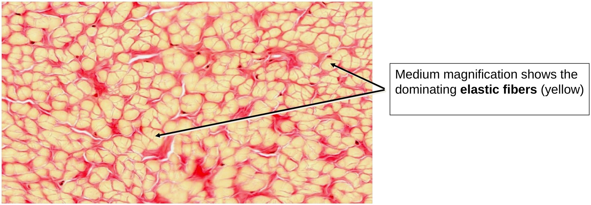
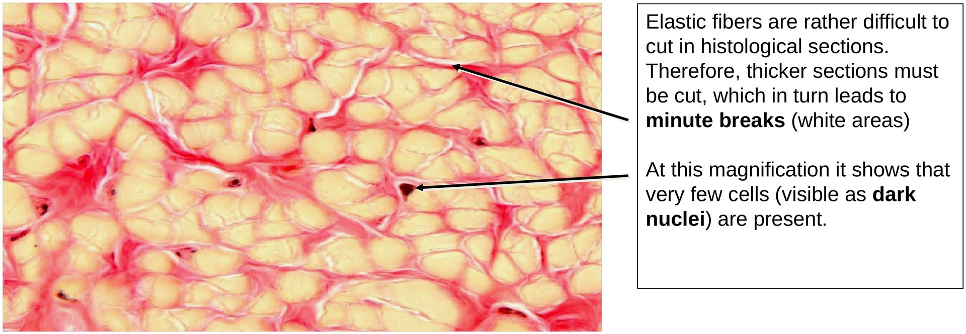
Elastic fibers are relatively difficult to process, so the sections in this preparation are slightly thicker than usual. Thicker sections cannot be focused as sharply under the microscope — or with a digital scanner — as thinner ones. With Van Gieson staining, collagen fibers appear red, whereas elastic fibers are stained yellow.
Objective of the Examination:
To study the structure of elastic ligaments and to recognize the differences between elastic ligaments and purely collagenous tendons. Additionally, to observe how collagen fibrils bundle and support the elastic fibers.
Special Features of the Specimen:
General Features (overview, low magnification): The cross-section of the nuchal ligament shows a distinctive pattern formed by the intermingling of elastic and collagen fibers.
Fine Structure (high magnification): The elastic fibers vary in diameter and appear yellow. Each elastic fiber is surrounded by red-stained collagen fibers, forming supporting sheaths. The dark-stained nuclei of fibroblasts can be observed adjacent to the fibers. Small blood vessels are present between the bundles of fibers.
Tasks:
• Estimate the proportion of cells relative to the total tissue volume of the nuchal ligament.
• Locate a small vessel supplying the ligament.
• At high magnification, examine the red-stained rims surrounding the elastic fibers — these correspond to collagen fibrils that form bundles enclosing the elastic fibers.
License
University of Basel
Downloads