ENDOCRINE ORGANS (ANATOMICAL MICROSCOPY)
9.7
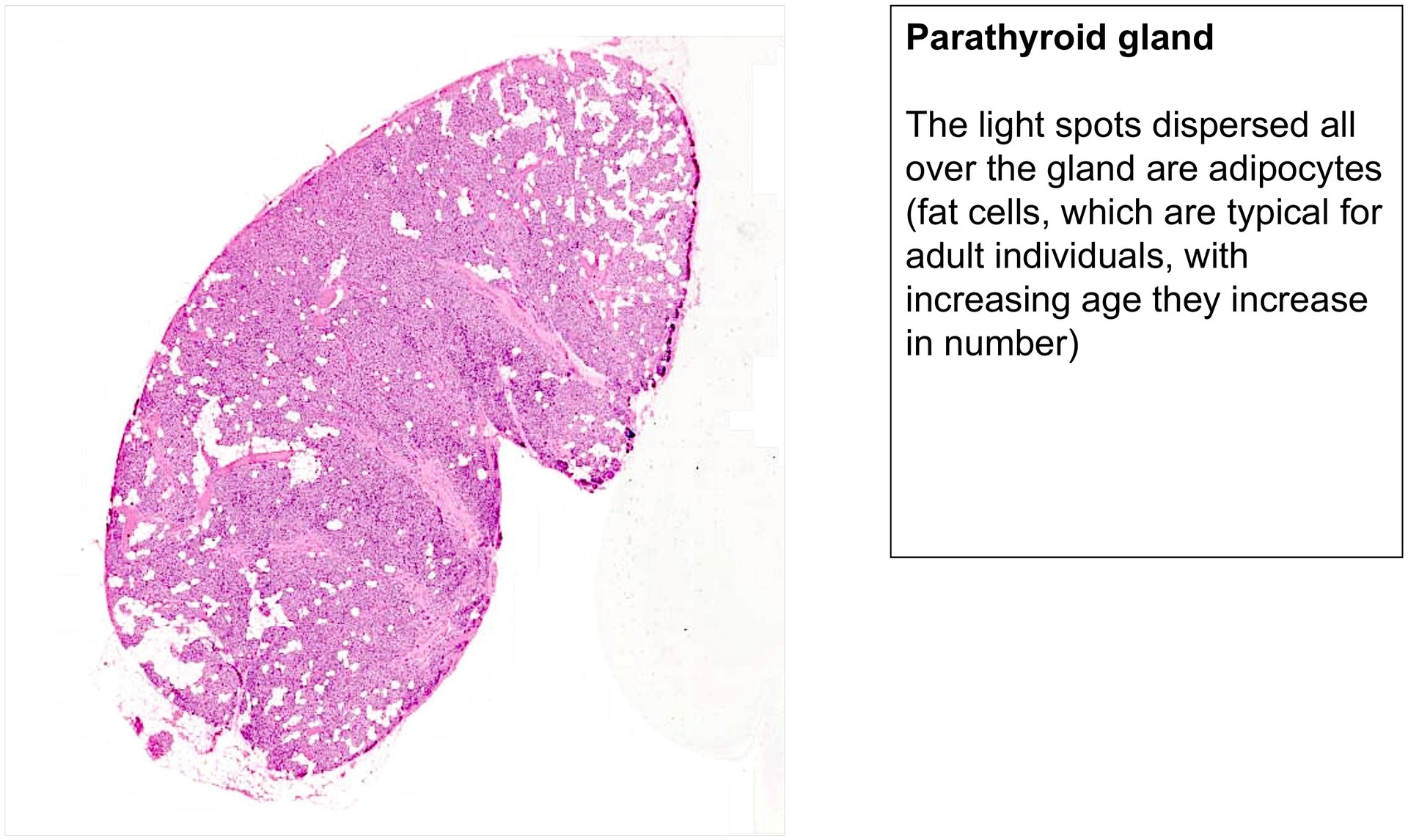
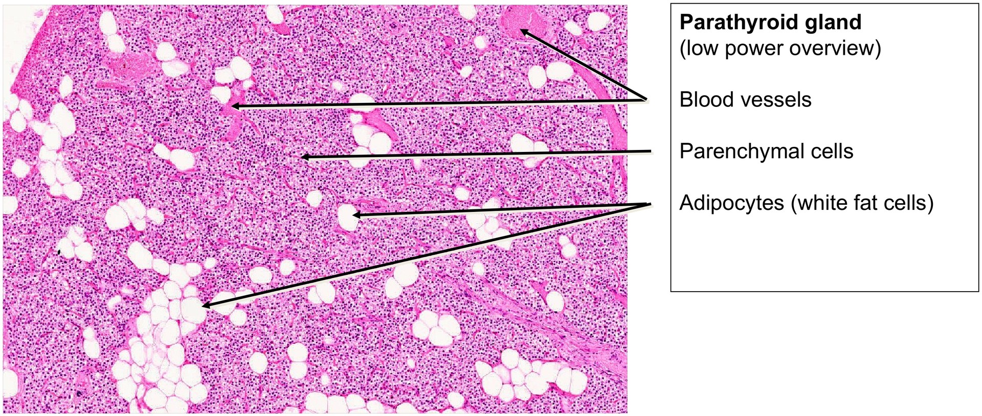
Parathyroid gland
Specimen Details:
Specimen Details:
Organ: Parathyroid gland
Origin: Human
Staining: Haematoxylin-Eosin (H&E)
Method and Specimen Description:
Routine histological section stained with H&E, providing an overview of the cellular and vascular architecture typical of endocrine tissue.
Objective of the Examination:
To understand the structure of the parathyroid gland as a prototype of endocrine organs, characterized by its epithelial cell organization and rich vascularization, both essential for efficient hormone release into the bloodstream.
Special Features of the Specimen:
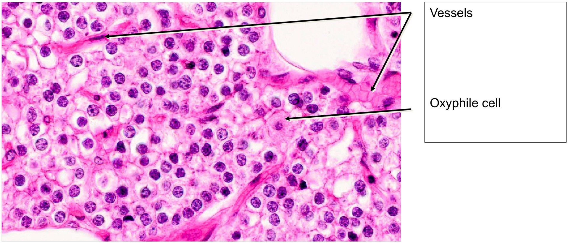
At first inspection, the parathyroid gland appears relatively inconspicuous. It is composed of tightly packed epithelial cells interspersed with an extensive capillary network, which is clearly visible in this preparation due to the H&E staining.
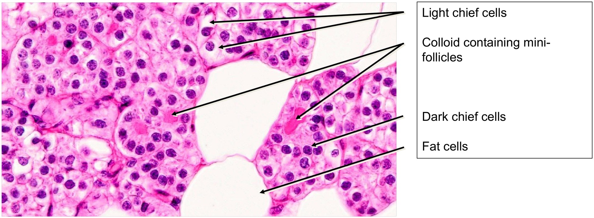
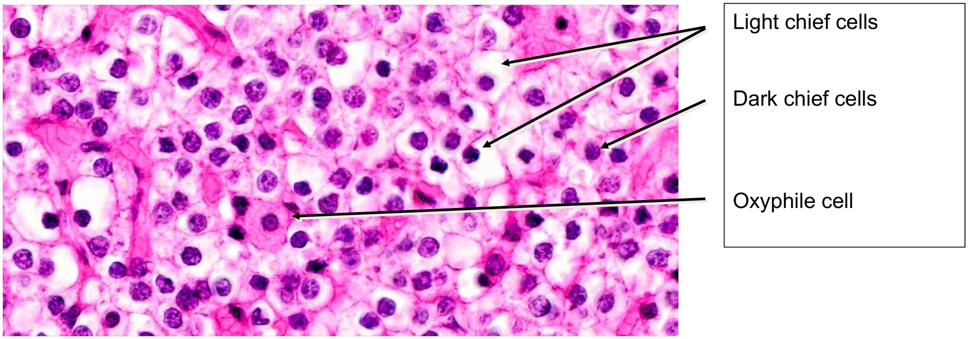
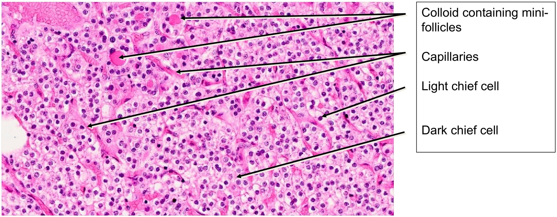
Three main cell types can be distinguished within the epithelial component: light chief cells, dark chief cells, and oxyphilic cells.
-
Light chief cells contain abundant glycogen in vivo, which is lost during fixation and processing, resulting in a pale cytoplasm.
-
Dark chief cells are thought to represent chief cells in a resting or depleted secretory state. Both types of chief cell synthesize and secrete parathyroid hormone (PTH), which regulates calcium and phosphate balance in the body.
-
Oxyphilic cells are larger, with intensely eosinophilic cytoplasm due to their high mitochondrial content. Their nuclei are typically small and pyknotic. Their exact function remains uncertain, although they may represent an inactive or degenerative stage of chief cells.
Another typical feature of the parathyroid gland is the presence of numerous adipocytes (fat cells) interspersed throughout the tissue, particularly in adult specimens. Their proportion increases with age. Small colloid-like accumulations are occasionally observed, forming mini-follicles, which can be easily overlooked at low magnification. These are non-functional and have no hormonal significance.
Tasks:
-
Identify the specimen based on its epithelial cell arrangement, rich vascularisation, and the presence of light and dark chief cells.
-
Examine the circumference of the specimen to determine whether it represents a section through the entire gland or only part of it. What can you infer about the overall size of the parathyroid gland?
-
Locate oxyphilic cells (reddish, homogeneous cytoplasm; small, pyknotic nuclei).
-
Assess the amount and distribution of adipocytes within the gland.
-
Identify which cells produce parathyroid hormone (PTH).
-
Search for mini-follicles containing colloid-like material and note their distribution.
License
University of Basel
Downloads