DIGESTIVE ORGANS: GASTROINTESTINAL TRACT (ANATOMICAL MICROSCOPY)
19.3
Duodenum
Preparation:
Preparation Details:
Organ: Duodenum
Origin: Human
Staining: Azan
Method and Specimen Description:
Normal histological section stained with Azan, which colors muscle, epithelium and erythrocytes red, and connective tissue blue.
Objective of the Examination:
To gain knowledge of the first segment of the small intestine, recognizing its characteristic Brunner’s glands and the layered organization typical of the gastrointestinal tract (GIT).
Special Features of the Preparation:
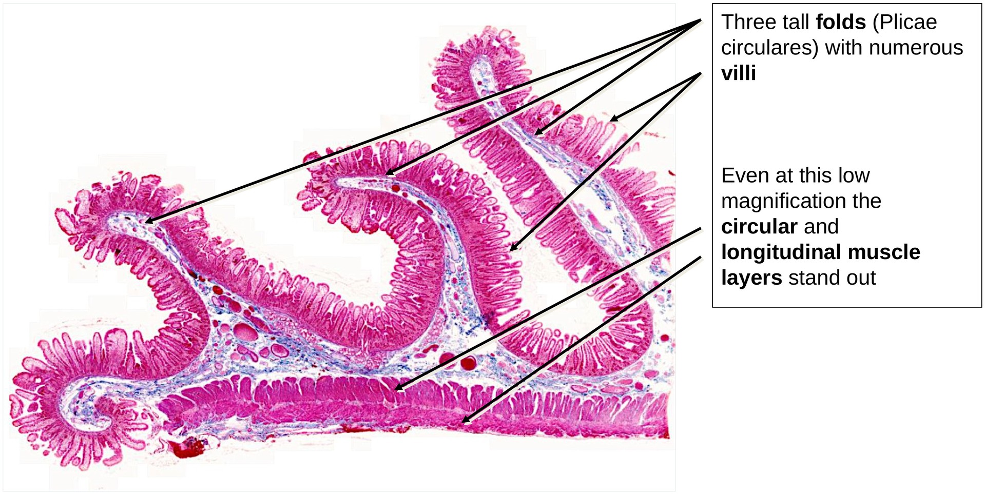
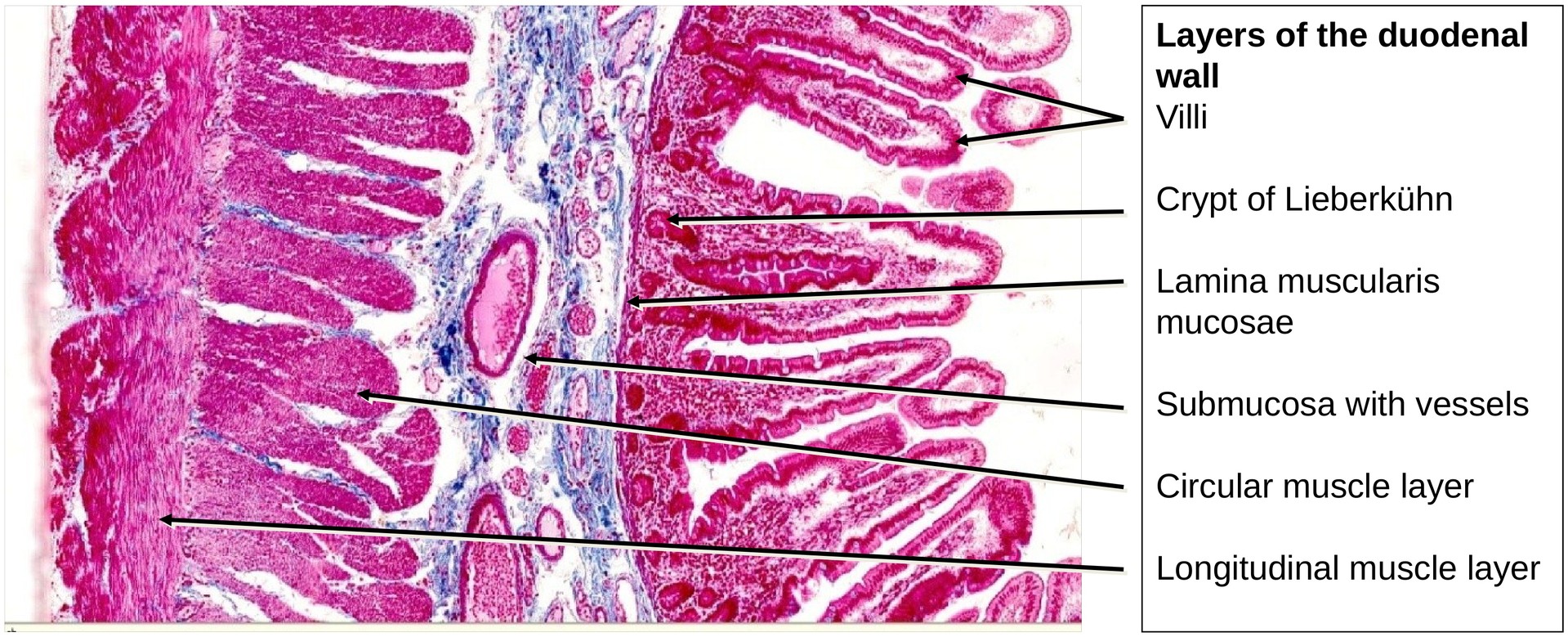
The duodenum follows the standard GIT wall structure, consisting of:
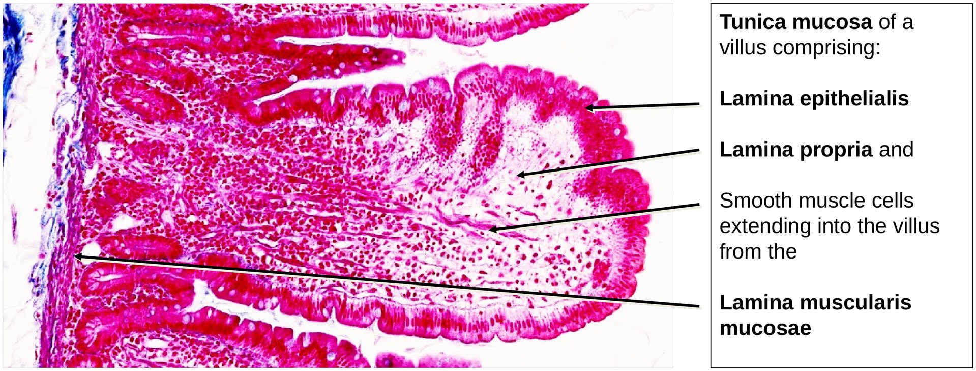
- Tunica mucosa (comprising the lamina epithelialis, lamina propria, and lamina muscularis mucosae)
- Tela submucosa (loose connective tissue)
- Tunica muscularis (inner circular and outer longitudinal smooth muscle layers)
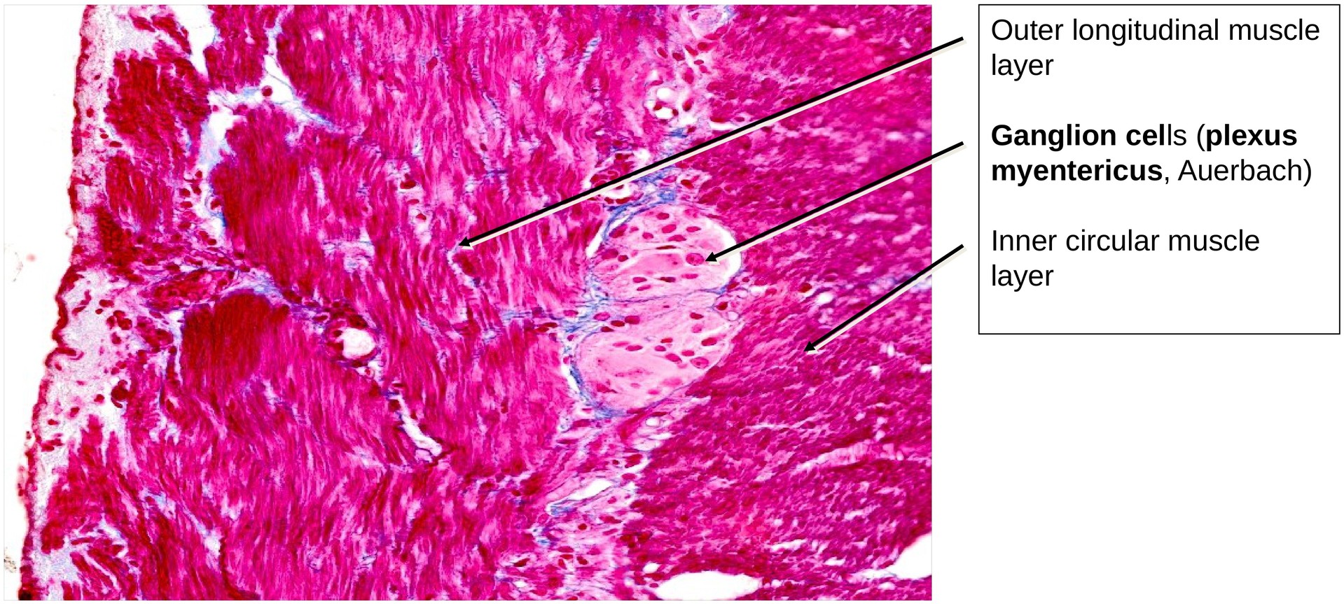
Within the submucosa lies the submucosal (Meissner’s) plexus, and between the muscle layers of the muscularis, the myenteric (Auerbach’s) plexus.
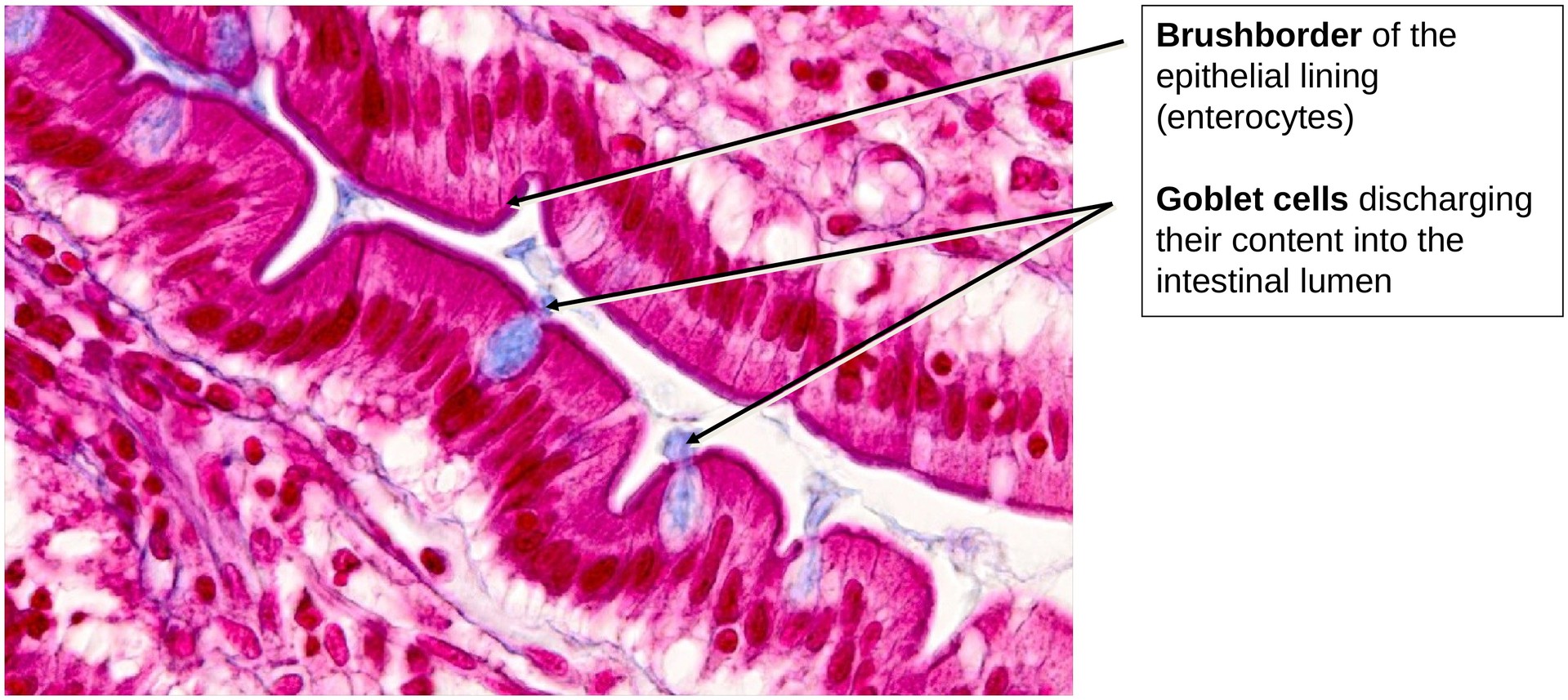
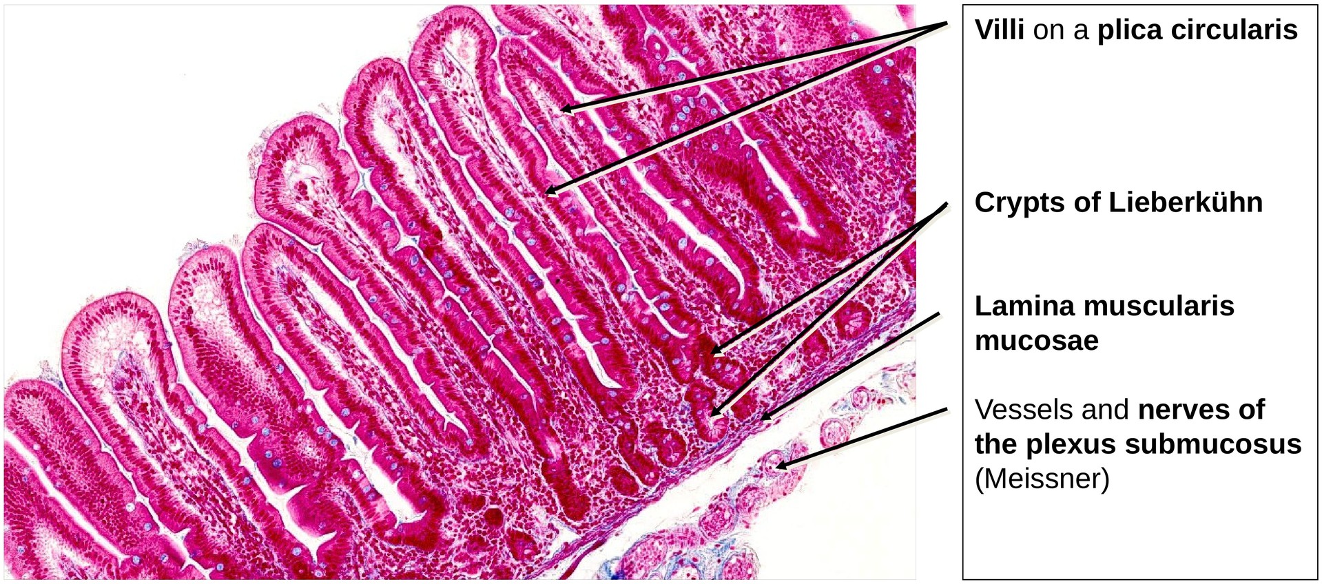
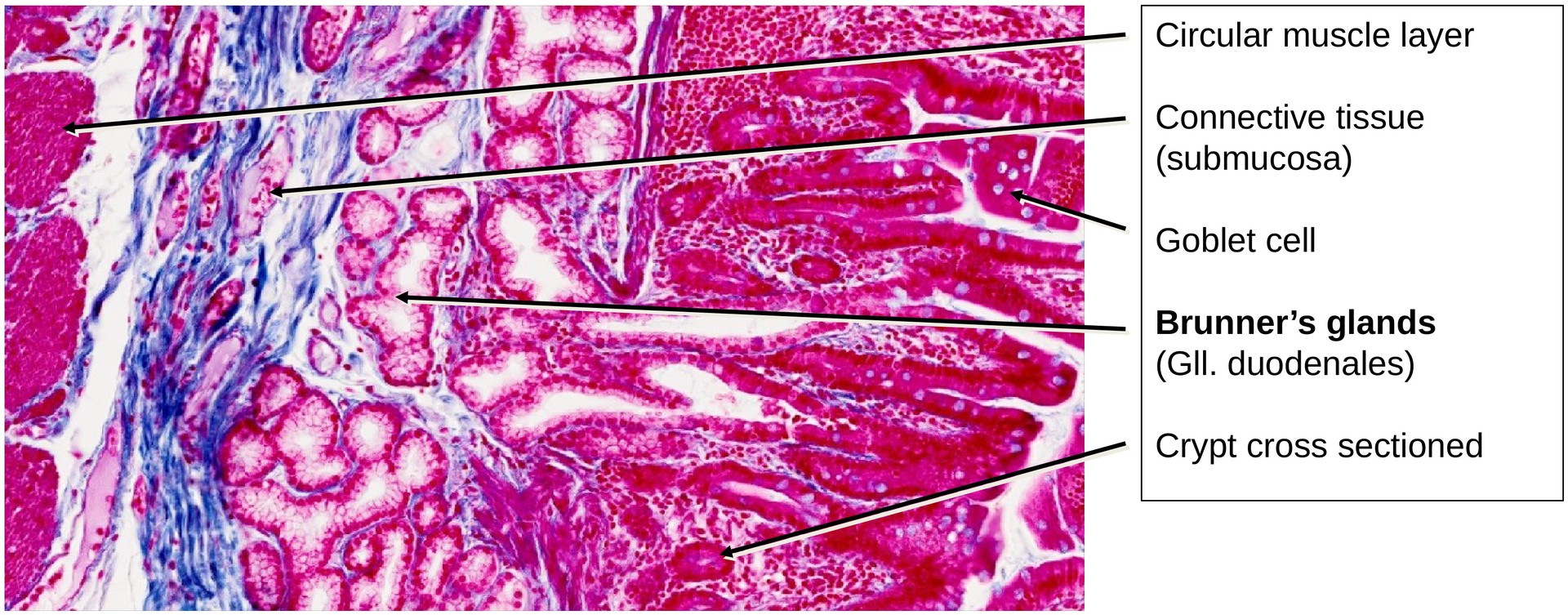
The mucosa forms tall plicae circulares (circular folds) that are covered by villi. The predominant epithelial cell type is the enterocyte, a tall columnar cell with a distinct brush border (microvilli) that enhances absorption. Interspersed among the enterocytes are numerous goblet cells, whose mucous content stains bluish and appears to interrupt the continuity of the brush border. Occasionally, goblet cells are visible in the process of active secretion.
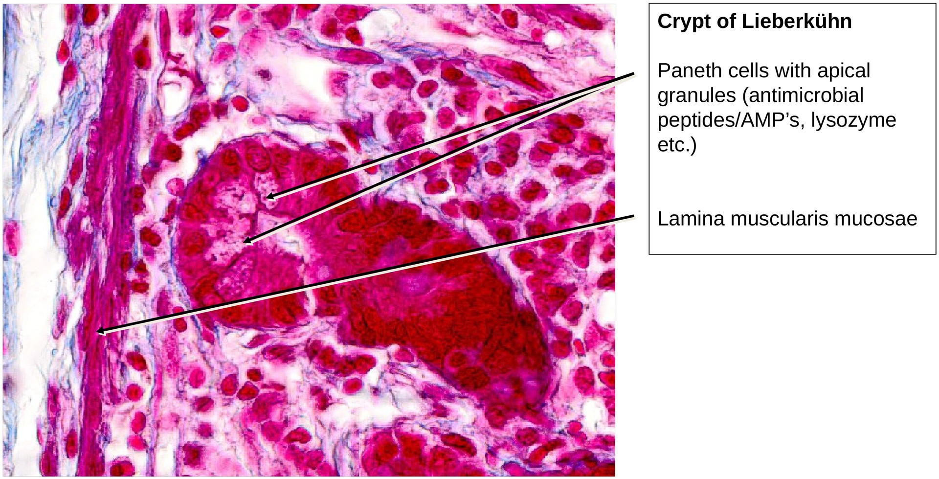
Between the villi lie the openings of the crypts of Lieberkühn (intestinal glands), which are relatively short with narrow lumina. Within these crypts, some cells show apical granules — these are Paneth cells, containing lysozyme and antimicrobial peptides, essential for mucosal defence.
The lamina muscularis mucosae extends into the base of the villi.
In the submucosa, Brunner’s glands are present — a defining feature of the duodenum. These glands secrete an alkaline, bicarbonate-rich mucus that neutralizes acidic gastric chyme and protects the intestinal epithelium.
The tunica muscularis consists of an inner circular and an outer longitudinal layer of smooth muscle. Between them, the myenteric (Auerbach’s) plexus can be identified, containing ganglion cells and nerve fibers. The submucosal (Meissner’s) plexus within the submucosa is less distinct in this preparation.
Tasks:
- Identify the general wall structure of the duodenum and its layers:
- Tunica mucosa (lamina epithelialis, lamina propria, lamina muscularis mucosae)
- Tunica muscularis (inner circular and outer longitudinal muscle layers)
- Locate the Brunner’s glands in the submucosa (between the lamina muscularis mucosae and the tunica muscularis).
- Identify components of the myenteric (Auerbach’s) plexus between the two muscle layers.
- Search for goblet cells among the enterocytes and note the presence of the brush border epithelium.
- Compare the length of the villi with that of the crypts.
- Identify Paneth cells within the crypts by their eosinophilic apical granules.
License
University of Basel
Downloads