CARTILAGE (GENERAL HISTOLOGY)
4.2
Fibrocartilage (intervertebral disc)
Specimen Details:
Specimen Details:
Organ: Intervertebral Disc
Origin: Human
Staining: Masson-Goldner
Method and Specimen Description:
The specimen was not demineralized prior to embedding. Instead, it was completely embedded in plastic after fixation and dehydration, allowing both soft and hard tissue components to be sectioned together. For this course, 5 µm-thick sections were prepared. Despite plastic embedding, strong shear forces during sectioning can cause wrinkles in some regions. The section was stained using Masson–Goldner, which differentially colors collagen, bone, and cytoplasmic elements.
Objective of the Examination:
To study the structure of fibrocartilage using the intervertebral disk as an example, and to distinguish between the annulus fibrosus and the nucleus pulposus.
Special Features of the Specimen:
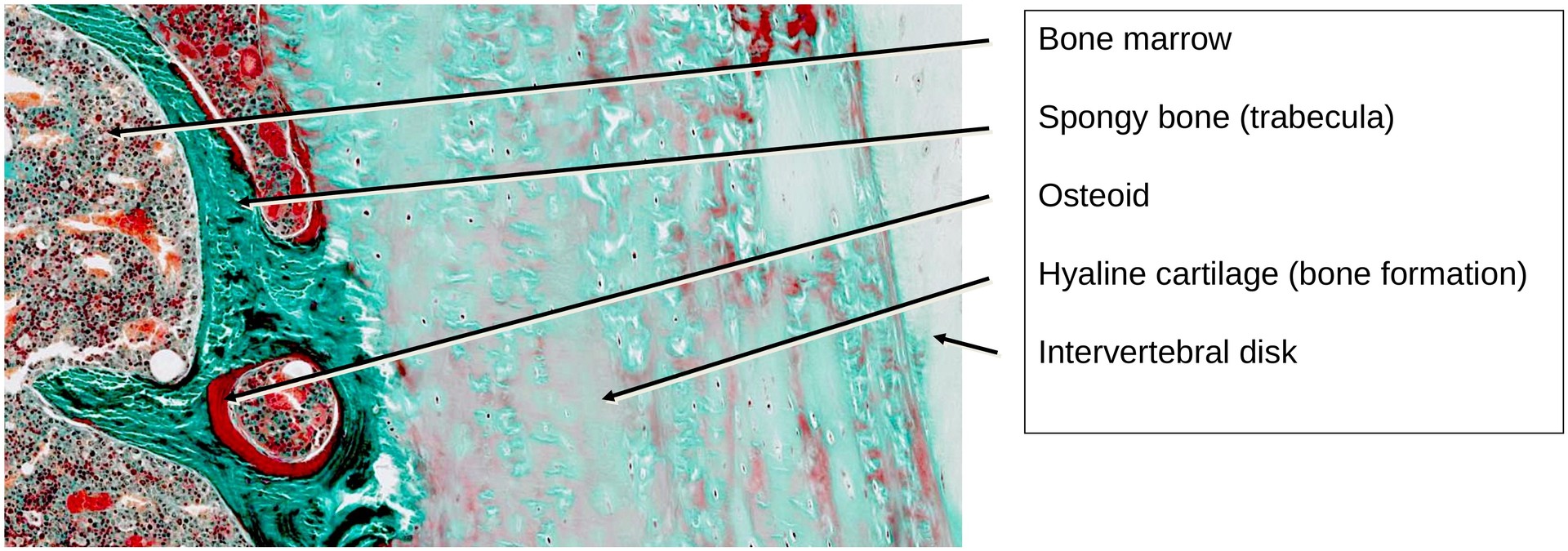
This section passes through a developing vertebral body, showing areas of spongy bone, hematopoietic bone marrow, and a cartilaginous zone involved in ossification, which will later form part of the synchondrosis.
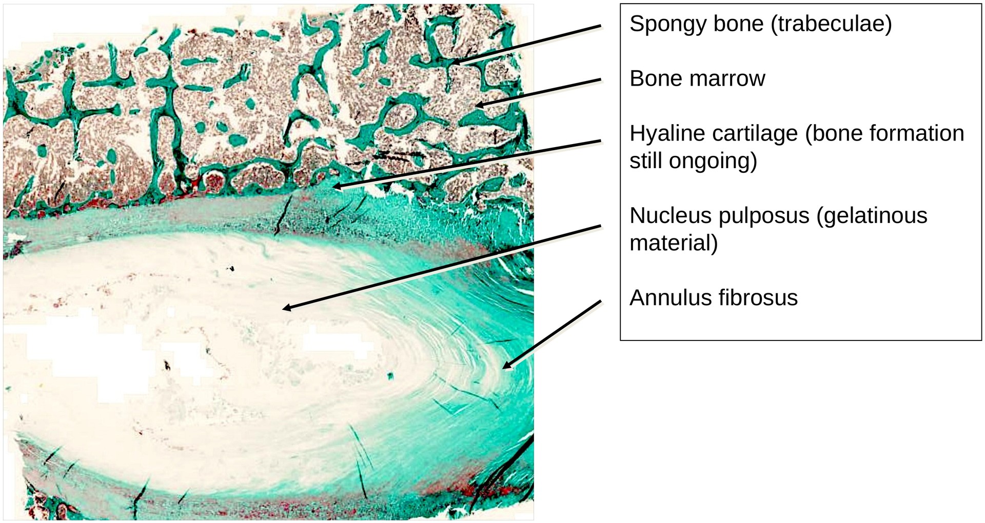
The intervertebral disk, as part of the mobile spine, is subjected to both tensile and compressive forces. It consists of:
-
The annulus fibrosus (outer region), made up of fibrocartilage, and
-
The nucleus pulposus (inner region), forming a gelatinous core.
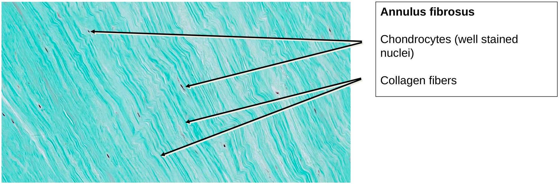
The annulus fibrosus structurally resembles a ligament, with collagen fibers arranged in roughly parallel bundles. In the body, the annulus fibrosus merges seamlessly with the anterior and posterior longitudinal ligaments, though these are not visible in this section. Due to the relatively low concentration of chondroitin sulphate, the collagen fibers in fibrocartilage are clearly visible and not masked, unlike in hyaline cartilage. The number of chondrocytes is comparatively low; these cells are responsible for synthesizing the extracellular matrix (ECM), consisting of amorphous ground substance and fibers.
In the center of the disk, remnants of the nucleus pulposus are visible. This gelatinous material possesses a high water-binding capacity, generating swelling pressure that enables the disc to absorb mechanical loads and maintain flexibility.
Due to this high water content, only small amounts of material remain after fixation — mainly precipitated protein residues and traces of glycosaminoglycans (GAGs).
Within the bony portion of the vertebral body, bone formation is still ongoing. Zones containing fresh osteoid (intensely red-stained), mineralized bone, and cartilaginous remnants are visible. The bone trabeculae exhibit the irregular architecture typical of woven bone, and hematopoietic marrow occupies the intertrabecular spaces.
Tasks:
• At low magnification, identify the annulus fibrosus and nucleus pulposus. How do these regions differ in appearance and structure?
• Observe the large quantity of extracellular matrix (ECM)—comprising fibers and amorphous ground substance.
• Identify chondrocytes, which appear as compressed dark lines, particularly within the annulus fibrosus.
• Describe the structure of the bone in the vertebral body.
• Determine which cell type typically lies adjacent to the red-stained osteoid areas.
License
University of Basel
Downloads