SKIN AND APPENDAGES (ANATOMICAL MICROSCOPY)
13.5
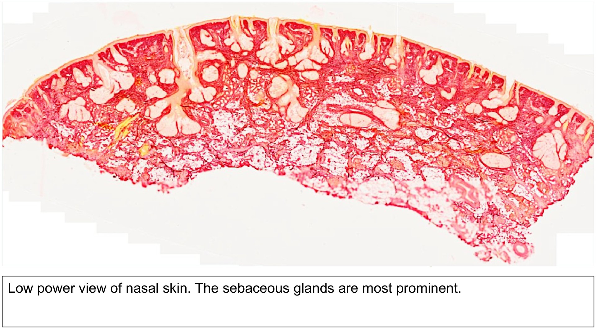
Nasal skin
Preparation:
Preparation details:
Organ: Nasal skin
Origin: Human
Staining: Van Gieson/Elastin
Method and Specimen Description:
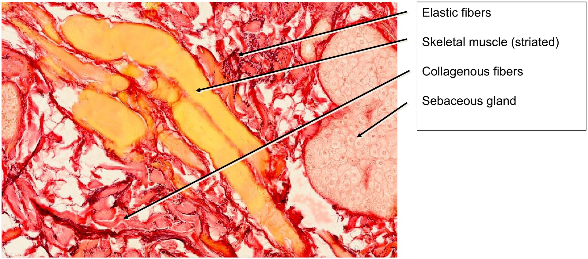
Normal histological section stained with Van Gieson/Elastin. In this staining, muscle, hair, and keratin appear yellow, connective tissue is red, and elastic fibers stain violet.
Objective of the Examination:
To study the structure of skin subjected to low mechanical stress.
Specific features of the preparation:
The nasal skin contains fine hairs, and thus displays all the typical layers of the skin:
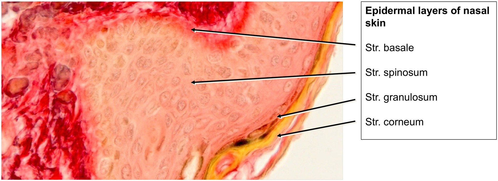
EPIDERMIS
- Stratum corneum
- Stratum granulosum
- Stratum spinosum
- Stratum basale
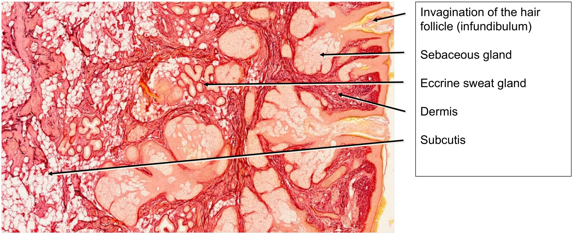
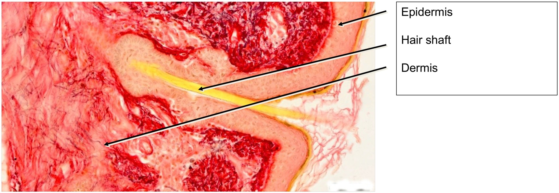
CORIUM (DERMIS) SUBCUTIS
The epidermis shows little interdigitation with the corium, which correlates with the low mechanical stress typical of this region.
Fine hairs are clearly visible, with their follicular invaginations extending down to the corium–subcutis boundary. The hair shafts stain yellow due to the keratin.
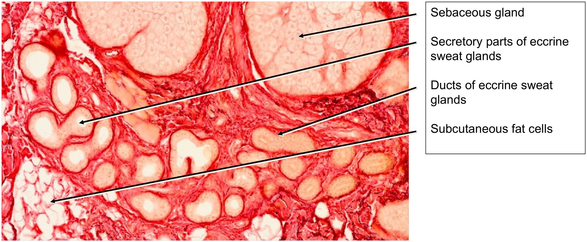
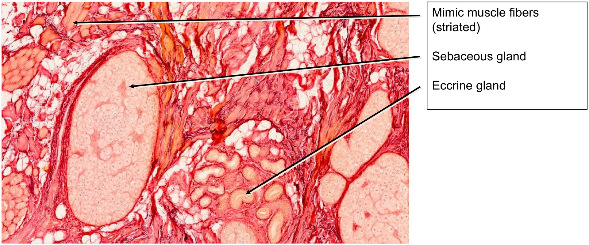
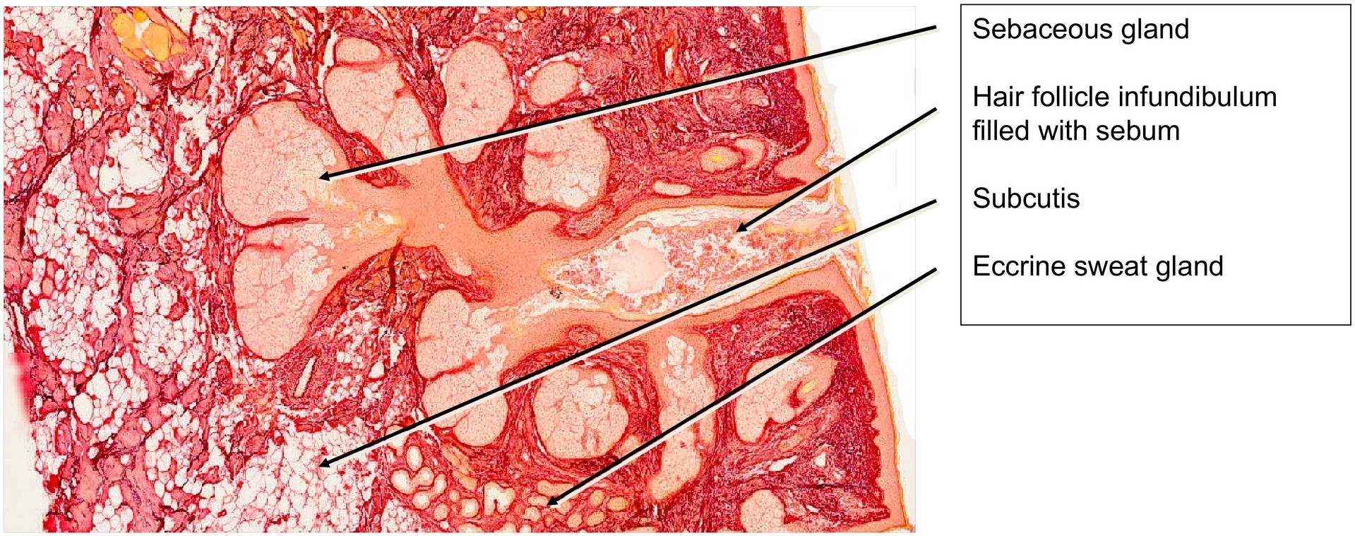
The sebaceous glands, which open into the hair follicles, are highly prominent. Their cytoplasm is largely filled with fat droplets, and the nuclei of the secretory cells are often degenerated, consistent with their holocrine secretion mechanism.
Arrector pili muscles are absent in this region.
In addition to sebaceous glands, a smaller number of eccrine sweat glands are present. These consist of:
- Secretory segments with a single-layered cuboidal to columnar epithelium, and
- Efferent ducts lined by a two-layered cuboidal epithelium. In this preparation, efferent ducts that traverse the epidermis are not observed.
The elastin staining highlights a dense network of elastic fibers within the corium. This concentration of elastic tissue, together with the firm attachment of the corium to the moderately fatty subcutis, explains the limited mobility of the nasal skin.
Tasks:
- Identify the layers of the skin (epidermis with its strata, corium, and subcutis).
- Locate hair follicles and determine in which layers they are found.
- Identify hairs within the follicles (note the yellow-stained keratin).
- Determine whether the cornification of the nasal skin is strong or weak and explain your reasoning.
- Observe and describe the sebaceous glands.
- Locate eccrine sweat glands and their efferent ducts. Where do these ducts normally open?
- Describe the distribution of elastic fibers and identify where they are most concentrated.
License
University of Basel
Downloads