ENDOCRINE ORGANS (ANATOMICAL MICROSCOPY)
9.3
Adrenal gland 1
Specimen:
SPECIMEN DETAILS:
Organ: Adrenal gland
Origin: Human
Staining: Hematoxylin Eosin (H&E)
METHOD AND SPECIMEN DESCRIPTION:
Routine histological section stained with the general Hematoxylin and Eosin (H&E) method.
OBJECTIVE OF THE EXAMINATION:
To study the structure and organization of the endocrine adrenal gland.
Special Features of the Specimen:
General:
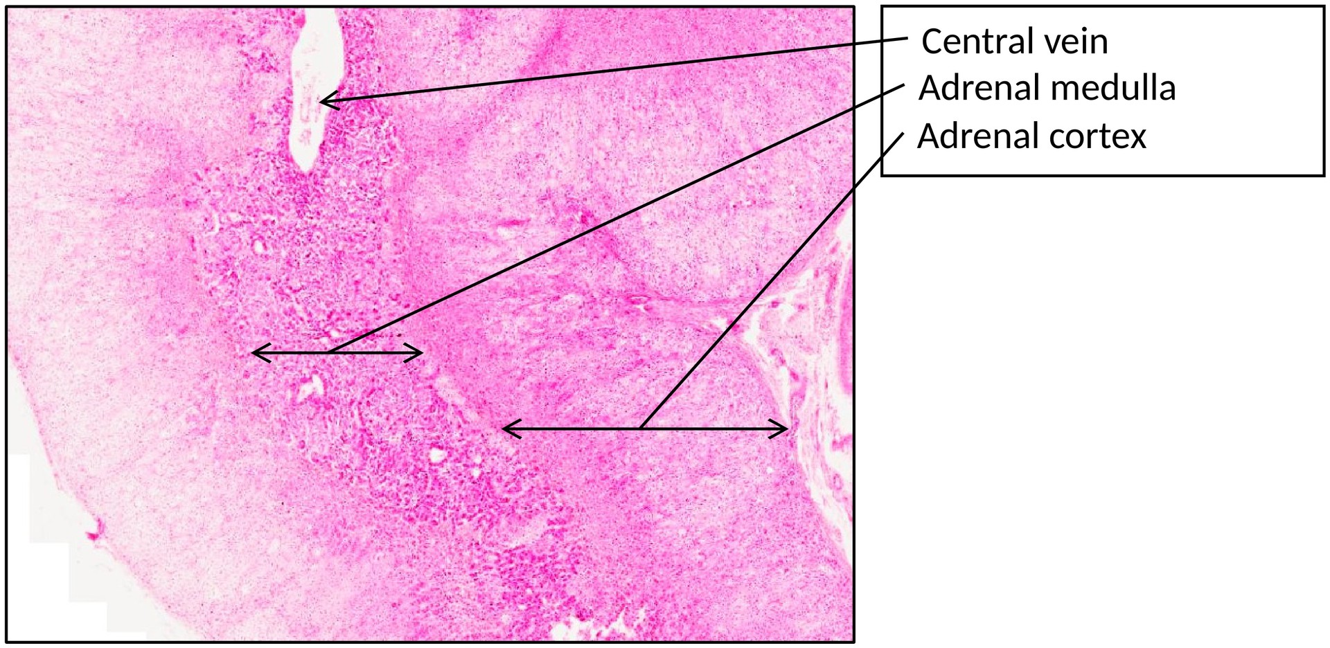
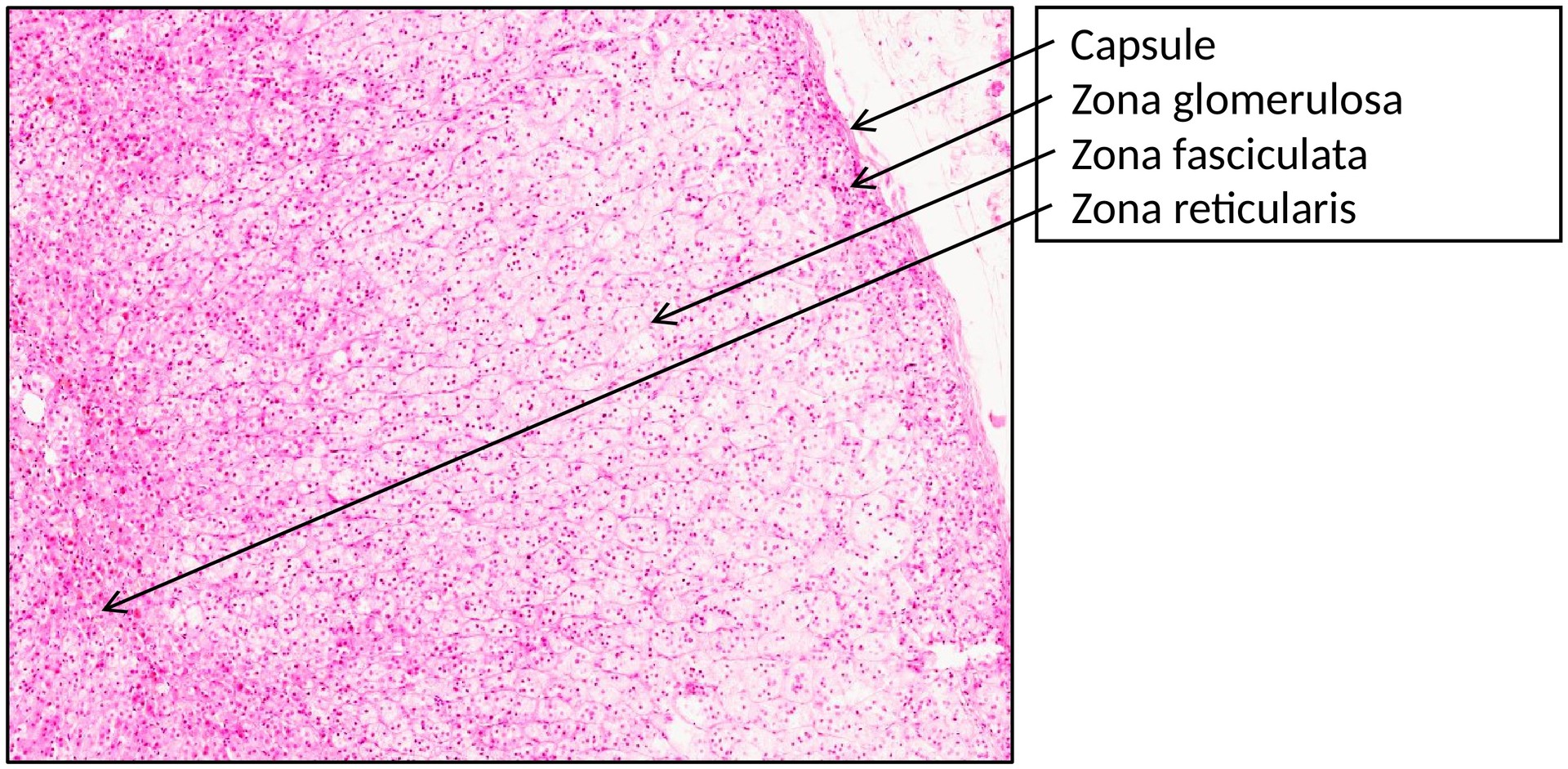
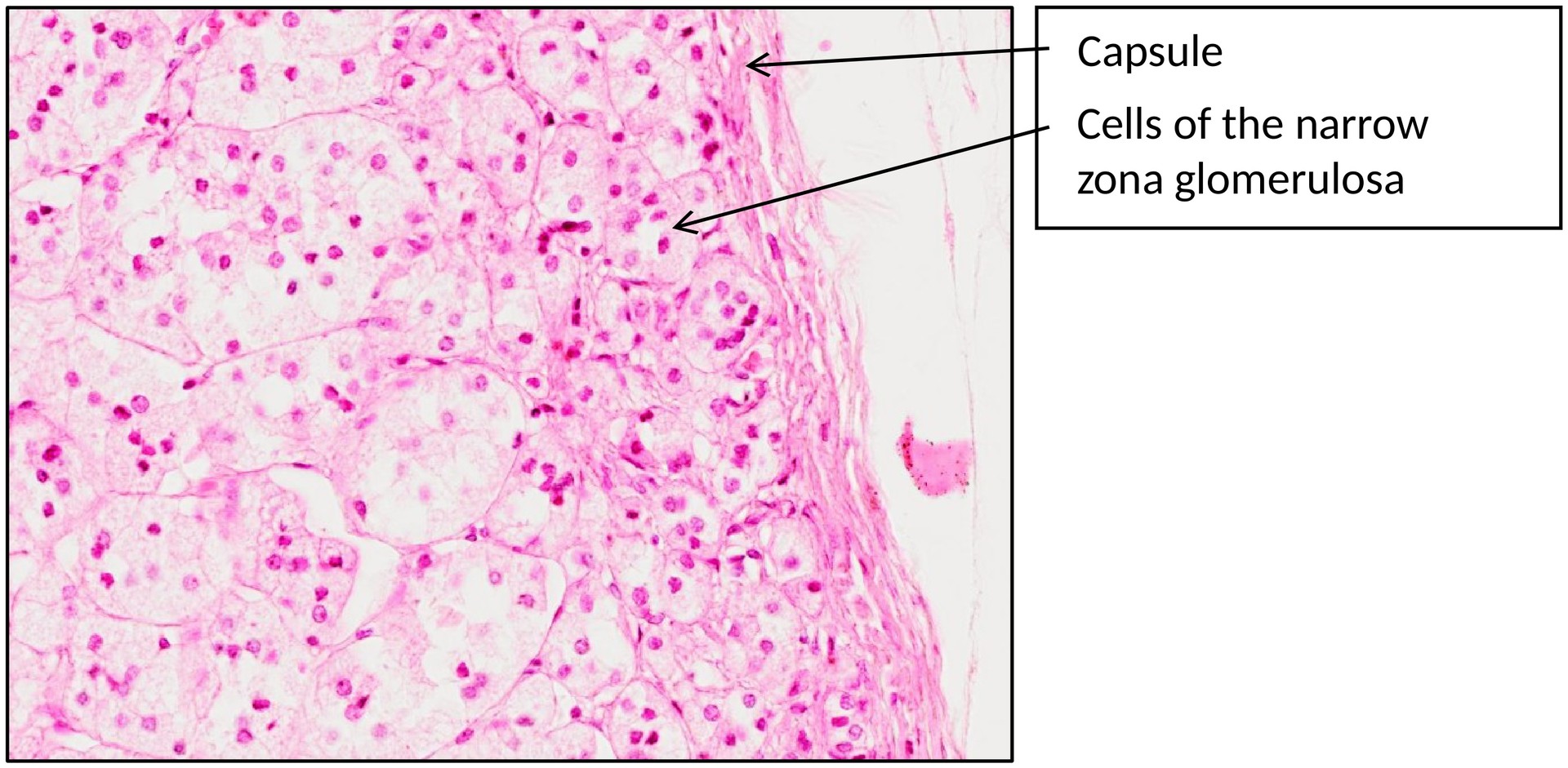
The adrenal glands occur as paired organs situated at the superior poles of the kidneys. Each gland is enclosed by a thin connective tissue capsule and consists of two morphologically and functionally distinct regions: the adrenal cortex and the adrenal medulla.
The adrenal cortex is divided into three concentric zones:
-
Zona glomerulosa – composed of small, rounded clusters of epithelial cells.
-
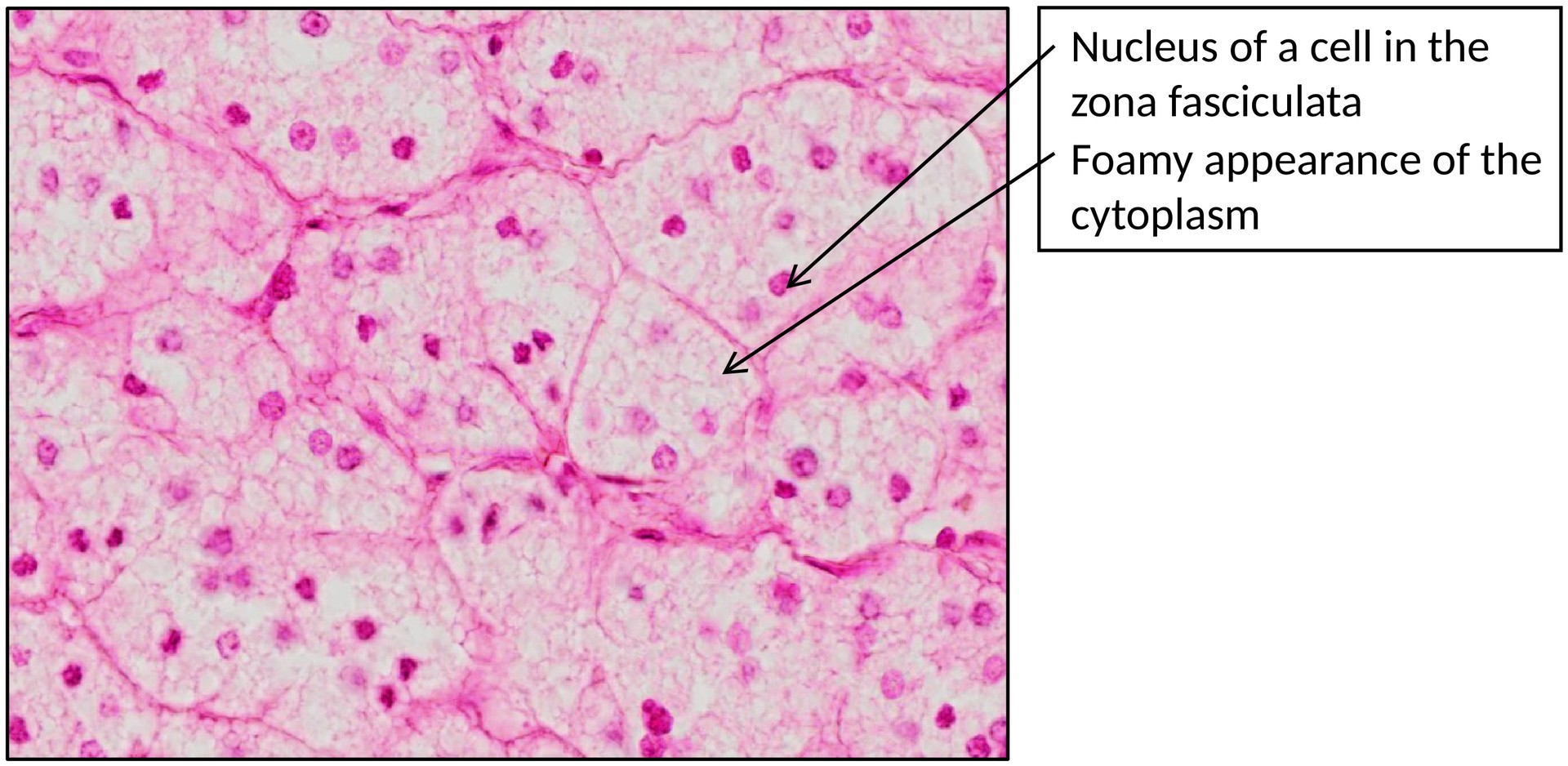
Zona fasciculata – formed by elongated cords of cells with pale, foamy cytoplasm due to numerous lipid droplets. These cords are interspersed with wide-lumen sinusoids.
-
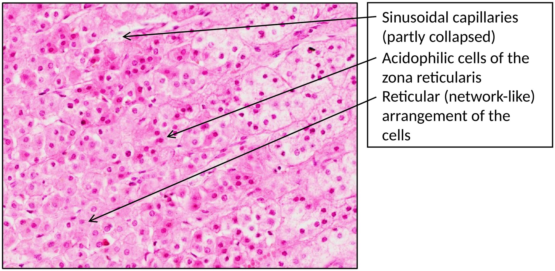
Zona reticularis – consists of smaller cells arranged in an irregular, network-like pattern. Their cytoplasm is more eosinophilic (acidophilic) and therefore stains red.
Each cortical zone synthesizes distinct steroid hormones:
-
Zona glomerulosa: produces mineralocorticoids (mainly aldosterone), which promote sodium reabsorption in the kidneys.
-
Zona fasciculata: secretes glucocorticoids (primarily cortisol), which regulate metabolism and facilitate adaptation to stress.
-
Zona reticularis: generates weak androgens (such as dehydroepiandrosterone, DHEA).
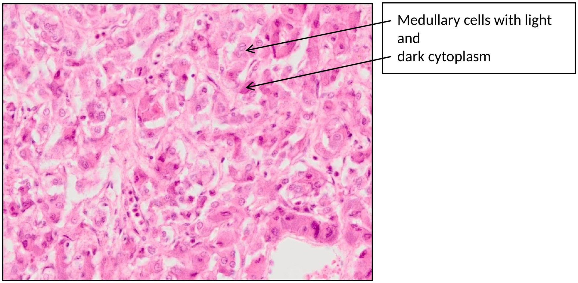
The adrenal medulla, derived from neural crest cells, is composed of groups of polygonal chromaffin cells. Their cytoplasm appears either darkly stained or pale and granular, depending on their catecholamine content. Approximately 80% of the cells produce adrenaline (epinephrine), while the remaining 20% secrete noradrenaline (norepinephrine).
The medulla also contains wide-lumen sinusoids and medullary veins with thick, smooth muscle walls. Blood from the cortical capillaries drains into the medulla, exposing the chromaffin cells to high concentrations of cortical steroids, which in turn stimulate the conversion of noradrenaline to adrenaline.
TASKS:
• Identify the division of the adrenal gland into cortex and medulla.
• Locate and distinguish the three cortical zones: zona glomerulosa, zona fasciculata, and zona reticularis.
• Observe the arrangement, staining characteristics, and morphology of the cortical cells and the accompanying sinusoidal capillaries.
• Identify the chromaffin cells and veins of the adrenal medulla.
• Consider the relationship between cortical and medullary circulation and its physiological significance.
License
University of Basel