GLANDS (GENERAL HISTOLOGY)
3.1
Pancreas, exocrine portion
Specimen Details:
Specimen Details:
Organ: Pancreas
Origin: Human
Staining: Hematoxylin - Eosin (H&E)
Method and Specimen Description:
Routine histological section of the pancreas stained with H&E, which allows clear differentiation of both the exocrine and endocrine components of the gland.
Objective of the Examination:
To study the structure of the purely serous exocrine portion of the pancreas and to recognize its defining histological features.
Special Features of the Specimen:
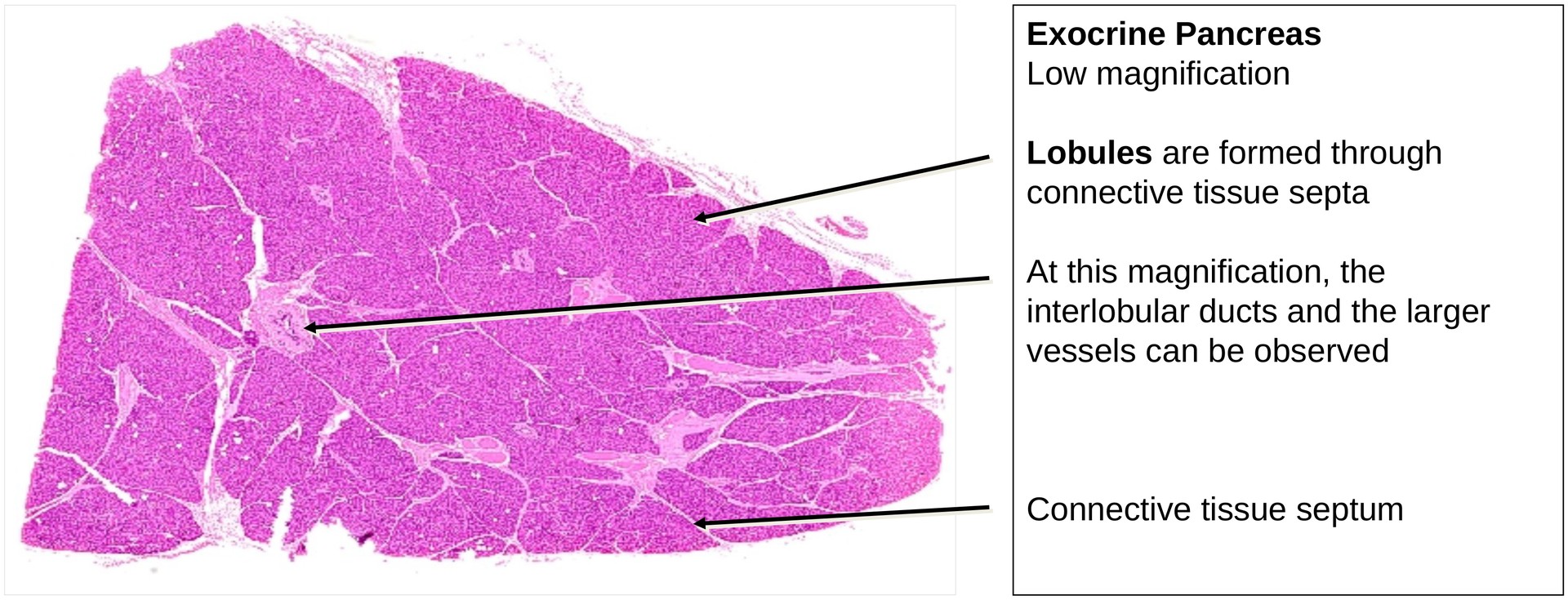
General (Low Magnification):
The dual functional character of the pancreas is evident even at low magnification. The tissue consists predominantly of the exocrine component, which appears relatively homogeneous and strongly stainable, interspersed with lighter-staining endocrine islets (islets of Langerhans).
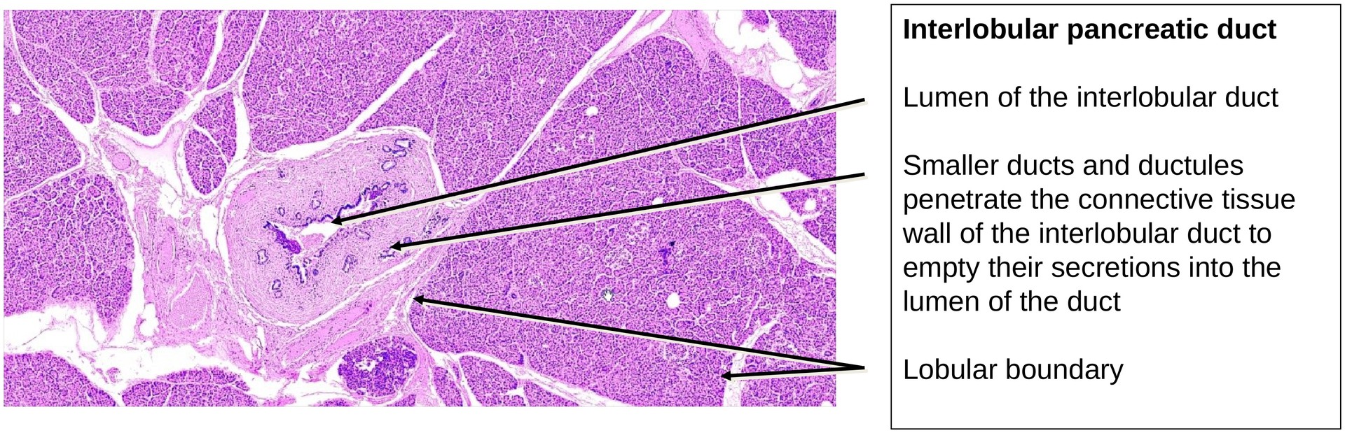
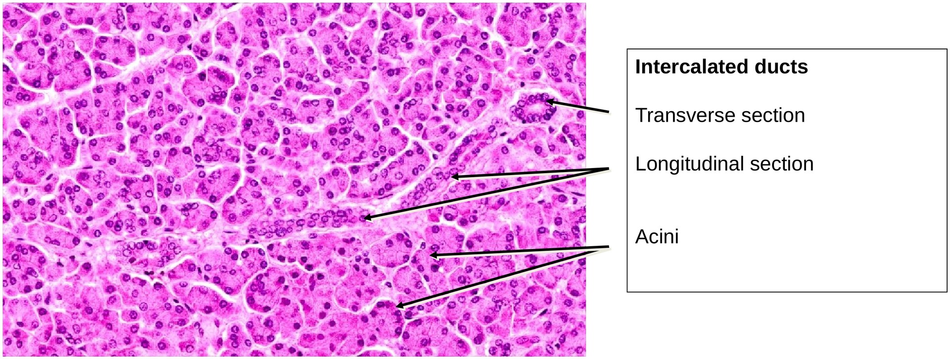
The exocrine portion is organized into lobules separated by connective tissue septa, within which run blood vessels and excretory ducts. Smaller intercalated ducts of the excretory system are visible within the lobules, whereas larger interlobular ducts are located within the connective tissue between lobules.
Medium to High Magnification:
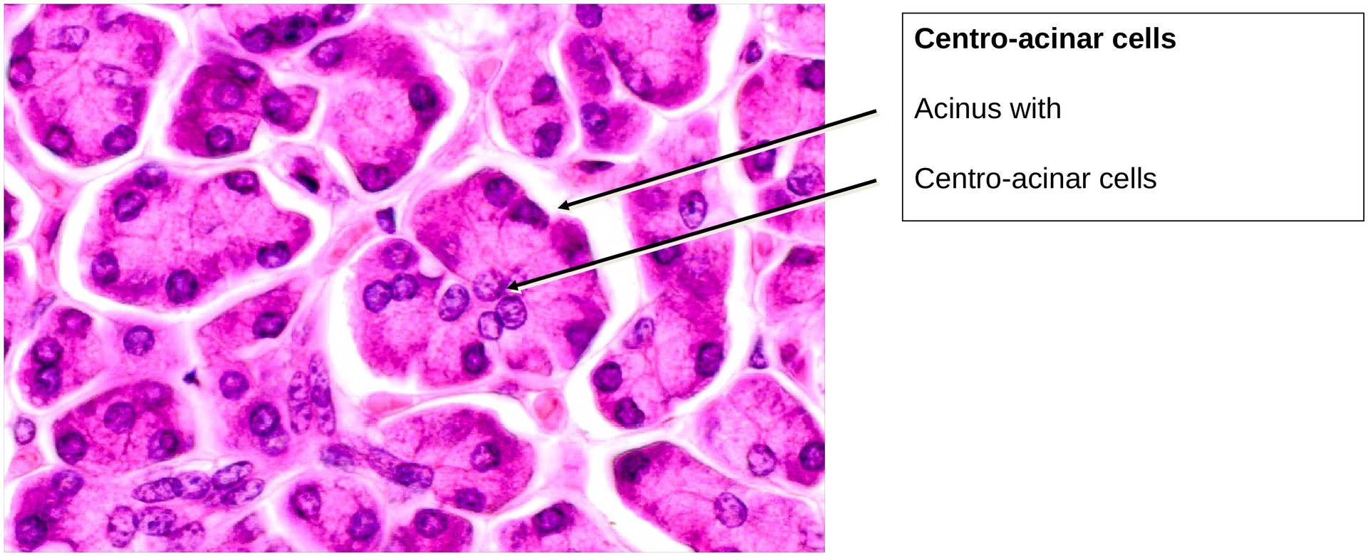
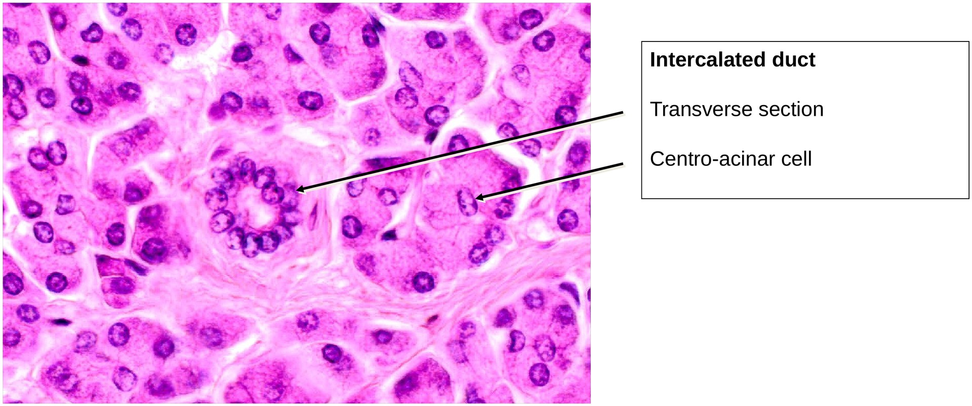
The secretory units (acini, singular acinus) are small and tightly packed, typical of serous glands. Each acinus is composed of pyramidal secretory cells with basophilic cytoplasm (rich in rough endoplasmic reticulum) and apical eosinophilic zymogen granules containing digestive enzymes.
A characteristic feature of the exocrine pancreas is the presence of centroacinar cells—pale-staining cells located centrally within the acini. These cells are continuations of the intercalated ducts that extend into the acinar lumen and are typical for pancreatic tissue.
Unlike many other exocrine glands, the pancreas lacks myoepithelial cells. The movement of secretions through the duct system is therefore driven by secretion pressure, not contractile activity.
Key Histological Features of the Exocrine Pancreas (for Differential Diagnosis):
-
Small, compact acini with basophilic cytoplasm and apical zymogen granules
-
Centroacinar cells visible within acini
-
Intercalated and interlobular ducts present
-
No striated (secretory) ducts
-
Absence of myoepithelial cells
-
Islets of Langerhans scattered between acini (endocrine component; A and B cells not distinguishable in H&E staining)
Tasks:
-
Obtain an overview of the lobular organization of the pancreas and locate a larger excretory (interlobular) duct within the connective tissue.
-
In the connective tissue surrounding the largest duct, identify smaller epithelial structures — these represent intercalated ducts or small vascular channels.
-
At high magnification, locate and describe centroacinar cells within acini.
-
Observe the structure of the secretory acini, noting cell shape, nuclear position, and cytoplasmic staining pattern.
-
Identify an intercalated duct and trace its connection to the acinar lumen if possible.
License
University of Basel
Downloads