FEMALE REPRODUCTIVE ORGANS (ANATOMICAL MICROSCOPY)
10.4
Uterine body 1
Specimen Details:
Specimen Details:
Organ: Uterine body
Origin: Human
Staining: Van Gieson
Method and Specimen Description:
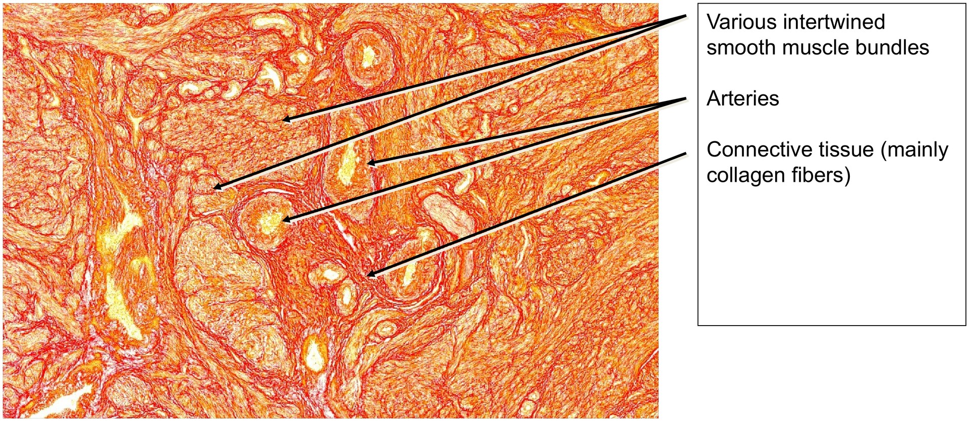
Routine histological section stained with Van Gieson, which stains connective tissue (collagen) red and smooth muscle yellow.
Objective of the Examination:
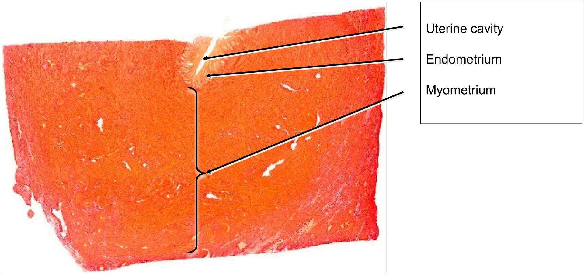
To understand the microscopic structure of the uterus, the organ with the most powerful smooth musculature in the female body, and to identify its two principal layers — the endometrium and the myometrium.
Special Features of the Specimen:
Because of the considerable thickness of the myometrium, which forms the major component of the uterine wall, only a partial section of the organ is shown. The specimen includes a small segment of the lateral wall of the uterine cavity, illustrating the typical arrangement of the endometrium, which consists of two layers: the stratum functionalis (functional layer) and the stratum basalis (basal layer).
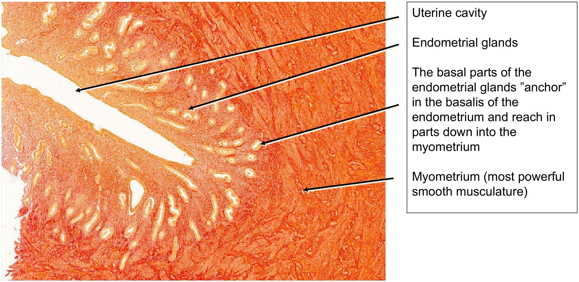
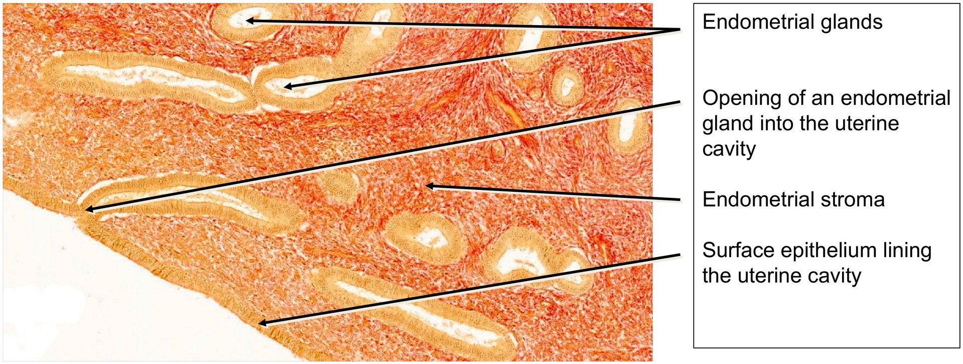
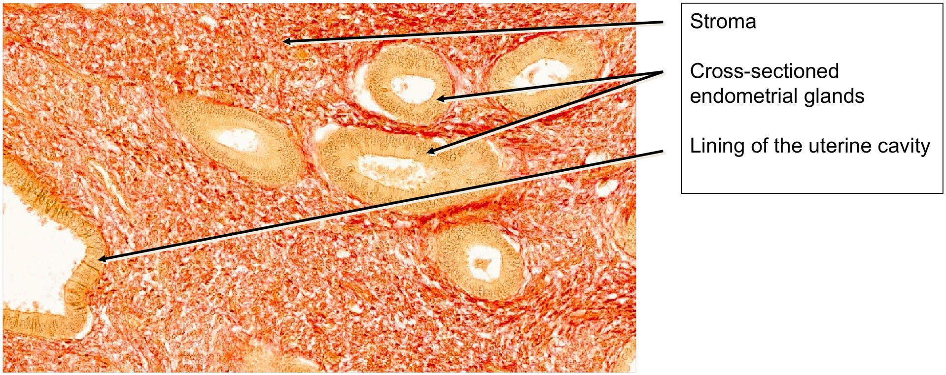
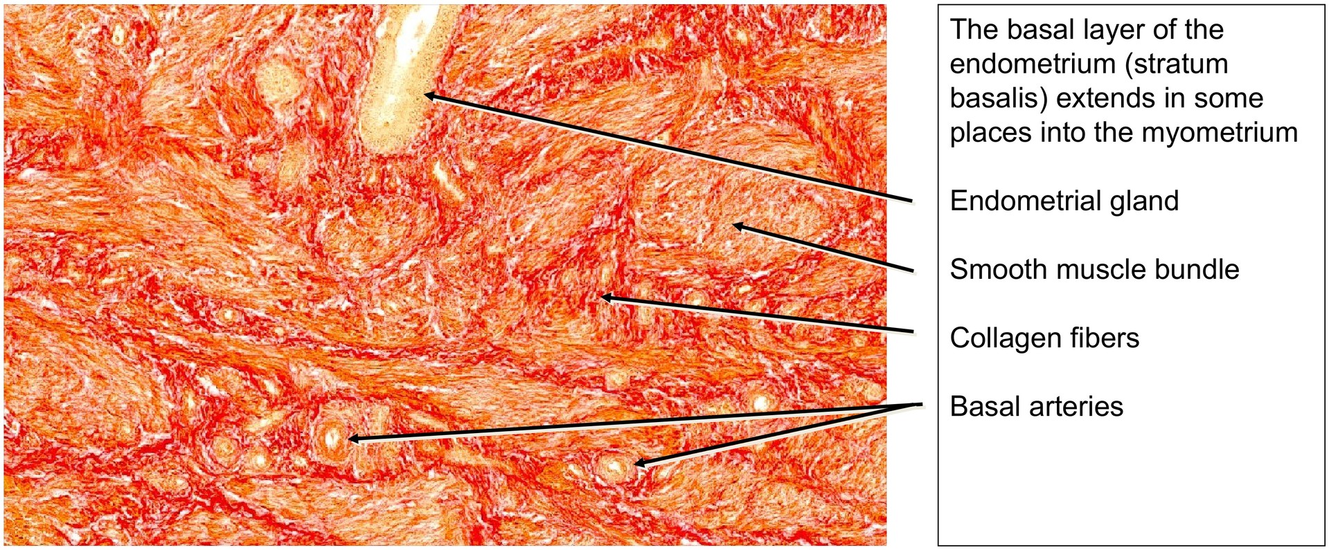
The endometrial glands, derived from the surface epithelium, extend deeply into the underlying tissue, sometimes reaching the myometrium. The endometrium, as a principal target organ of ovarian hormones, undergoes pronounced cyclical changes. In this section, the endometrium represents the late proliferative phase. At this stage, it has attained considerable thickness, which will further increase during the subsequent secretory phase.
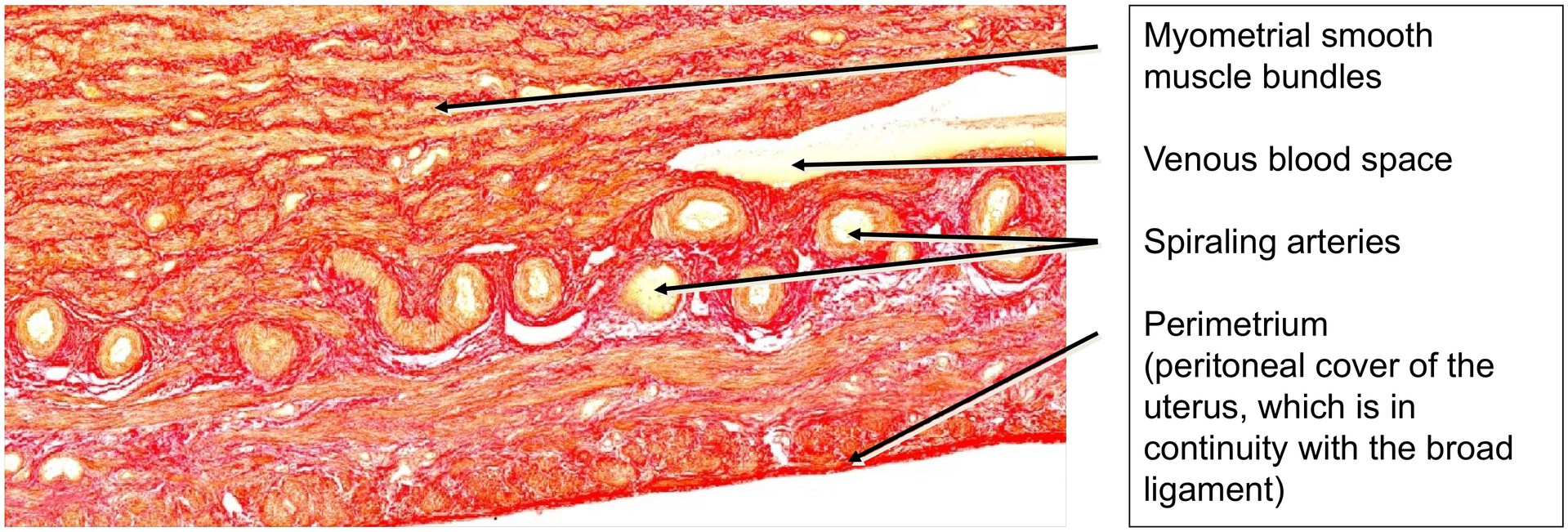
The spiral arteries, branching from the basal arteries, are still relatively undeveloped and not yet markedly coiled, a feature that becomes prominent in the secretory phase. The glandular epithelium shows no evidence of post-ovulatory changes — such as basal vacuolation or apocrine secretion of glycogen-rich cytoplasm — which are induced by progesterone.
The stromal tissue is only mildly edematous, and the morphological distinction between the stratum functionalis and stratum basalis is not clearly pronounced, being recognisable mainly by position rather than structure.
Tasks:
-
Delineate the endometrium from the myometrium at low magnification.
-
Examine the deep portions of the glands and observe how they extend into the myometrium — note their role as the regenerative source for the endometrium after menstruation.
-
Describe the morphology and course of the endometrial glands.
-
Locate spiral arteries within the endometrial stroma.
-
Examine the surface epithelium for its simple columnar structure and look for ciliated cells.
-
Trace the bundles of smooth muscle in the myometrium — what can be said about their arrangement and orientation?
-
Identify the connective tissue septa that separate the muscle bundles.
-
Determine the type of the larger vessels located within the myometrium.
-
Identify the outermost layer covering the myometrium in certain regions — is it adventitia or serosa?
-
At the junction between endometrium and myometrium, locate cross-sections of vessels and identify them (typically basal arteries).
License
University of Basel
Downloads