NERVOUS SYSTEM (ANATOMICAL MICROSCOPY)
16.7
Spinal cord, cervical
Specimen:
SPECIMEN DETAILS:
Organ: Spinal cord, cervical region
Origin: Human
Staining: Luxol Fast Blue/Cresyl Violet
METHOD AND SPECIMEN DESCRIPTION:
Luxol Fast Blue stains myelinated axons blue, highlighting white matter and phospholipid-rich myelin sheaths. Cresyl Violet, a basic dye, binds to nucleic acids, staining chromatin, nucleoli, and rough endoplasmic reticulum (Nissl bodies) blue-violet.
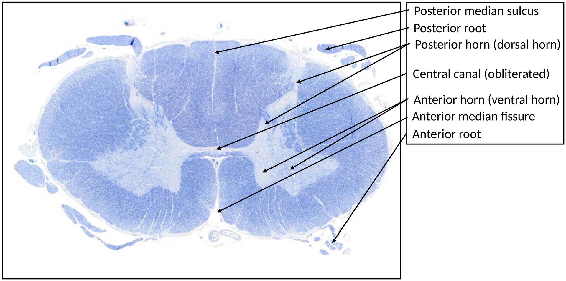
The spinal cord, like the vertebral column, is bilaterally symmetrical, consisting of two halves separated ventrally by the anterior median fissure. Unlike the cerebrum and cerebellum, the spinal cord has white matter externally and grey matter internally, the latter forming a butterfly-shaped structure. Motoneurons are located in the anterior (ventral) horn of the grey matter.
OBJECTIVE OF THE EXAMINATION:
To identify the distribution of grey and white matter, and to locate the motoneurons within the anterior horn of the cervical spinal cord.
Special Features of the Specimen:
In cross-section, the spinal cord displays the characteristic butterfly-shaped grey matter, comprising:
- Two narrow posterior (dorsal) horns,
- Two broad anterior (ventral) horns, connected by the grey commissure.
No lateral horns are present in the cervical region (these occur in thoracic segments). The central canal is located in the midline, often obliterated by proliferating ependymal cells.
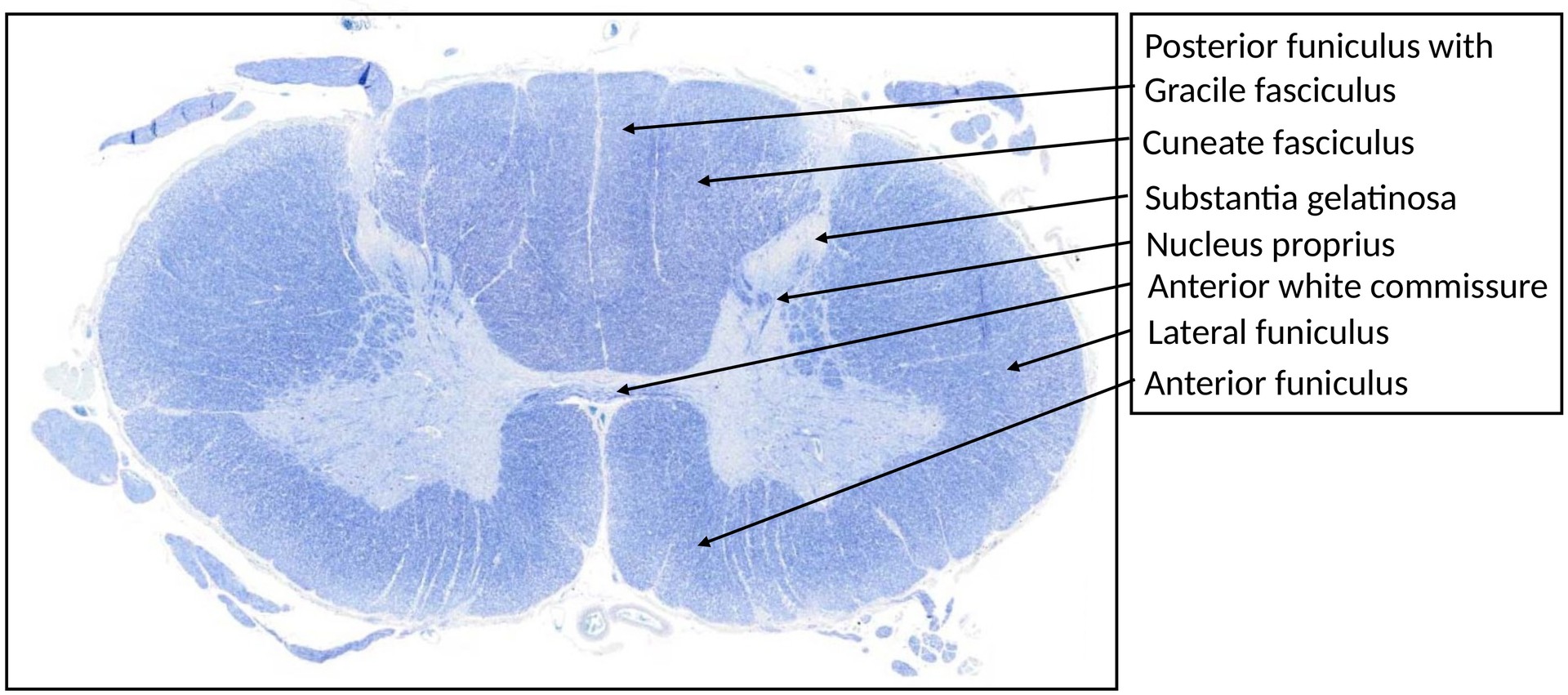
The surrounding white matter (substantia alba) is divided into:
- Posterior funiculus,
- Lateral funiculus, and
- Anterior funiculus.
The posterior funiculi widen progressively from caudal to cranial levels, and in the cervical cord can be clearly divided into the fasciculus gracilis and fasciculus cuneatus.
Core Areas of the Grey Matter:
- Substantia Gelatinosa:
- Contains many small, densely packed neurons.
- No myelin, hence unstained in blue.
- Nucleus Proprius:
- Contains myelinated fiber bundles.
- Cells are larger and more variable in shape, and less densely packed than in the substantia gelatinosa.
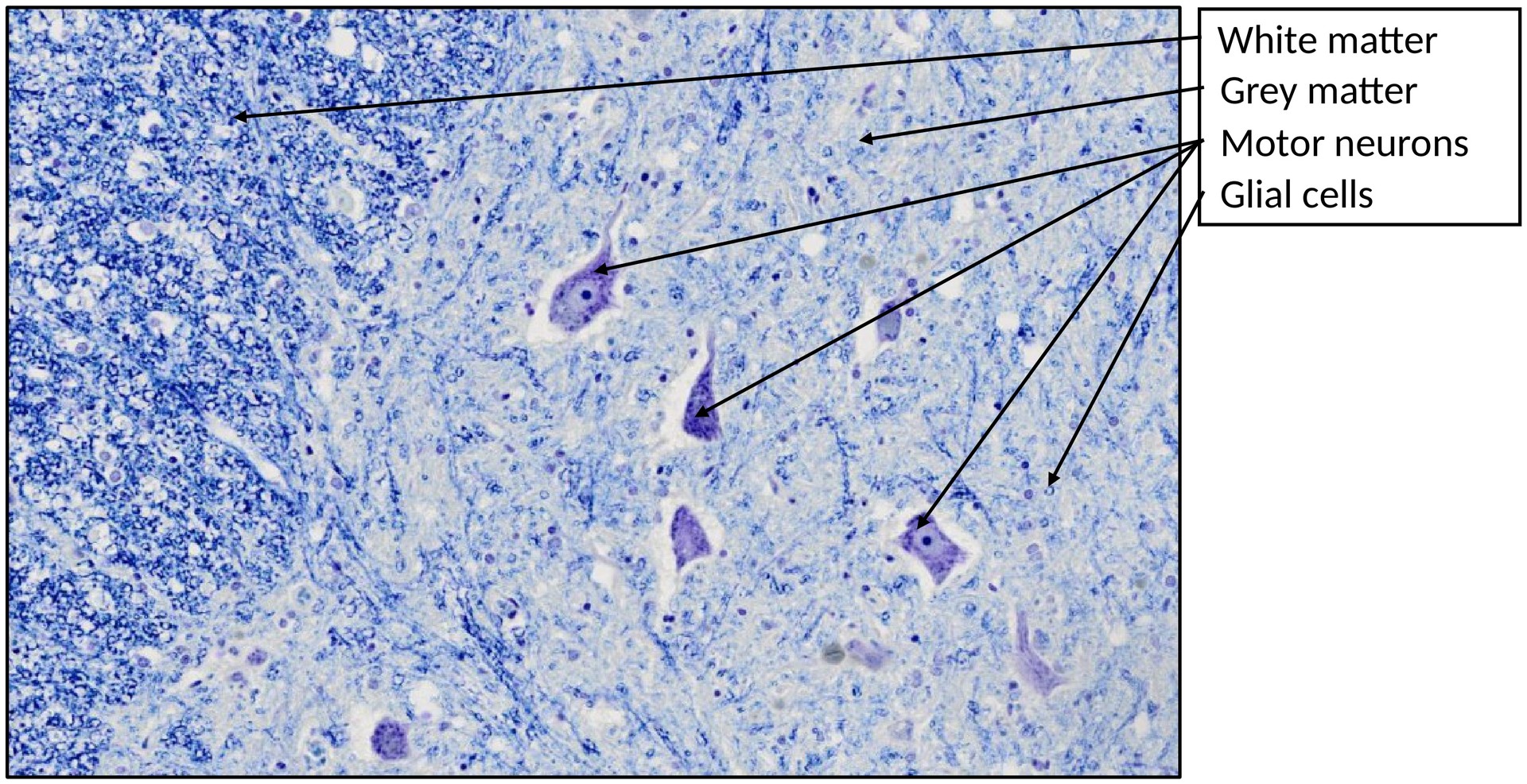
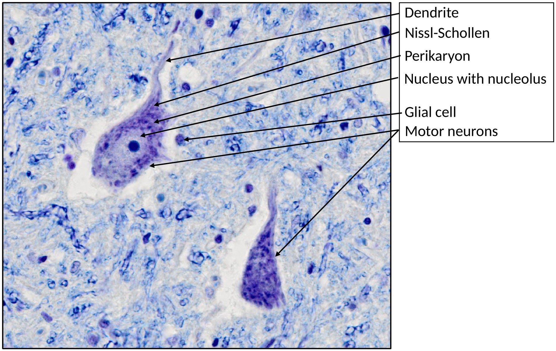
- Anterior Horn:
- Contains large multipolar motoneurons, arranged in clusters.
- Prominent nuclei and nucleoli, with abundant Nissl bodies extending into dendrites (but not into axons).
- Glial nuclei (oligodendrocytes and astrocytes) are visible between neurons, though indistinguishable from each other.
White Matter:
- Myelinated axons stain blue (if preserved); if myelin is dissolved, pale vacuolated “holes” appear.
- Large axons can appear as red or round structures.
- Glial cells (mainly oligodendrocytes and astrocytes) are present but difficult to differentiate.
Meninges:
The spinal cord is externally covered by the pia mater, a delicate connective tissue layer that sends thin extensions into the underlying tissue and merges with arachnoid trabeculae of the arachnoid mater.
TASKS:
- Identify the following structures:
- Grey matter
- White matter
- Central canal
- Anterior horn
- Posterior horn
- Within the anterior horn, identify motoneurons and their components:
- Nucleus
- Dendrites
- Axon
- Surrounding glial cells
- Which major fiber tracts are located in the lateral funiculus?
- Which major fiber tracts are located in the anterior funiculus?
License
University of Basel