DIGESTIVE ORGANS: ORAL CAVITY (ANATOMICAL MICROSCOPY)
18.9
Tooth Development II
Specimen:
Specimen Details:
Organ: Tooth germ (bell stage)
Origin: Rat
Staining: Azan
Method and Specimen Description:
In this preparation, bone and tooth hard tissues are already present in a partially mineralised state. To allow sectioning, the tissue was decalcified using acid or chelating agents before embedding and sectioning.
The subsequent Azan staining allows good differentiation of structures:
- Bone tissue of the mandible appears dark blue to black.
- Muscle tissue stains red.
- Connective tissue appears blue.
Objective of the Examination:
To understand tooth development during the bell stage, focusing on the formation of hard tissues by:
- Odontoblasts (forming dentine on the inner side), and
- Ameloblasts (forming enamel on the outer side).
Special Features of the Specimen:
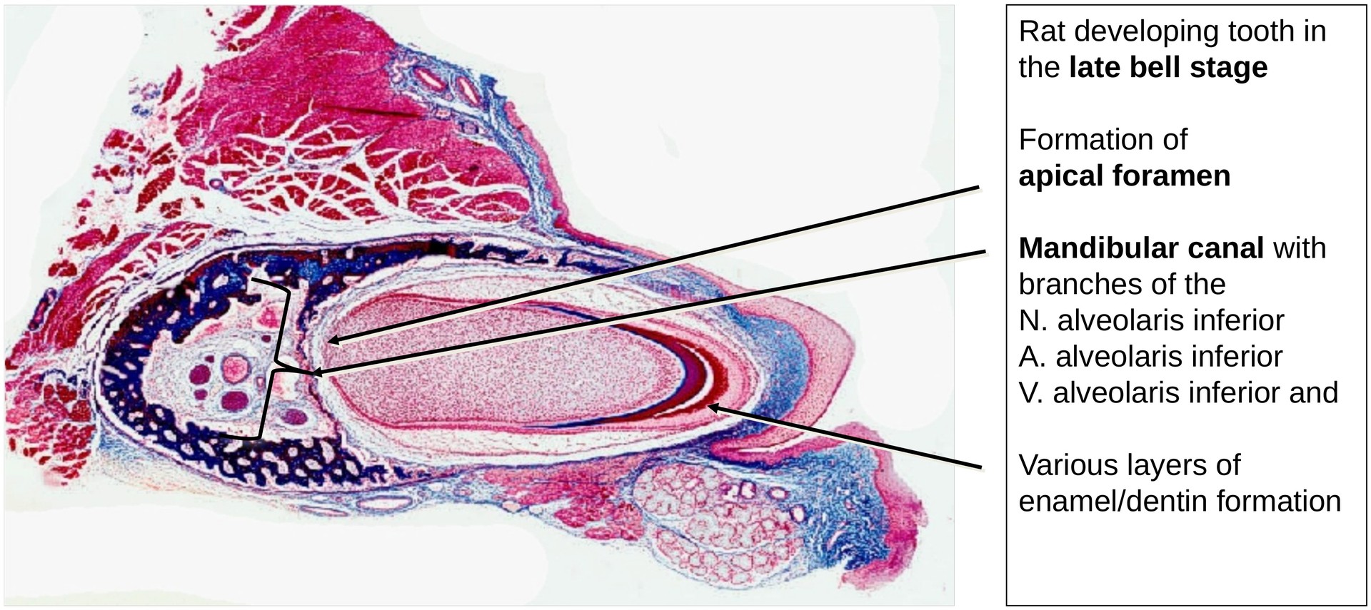
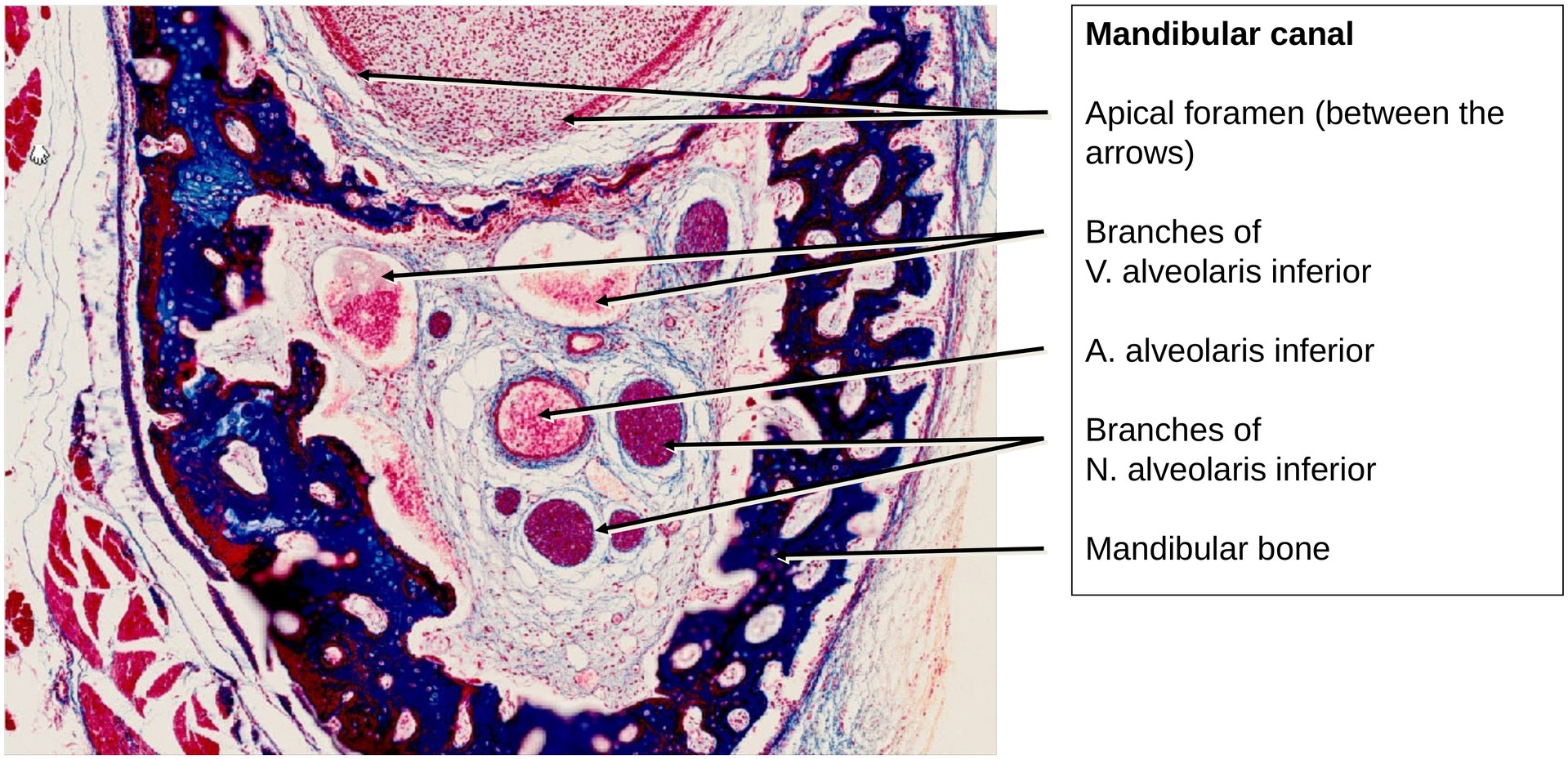
This section shows the mandible of a rat in a late bell stage of tooth development. The apical foramen is already beginning to form.
Within the mandible, the mandibular canal is visible, containing branches of the mandibular nerve and associated vessels. In the rat, this canal differs from that in humans in that it appears fully enclosed at this stage of development.
Tooth Crown Region (Biting Cusp Area):
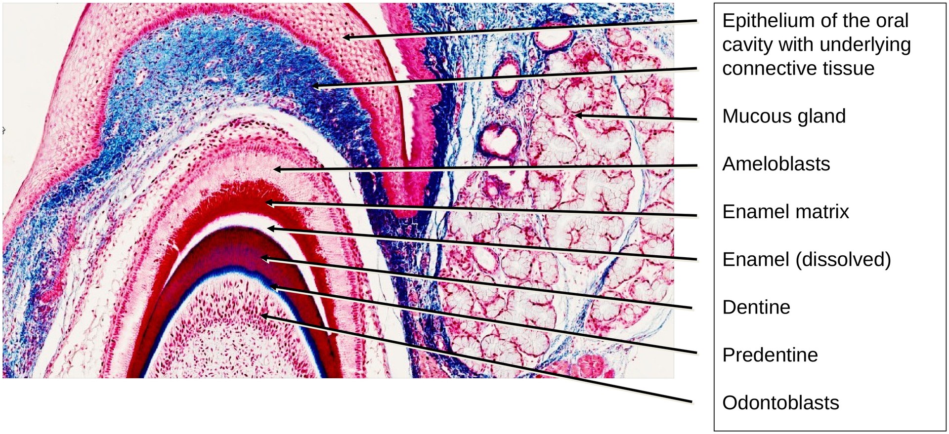
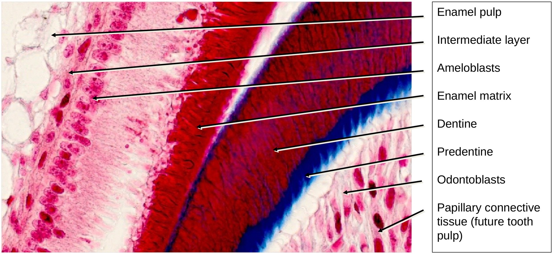
The most advanced differentiation occurs at the future biting cusp. Here, dentine and enamel formation are already active.
On the enamel side, derived from the inner enamel epithelium, the ameloblasts (adamantoblasts) demonstrate progressive differentiation:
- In the peripheral regions, they are short and undifferentiated.
- Toward the cusp, they become tall, columnar, and fully secretory.
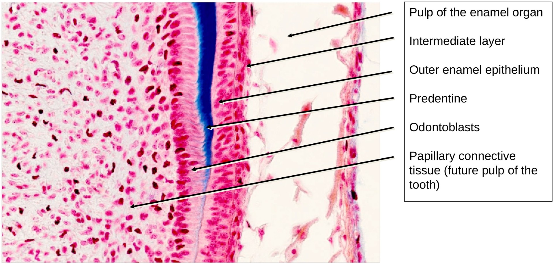
On the dentine side, within the tooth papilla, the odontoblasts show a corresponding gradient of differentiation — tall and secretory near the cusp, less developed toward the root region.
Between these two forming tissues, the predentine stains blue, representing unmineralized matrix. Once mineralized, it stains red, despite decalcification. The space seen between the enamel and dentine layers is not an artefact; it corresponds to the almost completely mineralized enamel, which contains only about 3% organic material. During decalcification, no stainable material remains here.
Tooth Root Region:
In the developing root area, the inner and outer enamel epithelium lie directly adjacent, separated only by the membrana preformativa. Here, the enamel organ begins to bend, marking the future apical foramen.
Adjacent Structures:
Immediately below the oral mucosa epithelium, a mucous gland is visible, representing a labial (or buccal) gland, complete with excretory ducts. The oral epithelium itself can be studied to determine its epithelial type.
Tasks:
Identify and analyse the following structures and relationships:
- In the region of the future biting cusp, identify:
- Ameloblasts (adamantoblasts)
- Partially mineralized enamel matrix
- Enamel layer (gap due to decalcification)
- Dentine (with dentinal tubules)
- Predentine (blue-stained, unmineralized)
- Odontoblast layer
- Papillary connective tissue (future pulp)
- Trace the ameloblasts (adamantoblasts) along the enamel organ towards the outer enamel epithelium.
- Observe how cell height and morphology change with the stage of differentiation.
- Locate the apical foramen, found where the inner and outer enamel epithelium bend together at the base of the enamel organ.
- Identify the branches of the inferior alveolar nerve and inferior alveolar vessels within the mandibular canal.
- Locate the labial gland beneath the oral mucosa epithelium and follow its excretory ducts.
- Examine the oral mucosa epithelium microscopically.
- What epithelial type is present?
- Determine the type of bone forming the mandible at this stage of development.
License
University of Basel
Downloads