DIGESTIVE ORGANS: GASTROINTESTINAL TRACT (ANATOMICAL MICROSCOPY)
19.5
Ileum, Human 1
Specimen:
Specimen Details:
Organ: Ileum
Origin: Human
Staining: Hematoxylin - Eosin (H&E)
Method and Specimen Description:
Routine histological section stained with an overview stain (H&E).
Objective of the Examination:
To understand the microscopic structure of the human ileum, its typical organization, and its differences from other regions of the gastrointestinal tract (GIT).
Special Features of the Specimen:
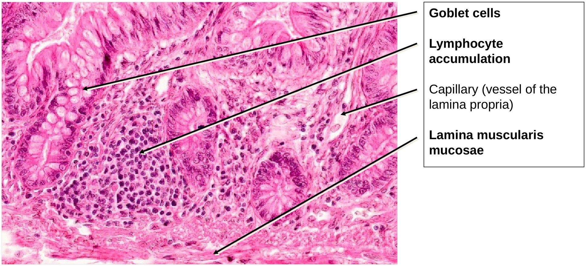
This specimen originates away from the antimesenteric side, and therefore Peyer’s patches are absent. Instead, scattered lymphocytic aggregates can be seen within the lamina propria. These form part of the mucosa-associated lymphatic tissue (MALT) — referred to specifically in the intestine as gut-associated lymphatic tissue (GALT).
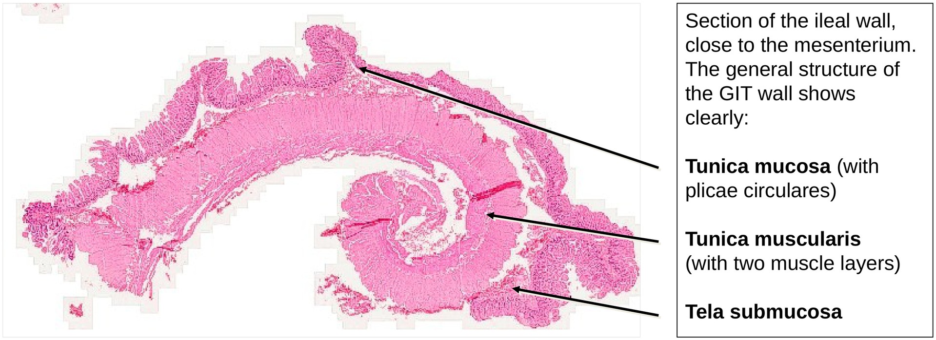
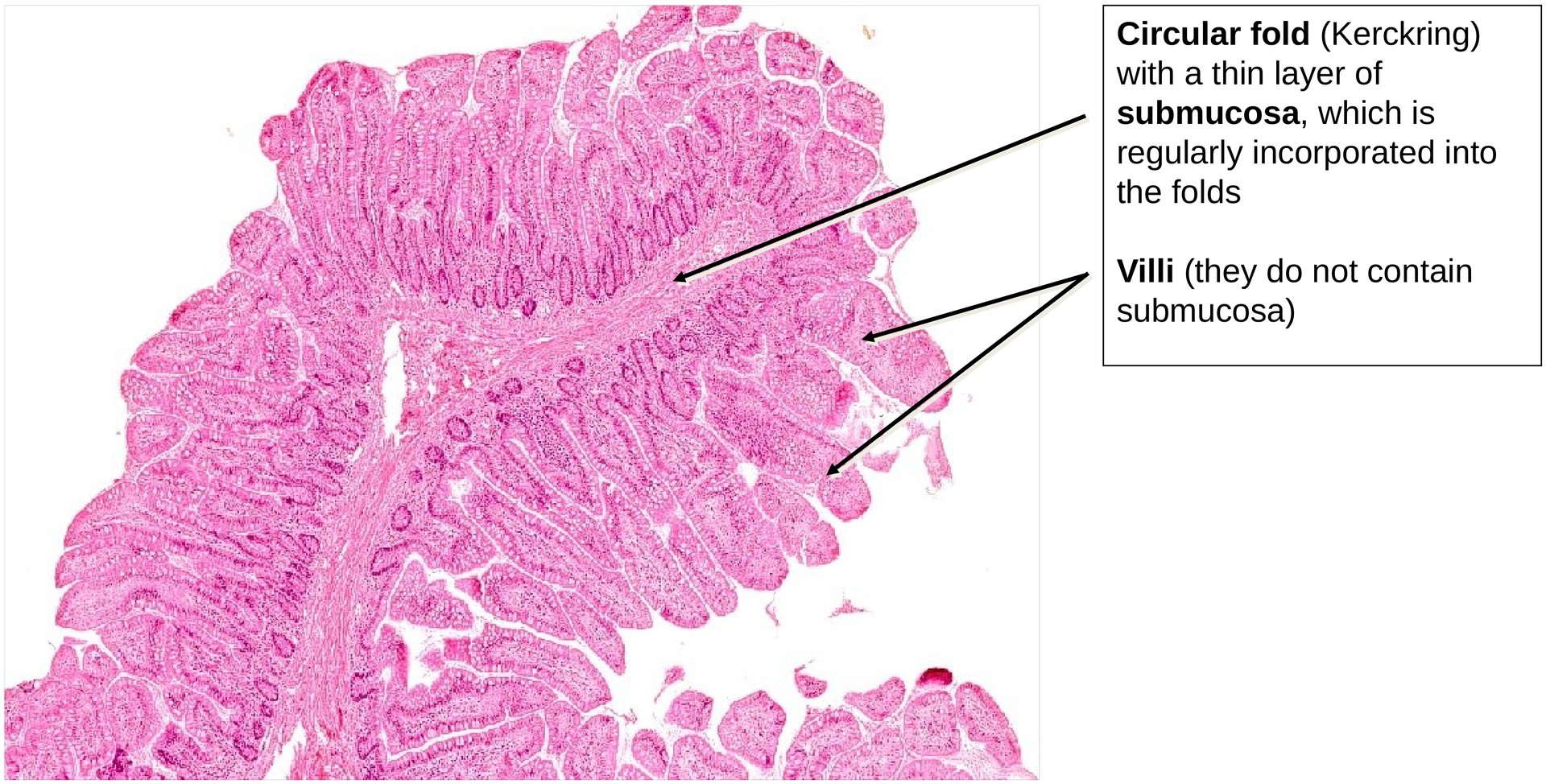
The general structural plan of the gastrointestinal tract is clearly visible, with all layers well preserved. The circular folds (plicae circulares) are lower here than in the duodenum or jejunum, indicating the transition toward the distal small intestine. The submucosa contains no glands, unlike the duodenum, which contains Brunner’s glands.
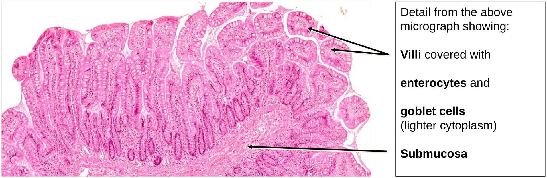
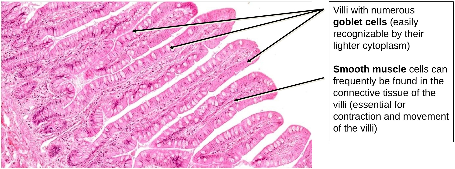
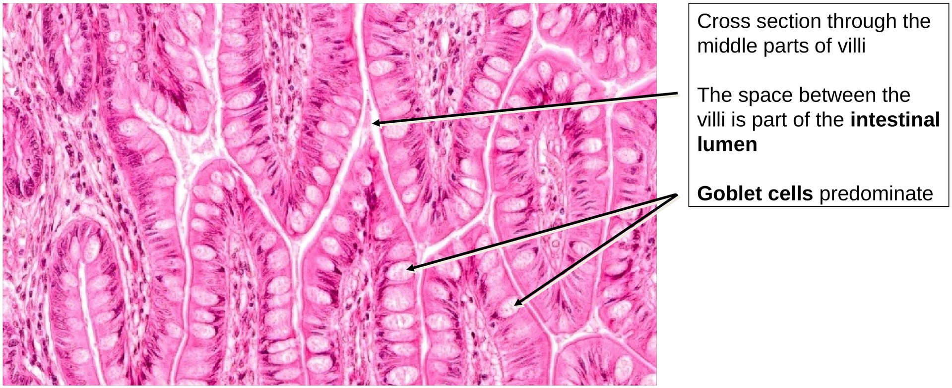
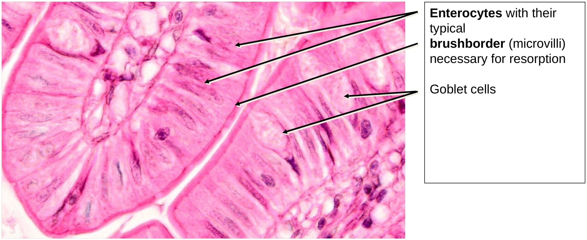
A high number of goblet cells is characteristic of the ileum, reflecting its increased mucus production for lubrication and protection. Between the goblet cells, the enterocytes display a distinct brush border (microvilli), indicating active absorption.
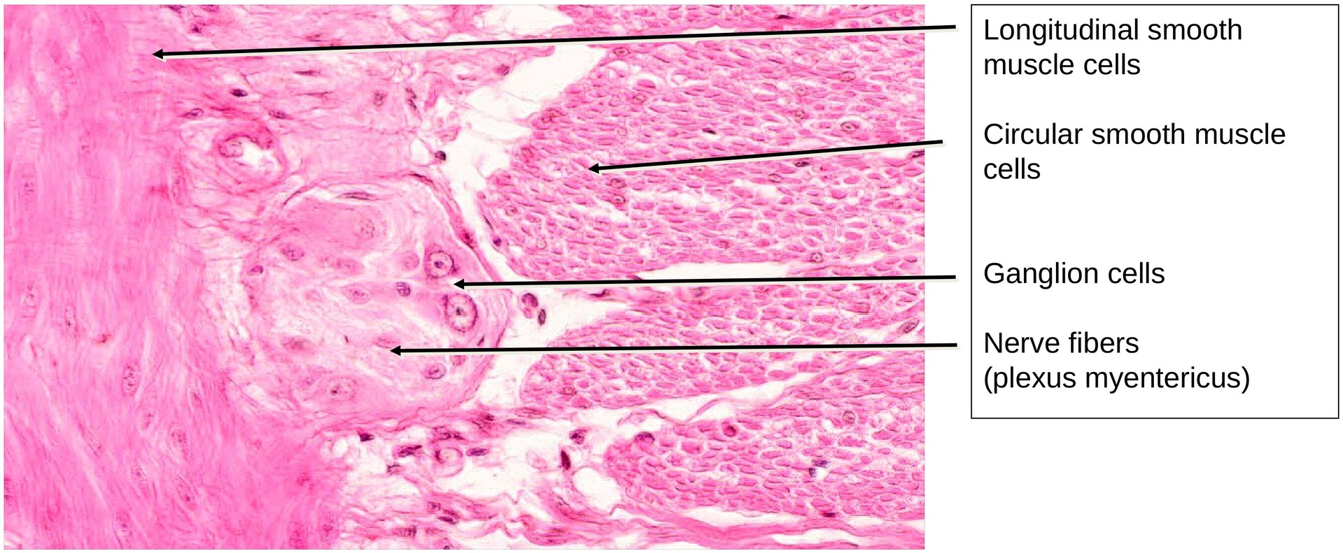
The lamina muscularis mucosae forms a clear boundary between the mucosa and the underlying tela submucosa. The tunica muscularis consists of the usual two smooth muscle layers:
- Inner circular muscle layer,
- Outer longitudinal muscle layer.
Between these layers lies the myenteric plexus (Auerbach’s plexus), which is clearly visible and contains ganglion cells and nerve fibers.
During fixation in formaldehyde, the longitudinal muscle contracted strongly. This caused the mucosa and epithelium to appear convex, whereas in the living state the intestinal wall normally shows a concave contour due to relaxation.
Tasks:
- Obtain an overview of the specimen and confirm its identification as part of the gastrointestinal tract based on its general wall structure.
- Distinguish between folds (plicae) and villi — folds involve both mucosa and submucosa, whereas villi are projections of the mucosa only.
- Identify the following layers:
- Tunica mucosa (with lamina epithelialis and lamina propria)
- Muscularis mucosae
- Tela submucosa
- Tunica muscularis (with circular and longitudinal layers)
- Locate the ganglion cells and nerve fibers of the myenteric plexus (Auerbach) between the muscle layers.
- Trace the muscle fibers of the lamina muscularis mucosae extending into the villi.
- Search for lymphocyte aggregates within the lamina propria, between the mucosa and the muscularis mucosae.
License
University of Basel
Downloads