URINARY ORGANS (ANATOMICAL MICROSCOPY)
12.5
Kidney, rabbit
Preparation:
Preparation details:
Organ: Kidney
Origin: Rabbit
Staining: PAS
Method and Specimen Description:
Normal histological specimen stained with PAS (Periodic acid–Schiff), which clearly highlights basement membranes, brush borders, and glycoprotein-rich structures in magenta tones.
Objective of the Examination:
To gain knowledge of the basic structural organization of the kidney—particularly the renal papilla, which also defines the typical cortical and medullary arrangement seen in the human multipapillary kidney—and to study the zones within the kidney. Detailed descriptions of the nephron components are provided in the two human kidney specimens.
Special features of the preparation:
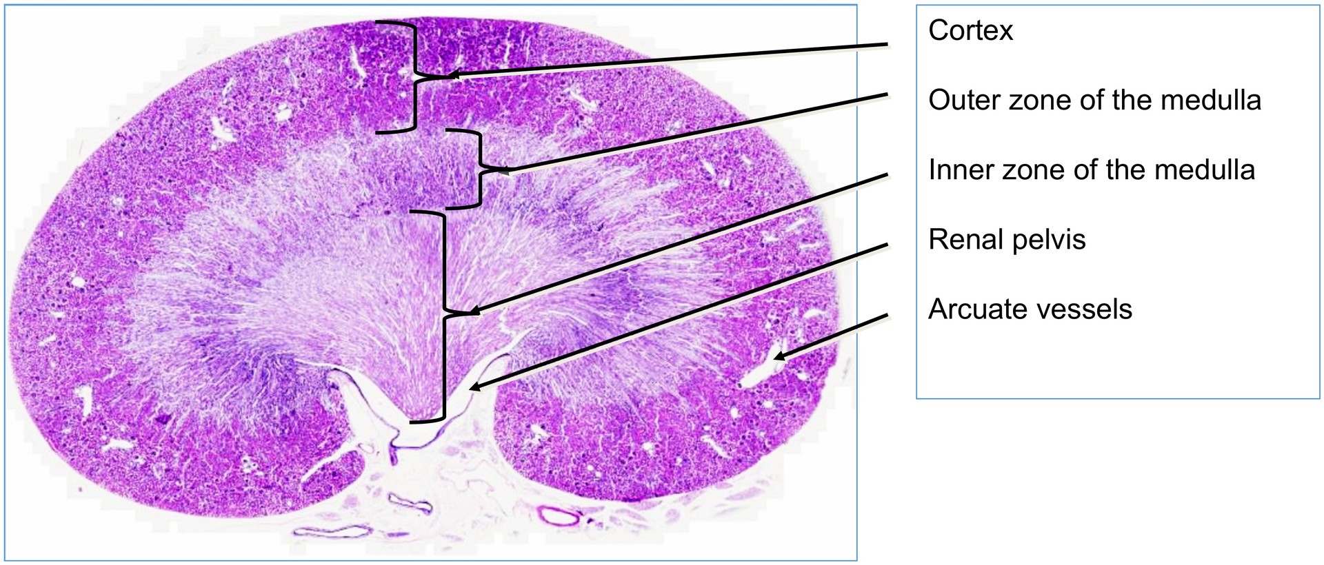
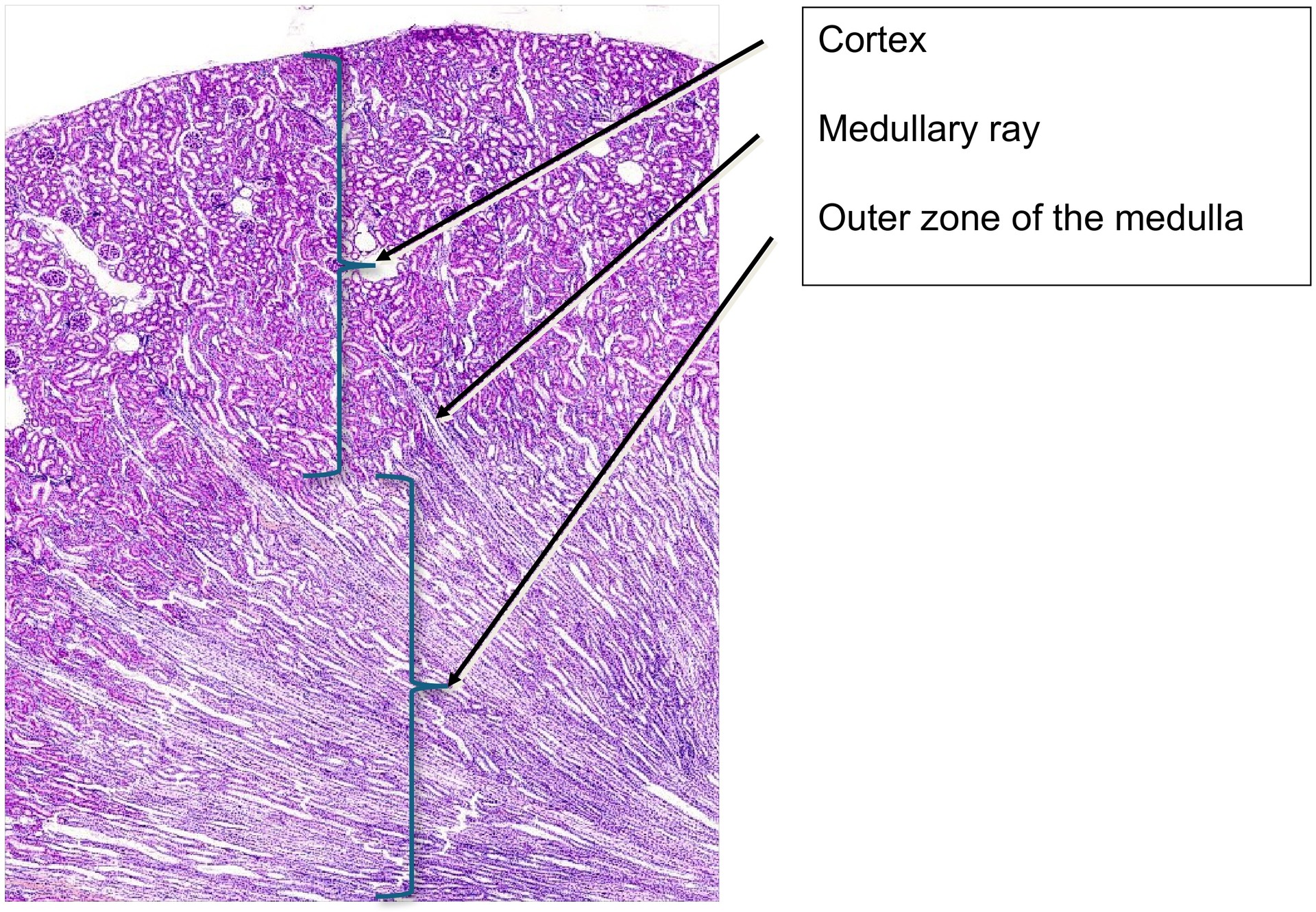
At low magnification, the two principal regions of the kidney—the cortex and medulla—can be clearly distinguished.
- The cortex contains the glomeruli and the convoluted segments of the nephron: the proximal and distal convoluted tubules.
- The medulla contains the straight portions of these tubules and the collecting ducts, which converge towards the papillary tip, forming the papillary ducts.
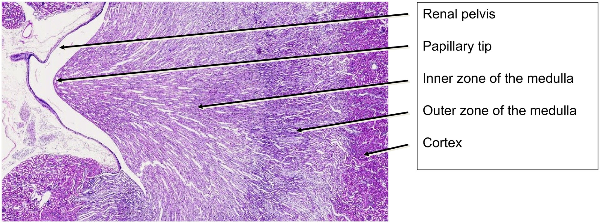
- The vasa recta are also present within the medulla. The medullary pyramid is surrounded by the cortex, and its papillary tip projects into and is enclosed by the renal pelvis.
Within the medullary pyramid, two zones can be distinguished even at low magnification:
- the outer zone, adjacent to the cortex, and
- the inner zone, extending towards the papilla. The outer zone is often further divided into an outer stripe (containing straight portions of the proximal and distal tubules and vessels) and an inner stripe (containing straight distal and intermediate tubules and vessels). This subdivision can be inferred in this specimen, though not distinctly observed.
At the corticomedullary junction, the arcuate vessels (vasa arcuata)—the arcuate artery and vein—are visible. The arcuate veins are identifiable by their larger lumina.
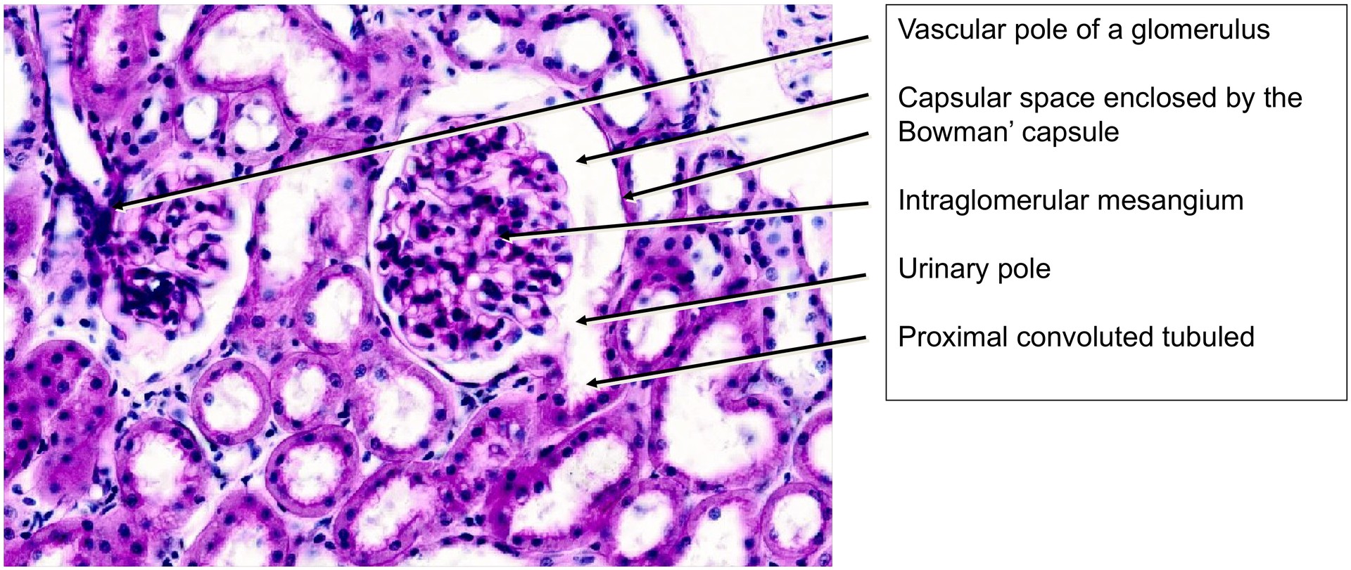
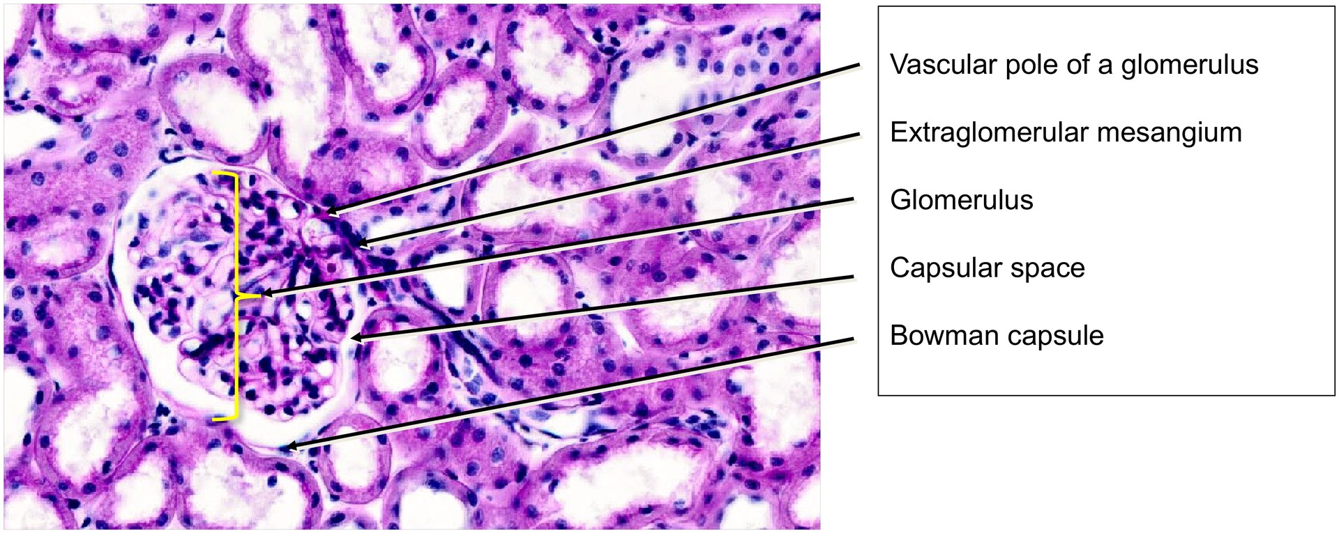
Within the cortex, the glomeruli are conspicuous. Depending on the plane of section:
- some display the vascular pole, where the afferent and efferent arterioles enter and exit,
- others show the urinary pole, where the proximal convoluted tubule begins.
The Bowman’s capsule surrounds the glomerulus, enclosing the capsular space. The parietal layer of the capsule is a thin epithelium, while the visceral layer consists of podocytes. The mesangium is also visible—both intraglomerular and extraglomerular.
The distinction between proximal and distal convoluted tubules is better demonstrated in the human kidney specimens (see “Kidney, Human 1”).
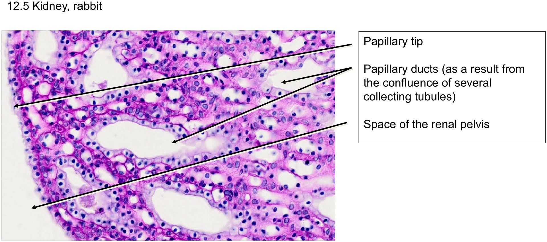
In the medullary papilla (towards the renal pelvis), the tissue contains mainly collecting ducts and papillary ducts, with some intermediate (thin) tubules—the thin limbs of the loop of Henle—and accompanying vasa recta.
At the papillary tip, the surface is lined by a simple squamous to cuboidal epithelium, which transitions into transitional epithelium (urothelium) in the fornices and renal pelvis. Urinary pores are not visible in this section.
Tasks:
- Identify the cortex, medulla, papillary tip, and renal pelvis at low magnification.
- In the cortex, locate glomeruli, the capsular space, and Bowman’s capsule.
- Find glomeruli showing a distinct urinary pole (adjacent proximal convoluted tubule) and a vascular pole (connection with afferent/efferent arterioles).
- Compare the structural organization of the cortex and medulla, and explain the differences in their histological appearance.
License
University of Basel
Downloads