SKIN AND APPENDAGES (ANATOMICAL MICROSCOPY)
13.3
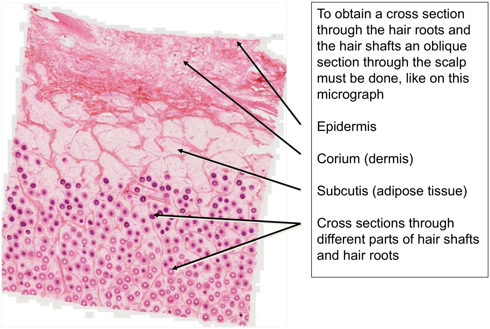
Scalp, axial cross section
Specimen:
Specimen Details:
Organ: Scalp
Origin: Human
Staining: Hematoxylin-Eosin (H&E)
Method and Specimen Description:
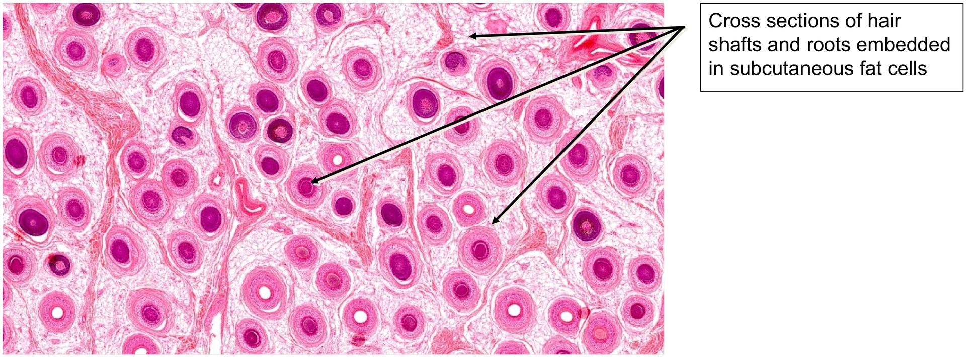
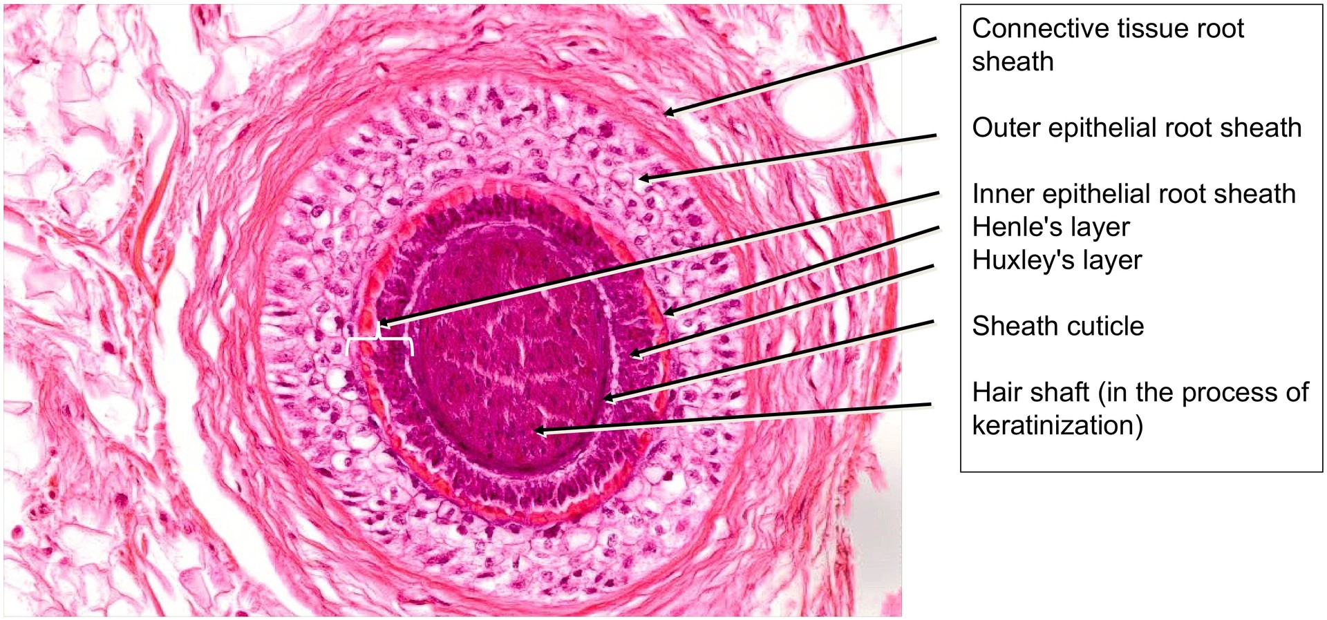
Oblique section through the scalp. Since the hairs are embedded obliquely within the scalp, this sectioning plane enables cross-sections of the hair shafts while simultaneously passing through all scalp layers — from the subcutis to the epidermis. In this way, all zones of the hair located within the scalp can be depicted, from the hair bulb to the already keratinized portion of the hair shaft.
Objective of the Examination:
To understand hair formation and recognize the individual layers of the hair at different developmental levels.
Special Features of the Specimen:
Depending on the level of section through the hair follicle, different developmental zones can be observed.
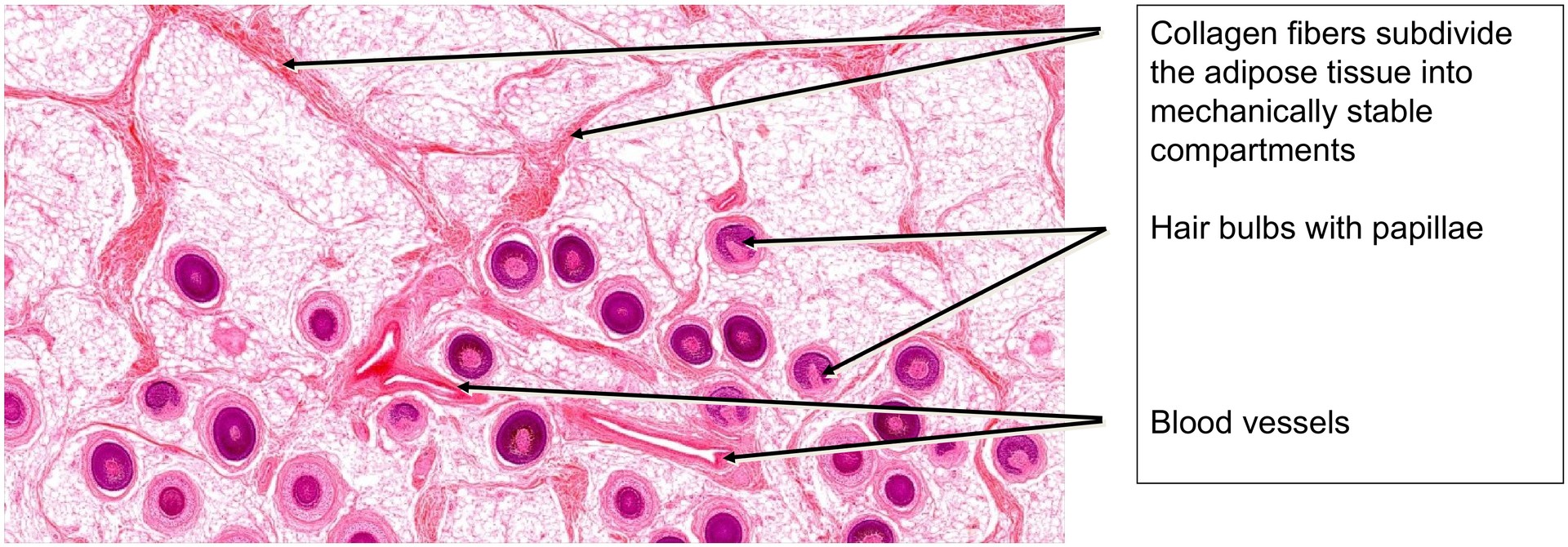
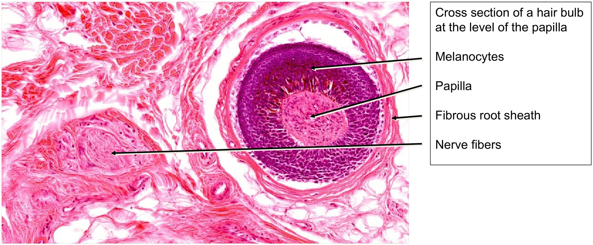
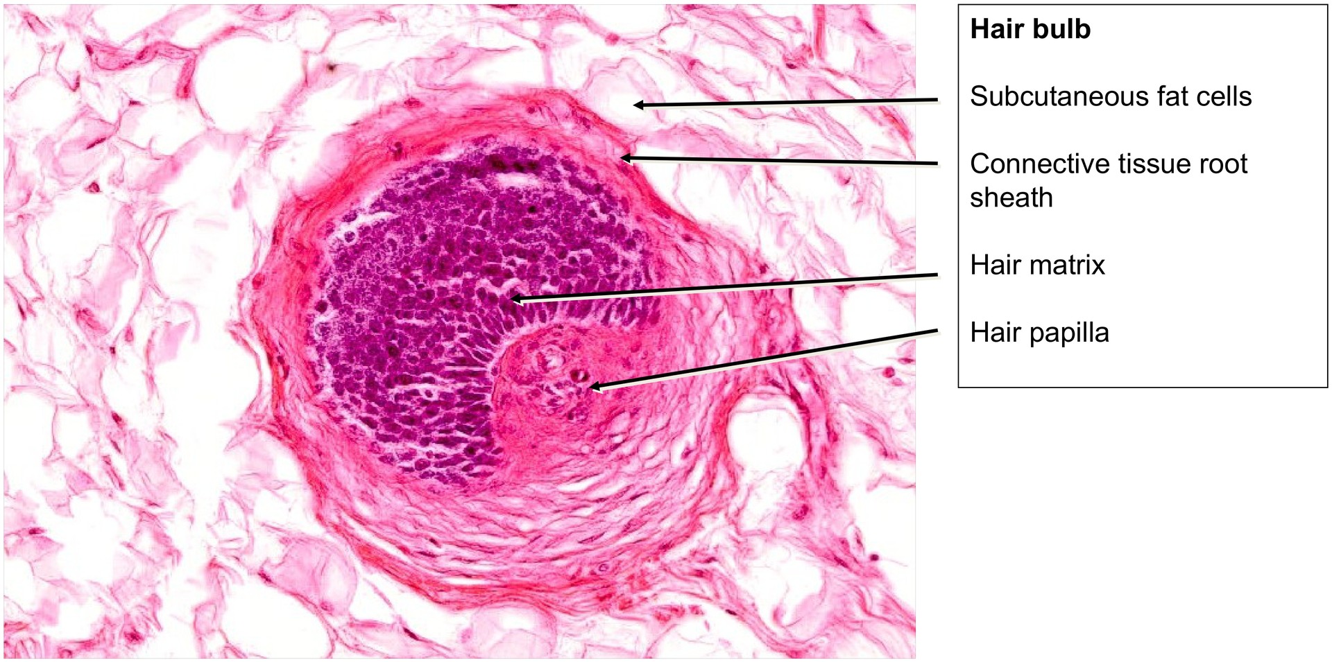
At the level of the hair bulb, the hair papilla can be seen in the center, surrounded by the germinal matrix, whose cells divide mitotically during hair formation and growth. These differentiating matrix cells give rise to the cells of the medulla, cortex, and cuticle, as well as the inner root sheath. Melanocytes lie between the matrix cells, transferring melanosomes to the developing cortical cells and thus determining hair pigmentation. The hair papilla, rich in capillaries, regulates the division behavior of the matrix cells and hair growth. Without a papilla, no hair growth occurs.
Moving upward toward the surface, the inner root sheath becomes fully differentiated, consisting of:
- an outer Henle layer, and
- an inner Huxley layer.
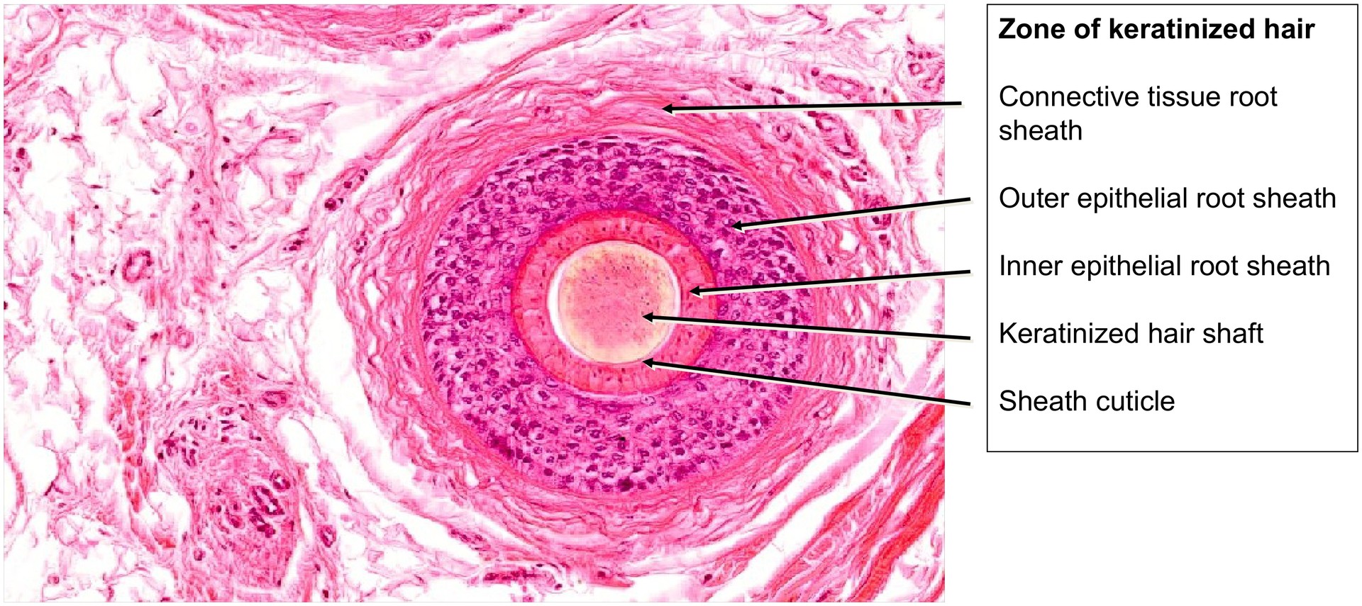
Beneath the Huxley layer lies the hair cuticle, followed by the cortex, and — in the central keratogenic zone — occasional cells of the medulla.
The epidermis invaginates deeply to form the hair follicle. Its layers largely correspond to those of the skin, except that within the follicle there is no keratinization and therefore no stratum granulosum.
Surrounding the epithelial root sheath (comprising inner and outer root sheaths) is the connective tissue root sheath. Between them lies a distinct basement membrane, relatively thick compared to other skin regions.
Tasks:
- Orient yourself in the specimen: identify the epidermis and subcutis.
- Determine where the hair bulbs are located.
- Distinguish the epithelial from the connective tissue root sheath.
- Locate a zone where the inner (epithelial) root sheath allows identification of its two layers: Henle (outer) and Huxley (inner).
- Search for melanocytes in the hair bulb region.
- Look for nerve fibers between the hair bulbs.
- Find a section through a hair papilla and, if possible, identify a capillary within the papilla.
License
University of Basel
Downloads