SENSORY ORGANS (ANATOMICAL MICROSCOPY)
17.2
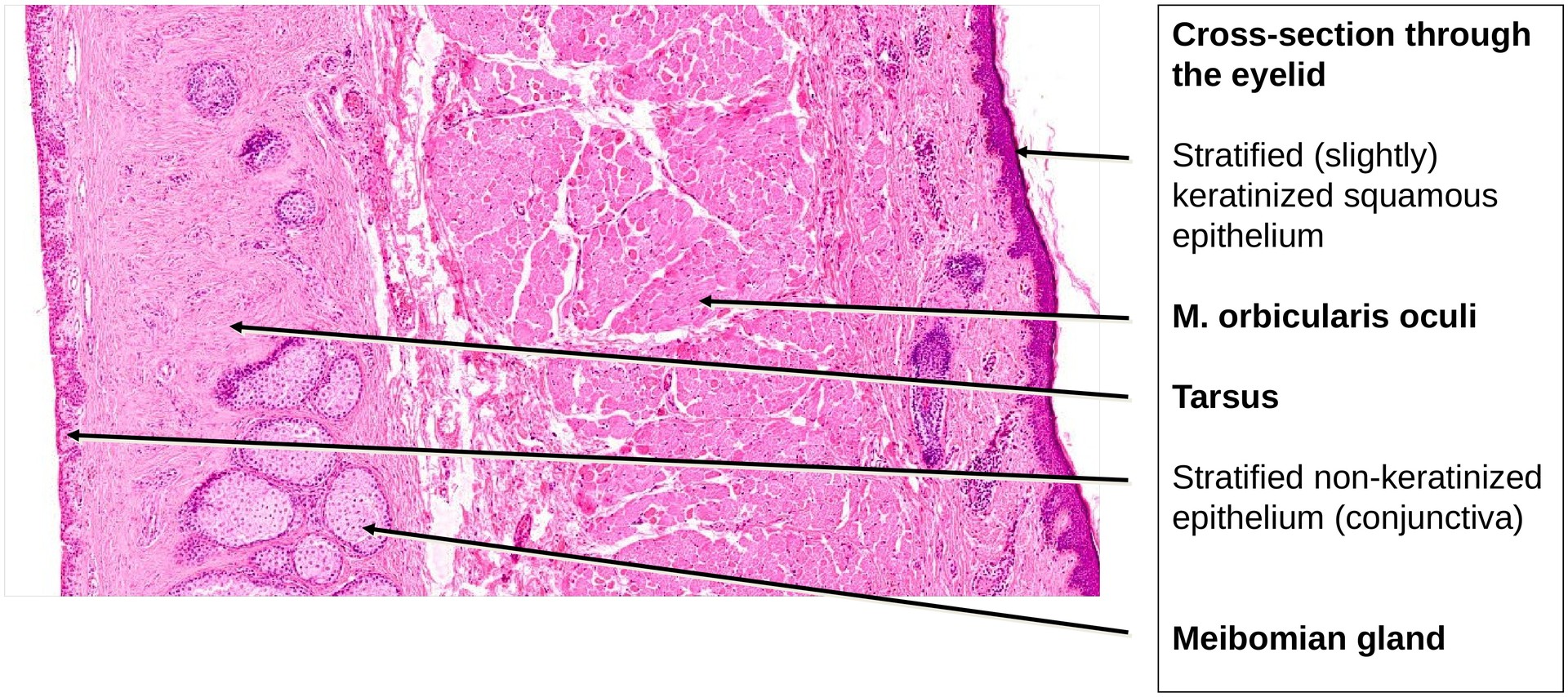
Eyelid
Specimen:
Specimen Details:
Organ: Eyelid
Origin: Human
Staining: Hematoxylin - Eosin (H&E)
Method and Specimen Description:
Routine histological preparation stained with hematoxylin and eosin, providing an overview of the different tissue layers and glandular components of the eyelid.
Objective of the Examination:
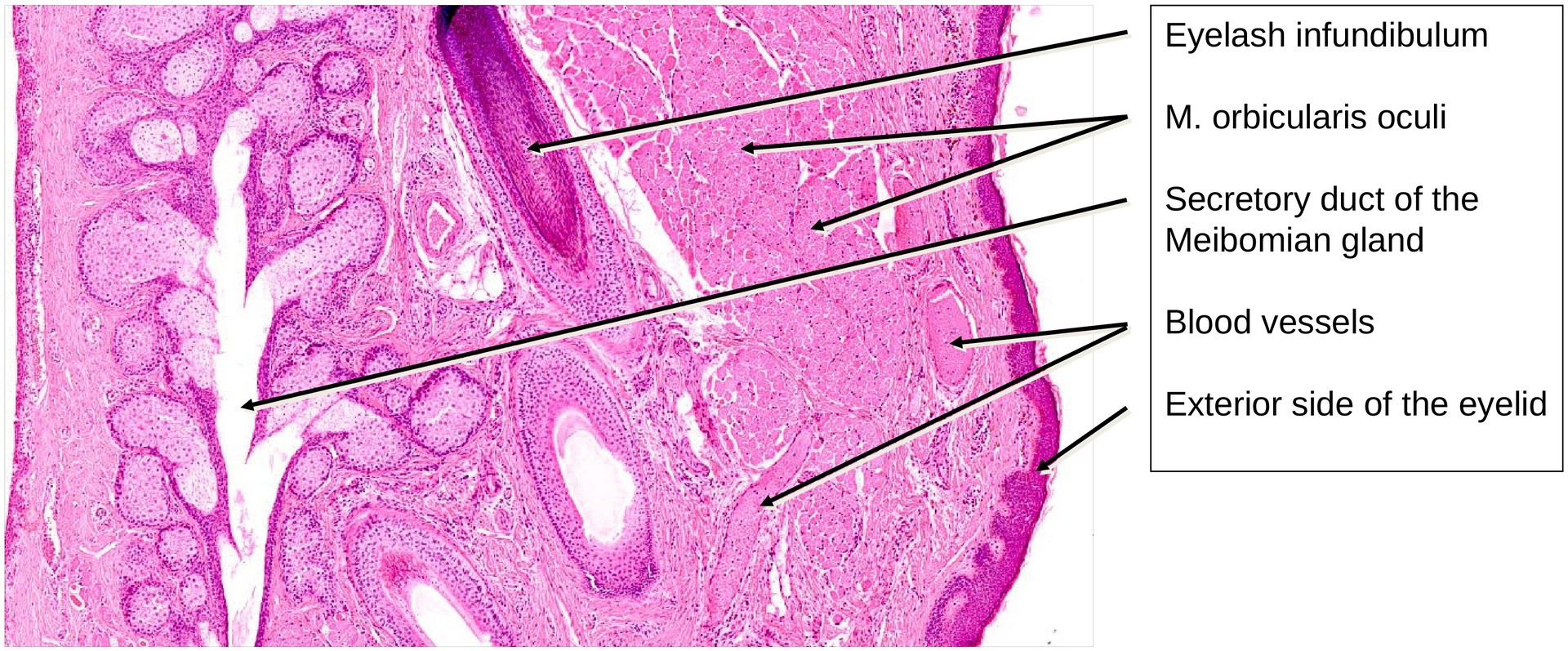
To study the structure of the tarsus as the supporting connective tissue of the eyelid, to identify the Meibomian glands (holocrine glands that open directly onto the lid margin), and to locate the orbicularis oculi muscle and related structures involved in eyelid function.
Special Features of the Specimen:
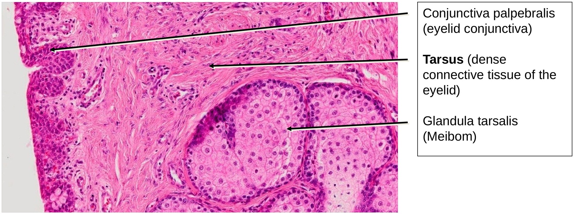
The eyelid is a movable skin fold that protects the eye and ensures uniform distribution of tears across the corneal surface. Its structural basis is the tarsus, a dense plate of collagenous connective tissue that provides firmness and shape to the lid. Embedded within the tarsus are the Meibomian glands (tarsal glands), which are modified sebaceous glands opening along the free eyelid margin.
These glands secrete a lipid-rich (oily) secretion that:
- lubricates the eyelid margin,
- forms a thin oily layer over the tear film,
- reduces evaporation of the aqueous phase of the tears, and
- helps maintain the hydration and transparency of the cornea.
Without this layer, the cornea would dry out and lose its optical clarity.
In addition to the prominent Meibomian glands, two other types of glands may be present:
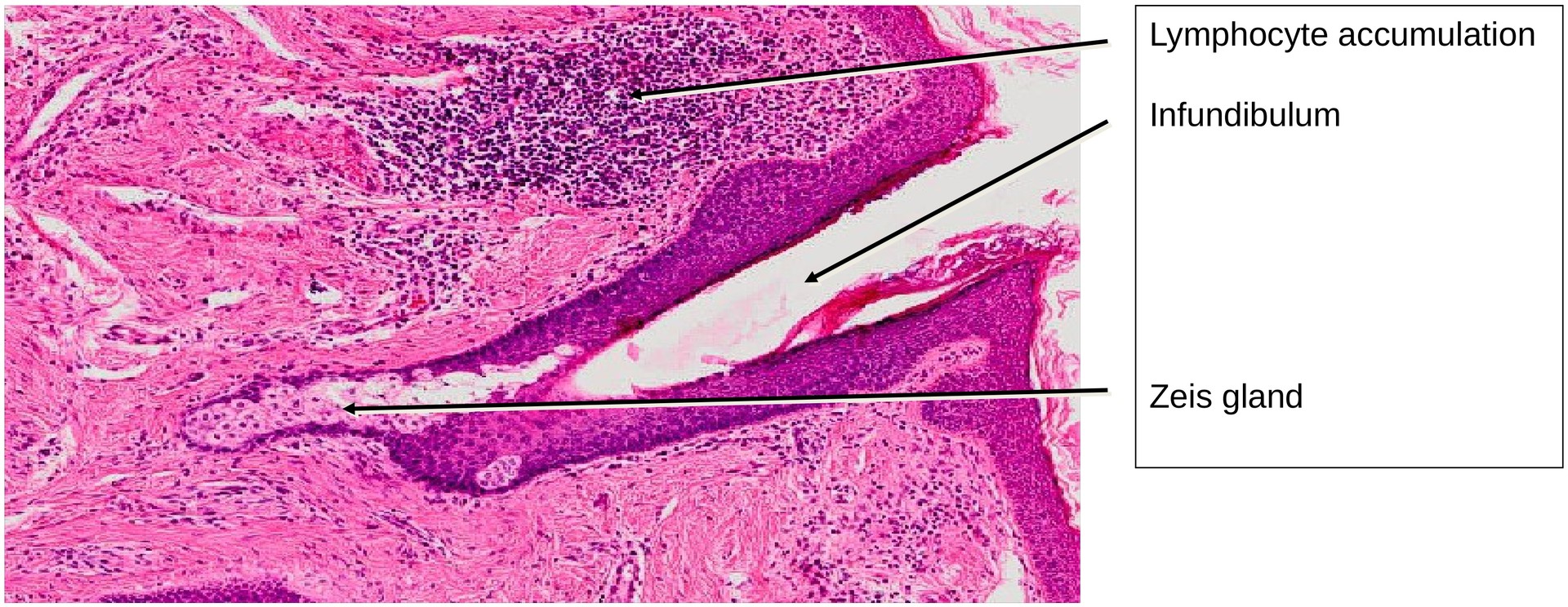
- Zeis glands: sebaceous glands that open into the hair follicles of the eyelashes to lubricate them.
- Moll glands: apocrine glands located near the follicles; their precise function remains incompletely defined and they are not visible in this specimen.
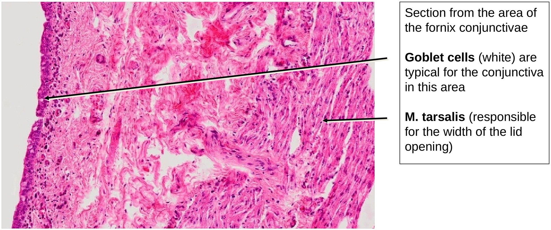
On the inner surface (facing the eyeball), the eyelid is lined by the palpebral conjunctiva (conjunctiva palpebralis) — a stratified non-keratinized epithelium containing occasional goblet cells. In the fornix conjunctivae (where the conjunctiva reflects from the eyelid onto the eyeball), goblet cells are relatively numerous and secrete mucins that stabilize the tear film.
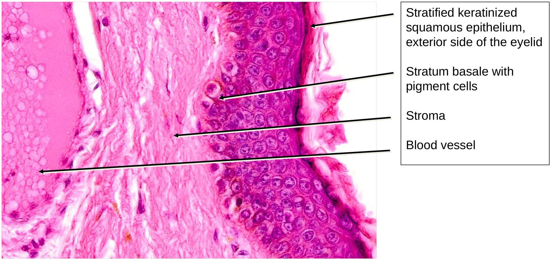
The outer surface of the eyelid is covered by stratified keratinized squamous epithelium, corresponding to normal skin. It contains small hair follicles and sweat, and sebaceous glands associated with the eyelashes.
Within the eyelid lies the orbicularis oculi muscle, composed of striated skeletal muscle fibres, responsible for closing the eyelids. The pars palpebralis of this muscle is identifiable in the eyelid section.
A small, specialized portion of this muscle, located at the lower edge of the lid between the hair follicles and the Meibomian glands, is the Riolan muscle. It also consists of striated fibers and functions to compress the Meibomian glands, facilitating secretion of their oily content onto the lid margin.
At the inner aspect, near the conjunctiva, a thin layer of smooth muscle fibers can be recognized — the tarsal muscle (Müller’s muscle). This muscle is sympathetically innervated and contributes to maintaining the width of the palpebral fissure. The tendon of the levator palpebrae superioris muscle, responsible for elevating the upper eyelid, also inserts into this region.
At the lower margin of the lid, a local accumulation of lymphocytes may be observed, indicating a minor immune response in the conjunctival or dermal tissue.
Tasks:
Identify the following structures and answer the corresponding questions based on the specimen:
- Tarsus and Meibomian glands — where do the ducts of these glands open?
- Hair follicles — which glands open into them?
- What are the differences between the inner and outer surfaces of the eyelid? Identify both histologically.
- Locate the tarsal (Müller’s) muscle — what is its function?
- Identify the Riolan muscle — to which muscle type does it belong, and what is its function?
License
University of Basel
Downloads