DIGESTIVE ORGANS: GASTROINTESTINAL TRACT (ANATOMICAL MICROSCOPY)
19.2
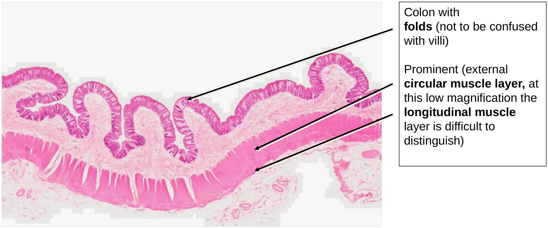
Colon
Specimen:
Specimen Details:
Organ: Colon
Origin: Human
Staining: Hematoxylin - Eosin (H&E)
Method and Specimen Description:
Normal histological section of the human colon, stained with the H&E method for general tissue visualization.
Objective of the Examination:
To understand the microscopic structure of the colon as part of the large intestine, and to recognize its characteristic features in comparison with other segments of the gastrointestinal tract.
Special Features of the Specimen:
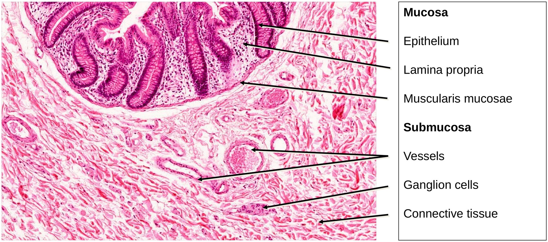
Although the colon follows the general structural plan of the gastrointestinal tract (GIT), it possesses several distinctive features.
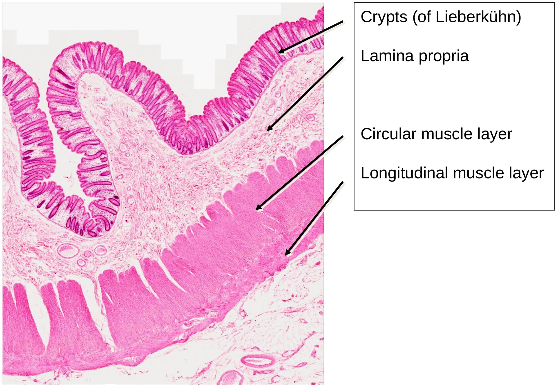
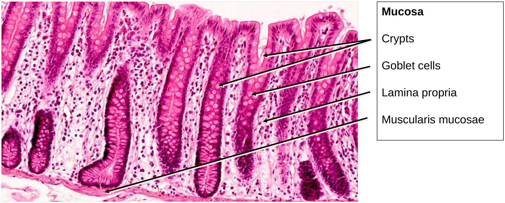
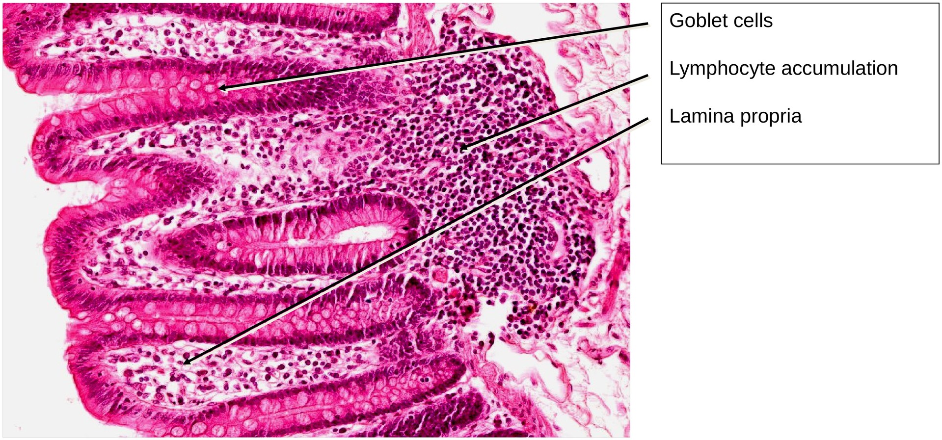
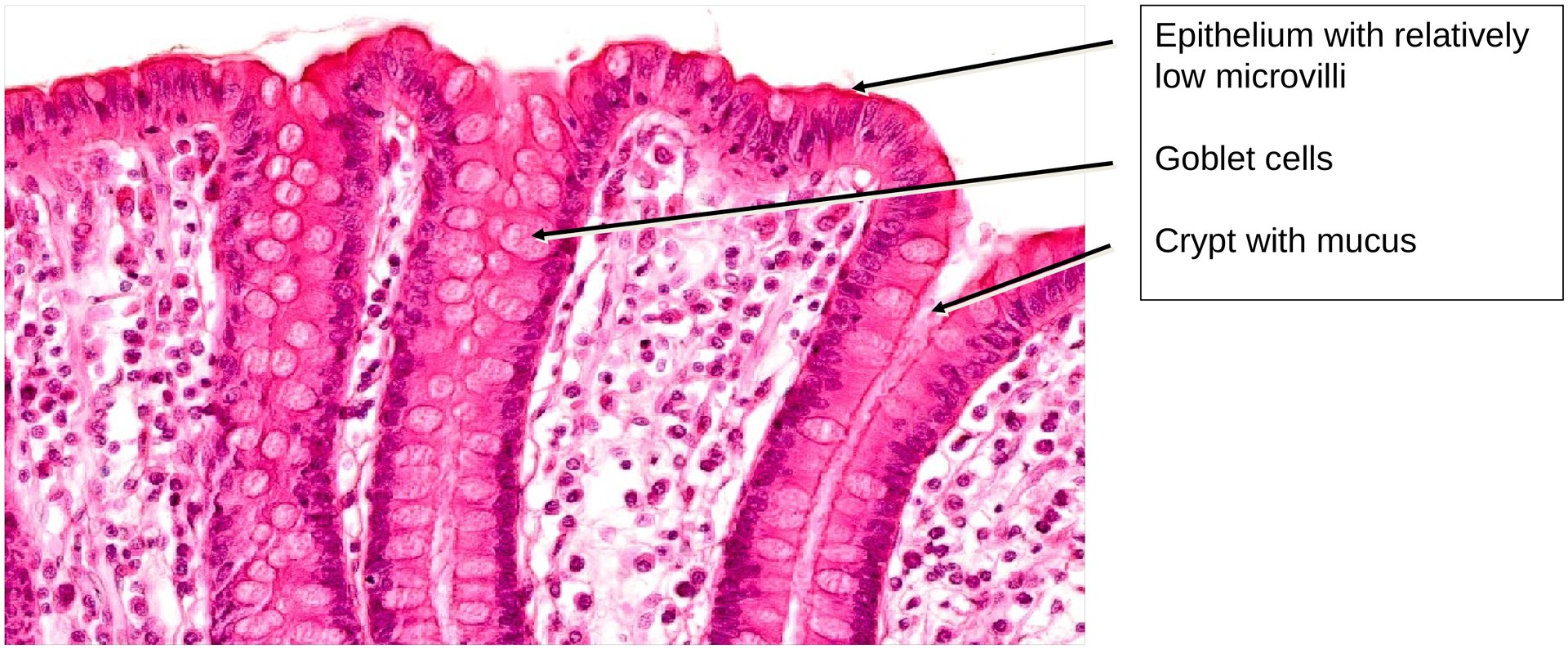
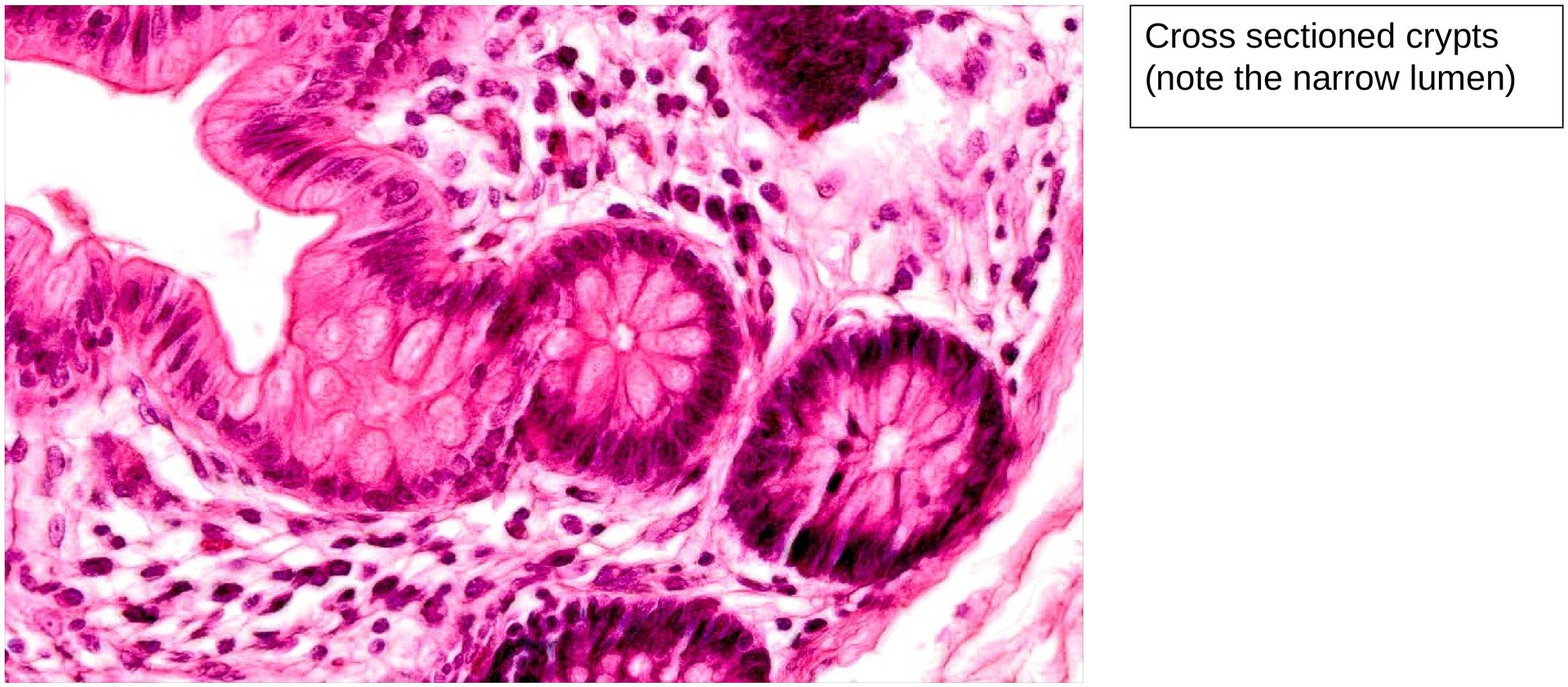
Unlike the small intestine, the colon lacks villi and instead contains straight tubular glands, known as crypts of Lieberkühn. The number of goblet cells is markedly higher than in other intestinal regions, reflecting the organ’s function in mucus secretion and lubrication of fecal material.
The epithelium is a simple columnar type, consisting of absorptive enterocytes interspersed with numerous goblet cells, which extend deep into the crypts. The enterocytes primarily facilitate water reabsorption through active sodium transport coupled with passive osmotic water movement.
Within the lamina propria, lymphocytes and plasma cells are abundant, often forming small aggregates, particularly near the crypt bases.
Beneath the mucosa lies a thin muscularis mucosae, composed of smooth muscle fibers running parallel to the mucosal surface.
The submucosa contains connective tissue, blood vessels, and elements of the submucosal (Meissner’s) plexus.
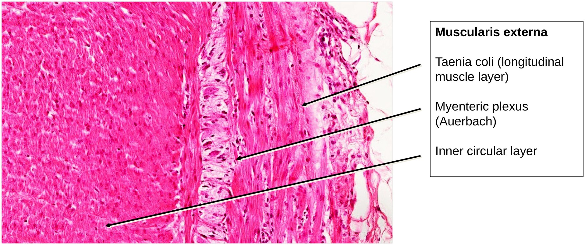
The muscularis externa consists of an inner circular and outer longitudinal muscle layer. The circular layer is relatively thick and continuous, whereas the outer longitudinal layer is organized into three distinct taeniae coli. In this specimen, one taenia coli has been sectioned.
Between the two muscle layers lies the myenteric (Auerbach’s) plexus, which contains ganglion cells and nerve fibers.
Tasks:
- Locate the crypts of Lieberkühn and identify the numerous goblet cells within them.
- Compare the epithelium with that of the small intestine (duodenum, jejunum, ileum).
- Note that microvilli are present but less prominent in the colon.
- Examine the muscularis mucosae and describe its structure and continuity.
- Differentiate between the inner circular muscle layer and the outer longitudinal layer (taenia coli).
- Locate and identify components of the myenteric plexus (ganglion cells and nerve fibres) situated between the muscle layers.
License
University of Basel
Downloads