FEMALE REPRODUCTIVE ORGANS (ANATOMICAL MICROSCOPY)
10.9
Placenta 1
Specimen:
SPECIMEN DETAILS:
Organ: Placenta
Origin: Human
Staining: Hematoxylin Eosin (H&E)
METHOD AND SPECIMEN DESCRIPTION:
Histological section of a mature human placenta, prepared with standard H&E staining for general tissue structure.
OBJECTIVE OF THE EXAMINATION:
To study the structure and components of the mature placenta, including the foetal and maternal portions and the placental barrier involved in maternal–fetal exchange.
Special Features of the Specimen:
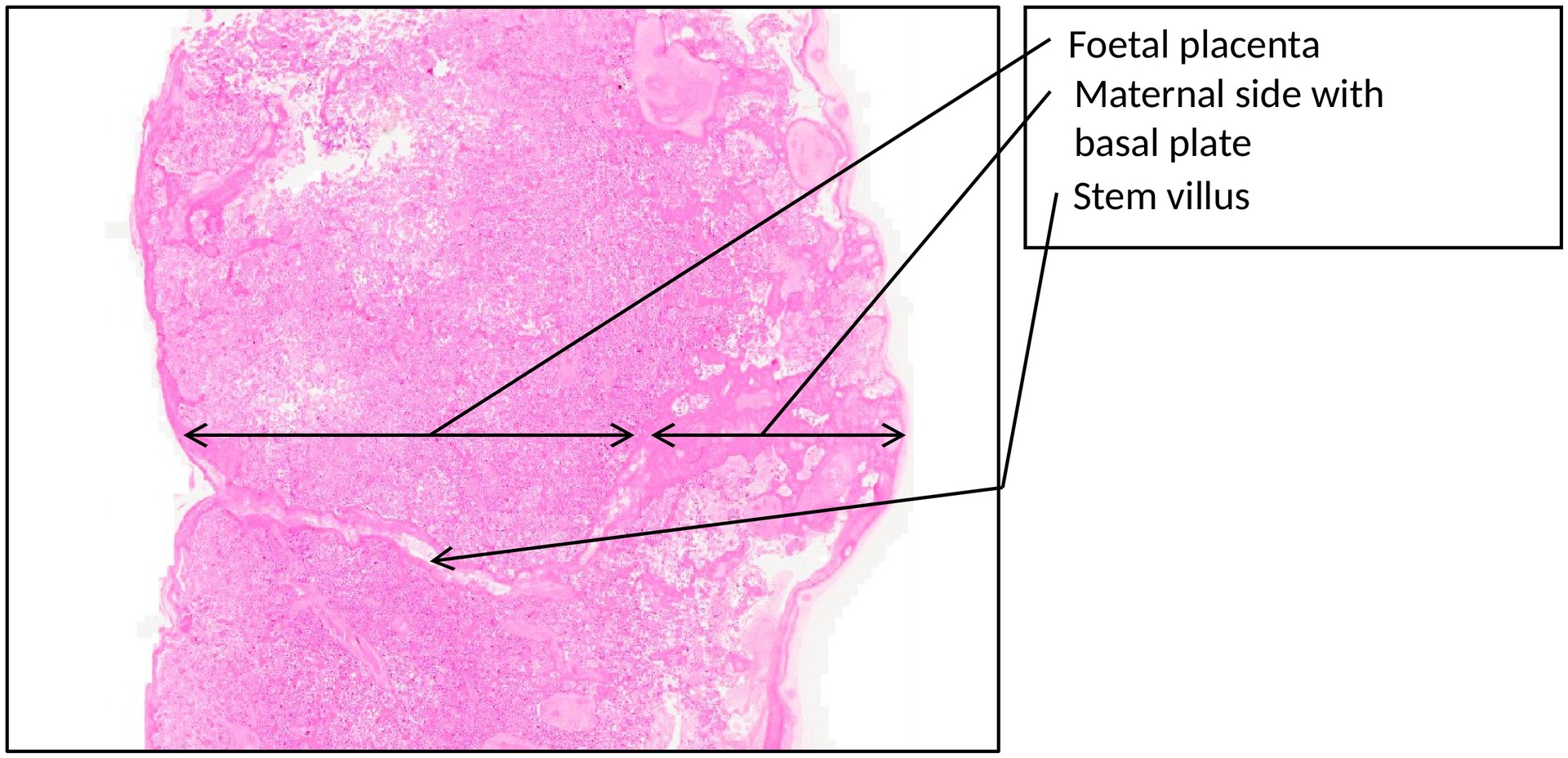
The placenta comprises two principal components:
-
the fetal (embryonic) component, derived from the chorion, and
-
the maternal component, derived from the decidua basalis (the functional layer of the endometrium following implantation).
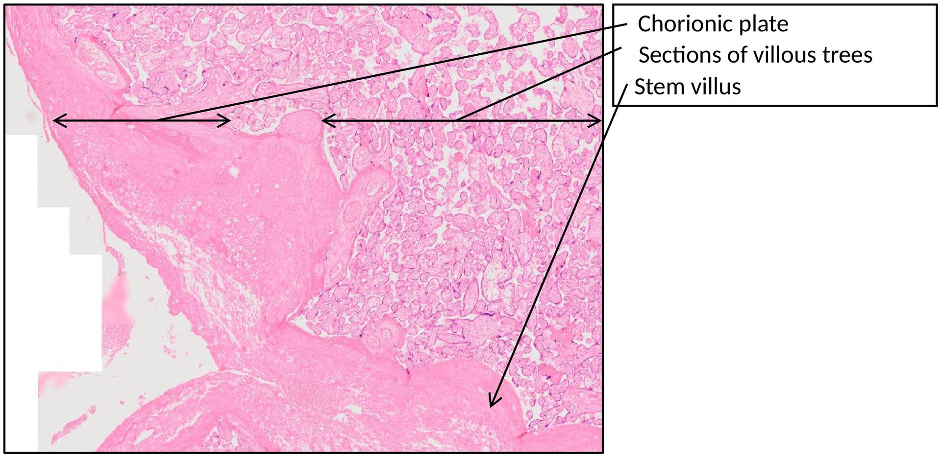
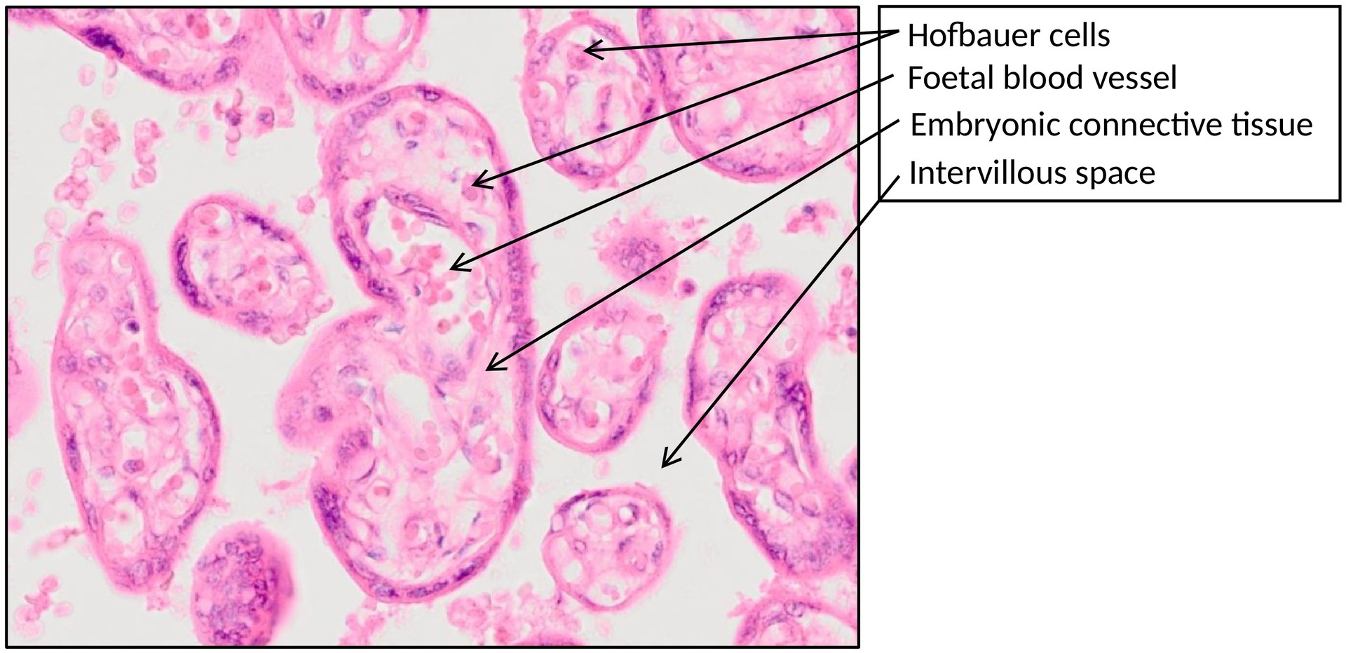
It is organized into two plates—the chorionic plate (fetal side) and the basal plate (maternal side)—between which lie extensive chorionic villous trees bathed in maternal blood.
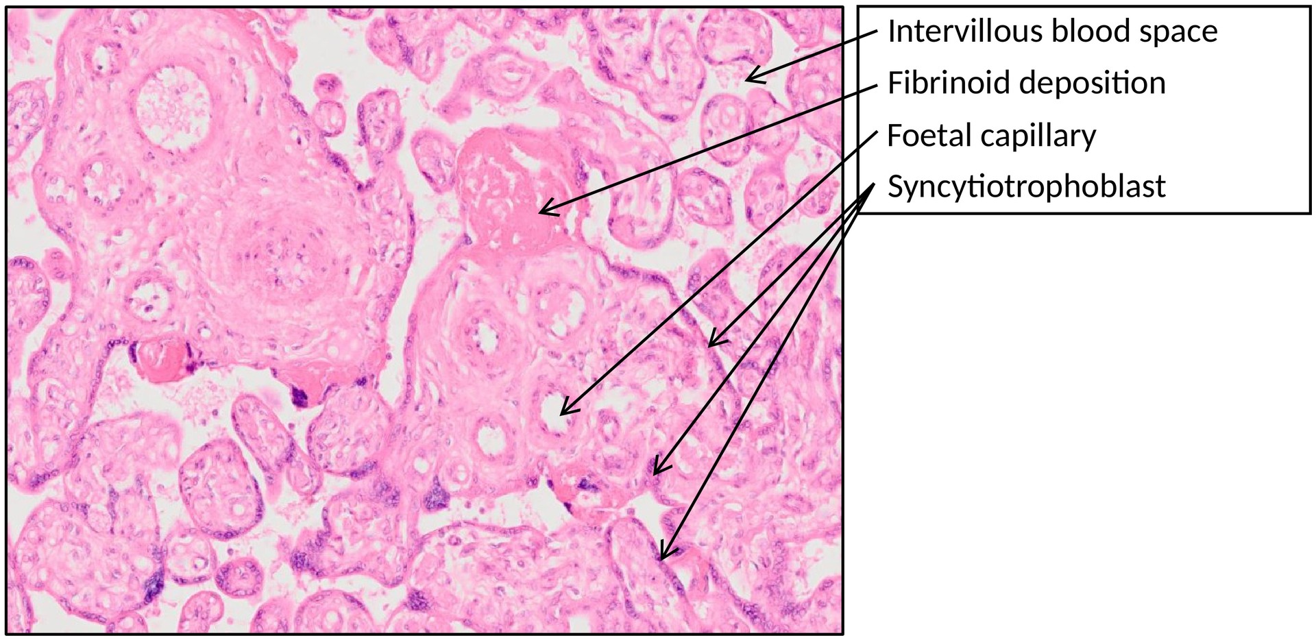
The chorionic villi contain fetal blood vessels, embedded within embryonic connective tissue (mesenchyme), and are externally covered by two layers of trophoblast:
-
an inner cytotrophoblast (Langhans’ cells), and
-
an outer syncytiotrophoblast, a multinucleated layer in direct contact with maternal blood in the intervillous spaces.
This arrangement enables the exchange of oxygen, carbon dioxide, nutrients, and waste products between maternal and fetal circulations.
The placental barrier in the mature placenta is formed by:
-
the syncytiotrophoblast,
-
the cytotrophoblast (which may be discontinuous late in pregnancy),
-
the basement membrane of the trophoblast,
-
the connective tissue core of the villus, and
-
the basement membrane and endothelium of the foetal capillaries.
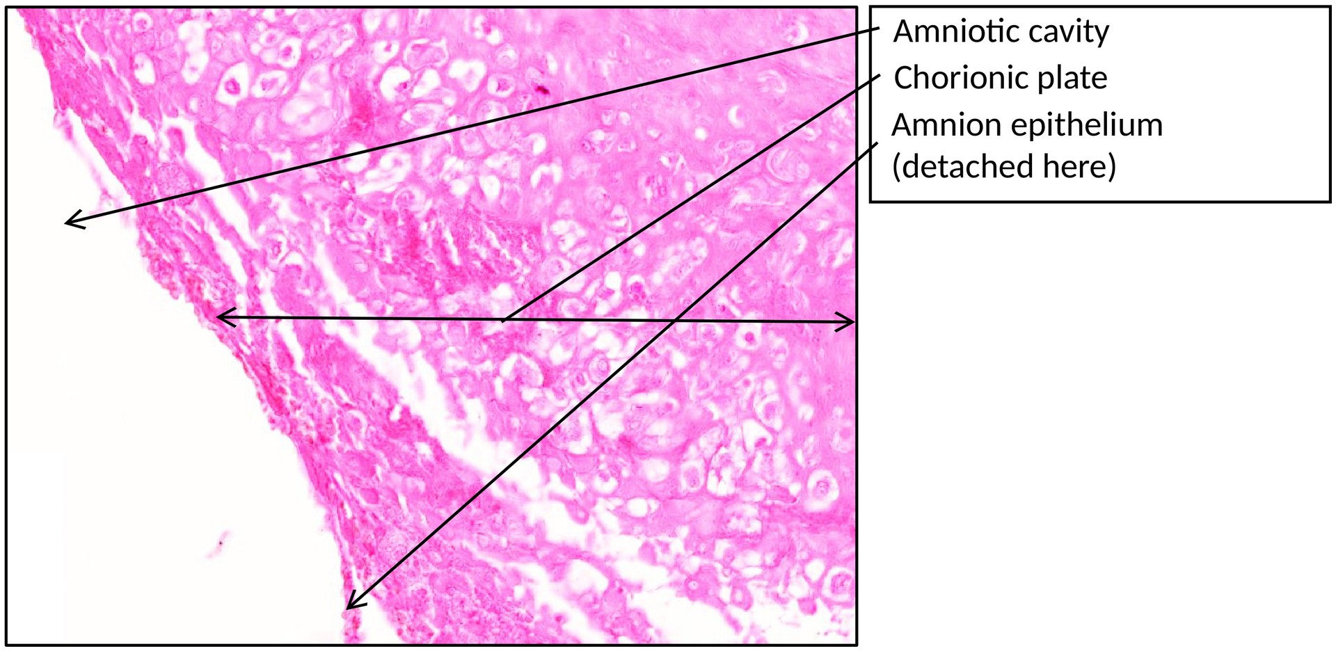
On the fetal side, the placenta is covered by a single layer of cuboidal amniotic epithelium, which forms the surface of the chorionic plate.
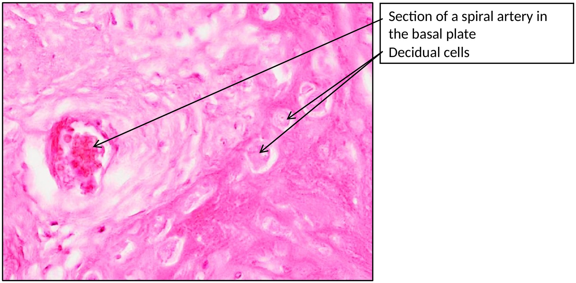
The maternal side appears irregular and trabeculated due to the decidua basalis, which contains large, pale decidual cells derived from the endometrial stroma.
Within the stroma of the villi, small macrophage-like cells known as Hofbauer cells can be found. These play a role in immune modulation and villous remodeling.
Maternal blood circulates freely through the intervillous spaces, bathing the villi and enabling exchange without direct contact between maternal and foetal blood.
TASKS:
-
Identify the foetal and maternal sides of the placenta.
-
Locate the amniotic epithelium and the chorionic plate on the fetal side.
-
Examine the chorionic villi and identify:
-
foetal blood vessels,
-
embryonic connective tissue, and
-
the surrounding syncytiotrophoblast.
-
-
Identify the intervillous spaces filled with maternal blood.
-
Locate the maternal basal plate containing large decidual cells.
-
Find and identify Hofbauer cells within the villous stroma.
License
University of Basel