CONNECTIVE TISSUE (GENERAL HISTOLOGY)
2.4
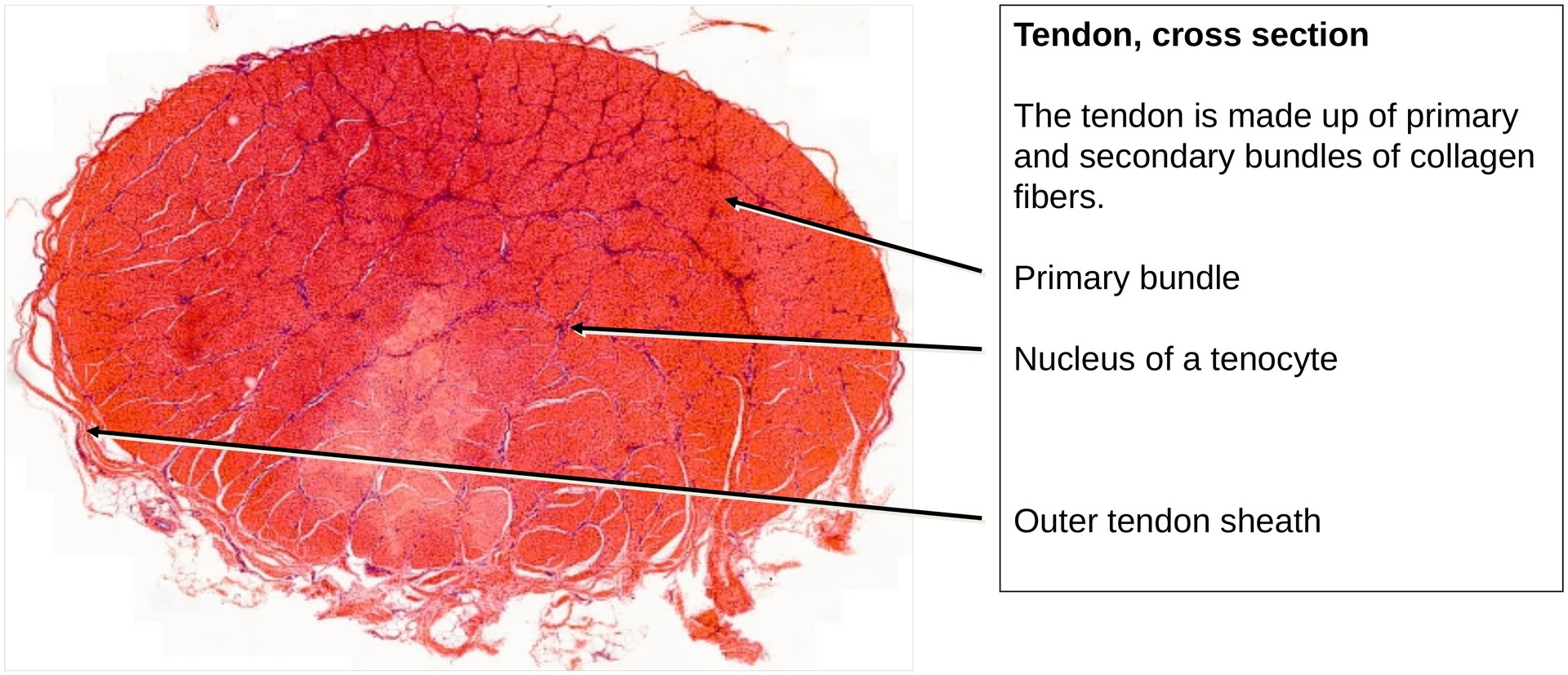
Tendon, transverse section
Specimen:
Specimen Details:
Organ: Tendon
Origin: Human
Staining: Hematoxylin - Chromotrope
Method and Specimen Description:
Because of their tough and fibrous consistency, tendons must be embedded in celloidin to obtain suitable sections. The minimum section thickness is approximately 25 µm, which limits the ability to achieve complete optical sharpness when scanning. Consequently, not all areas of the specimen appear equally well-focused, and fine cracks are common artefacts of this preparation method. The Hematoxylin–Chromotrope stain allows clear differentiation between the collagen fiber bundles and the tendon sheath cells (tenocytes).
Objective of the Examination:
• To study dense regular connective tissue using the tendon as an example.
• To understand that the extracellular matrix (ECM) forms the predominant component of tendinous tissue.
• To recognize the hierarchical bundling principle of collagen fibers, which ensures tensile strength and elasticity.
Special Features of the Specimen:
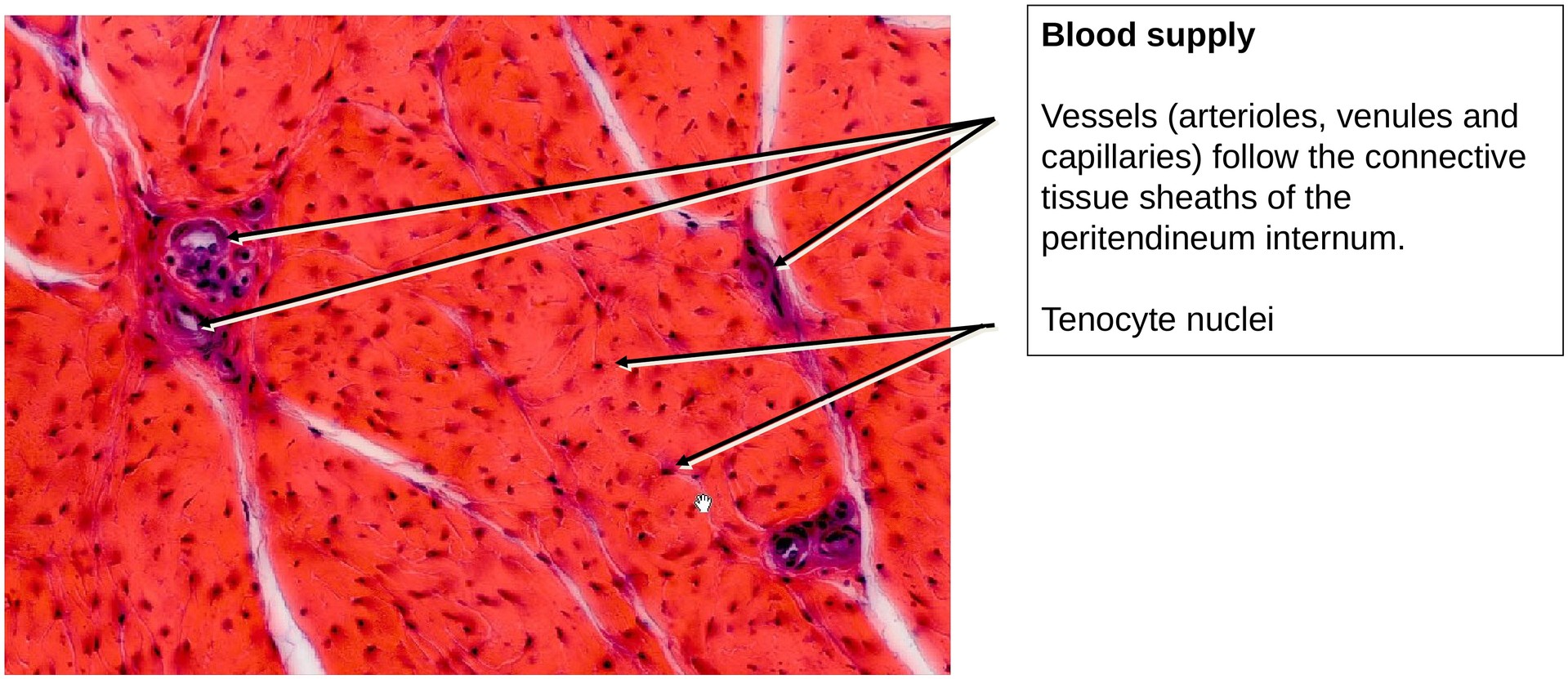
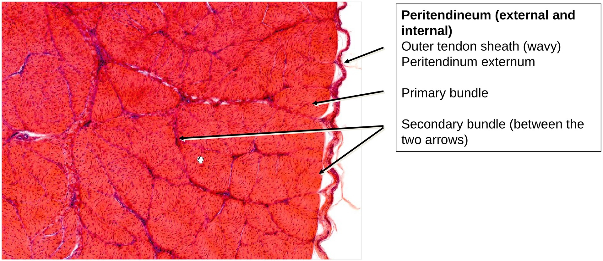
Even at low magnification, the bundled organization of the tendon is evident. The smallest collagen bundles (primary bundles) are enclosed by delicate collagenous sheaths, which unite to form secondary bundles and, ultimately, the entire tendon. Within these sheaths, small blood vessels and nerve fibers can be observed.
The tenocytes—the elongated fibroblast-like cells of the tendon—are located between adjacent collagen fiber bundles. Their elongated, flattened nuclei often appear slightly spiral due to the wavy (curled) course of the collagen fibers.
Tasks:
-
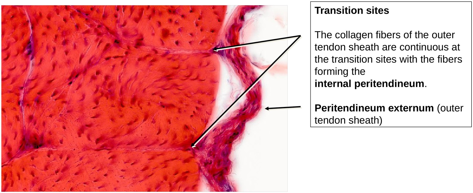
Examine the outer tendon sheath (peritendineum externum), which appears slightly wavy in this specimen.
-
Identify the transitions between the external peritendineum and the internal peritendineum, which envelops the primary and secondary collagen bundles.
-
At certain transition sites, these layers are continuous, indicating structural continuity of the collagen fibers.
-
Distinguish between primary bundles (fasciculi primarii) and secondary bundles (fasciculi secundarii).
-
Observe the nuclei of tenocytes, noting how they follow the slightly undulating course of the collagen fibres.
-
Locate small blood vessels within the internal peritendineum, which provide the limited vascular supply to the tendon.
-
License
University of Basel
Downloads