MALE REPRODUCTIVE ORGANS (ANATOMICAL MICROSCOPY)
11.7
Epididymis
Specimen Details:
Specimen Details:
Organ: Epididymis
Origin: Human
Staining: Van Gieson
Method and Specimen Description:
Normal histological specimen stained with Van Gieson.
In this staining, connective tissue (collagen) and epithelial cell nuclei appear red, while erythrocytes and smooth muscle cells stain yellow.
Objective of the Examination:
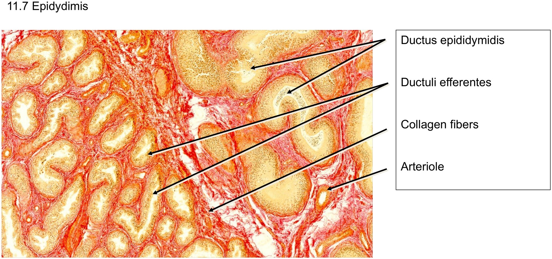
To study the duct systems of the human epididymis, particularly the ductuli efferentes and the ductus epididymidis, and to understand their epithelial differences and functional organization.
Special Features of the Specimen:
At low magnification, two features are immediately evident:
-
The duct system consists of two distinct epithelial types.
-
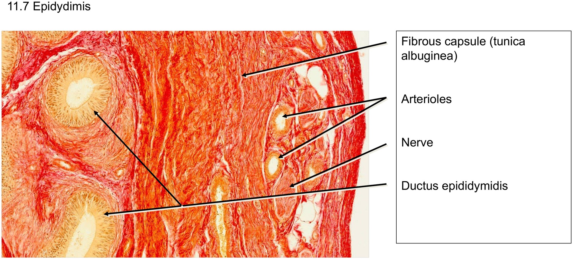
The epididymis is enclosed by a dense fibrous capsule, which is a continuation of the tunica albuginea of the testis.
The tunica albuginea here exhibits a tendon-like appearance, composed of dense, parallel collagen fibers interspersed with numerous blood vessels.
After spermatozoa leave the rete testis in the mediastinum testis, they pass through the ductuli efferentes into the ductus epididymidis, where they are stored and matured until ejaculation.
Because spermatozoa are immotile in the acidic environment of the epididymis and must conserve their limited energy reserves, passive and contractile transport mechanisms are required to move them onward.
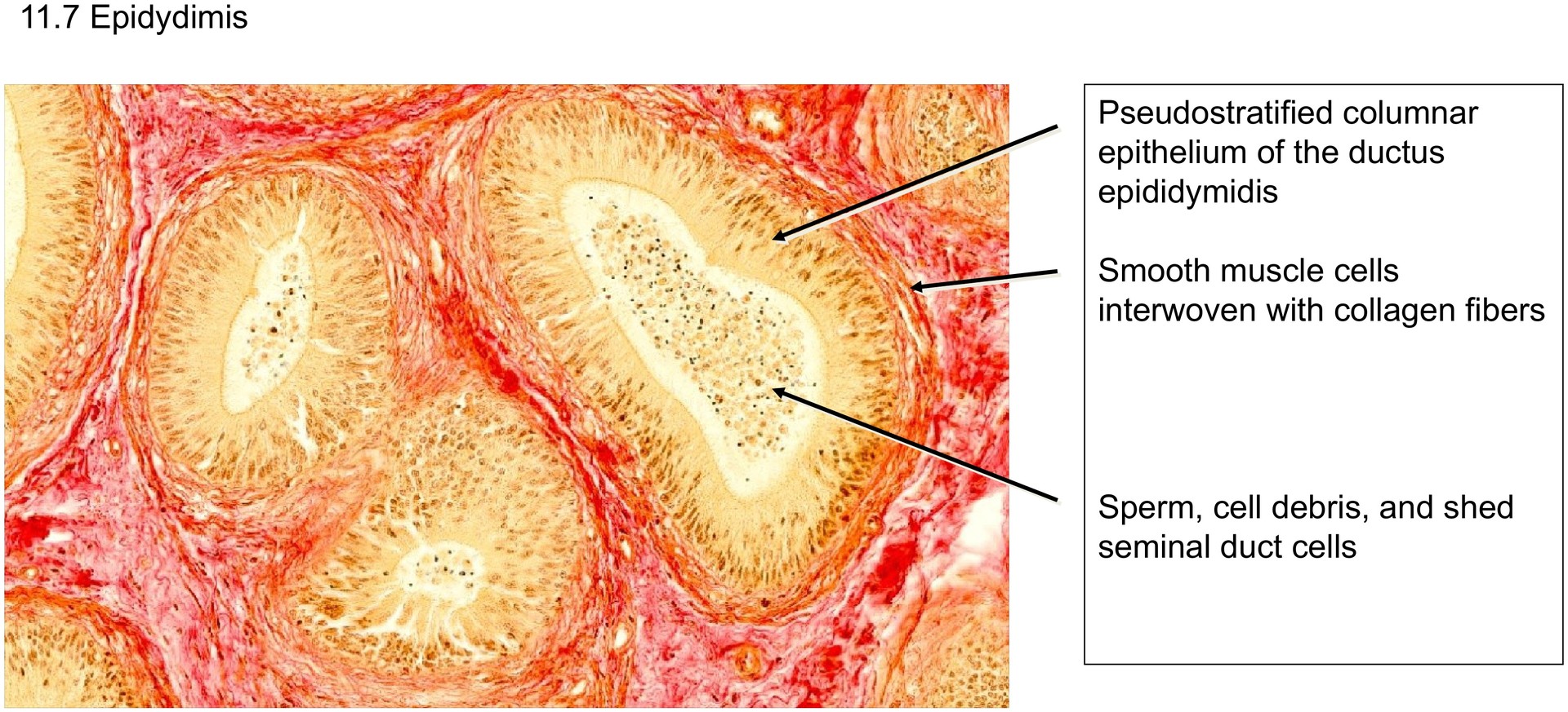
In the ductus epididymidis, this role is performed by smooth muscle cells, while in the ductuli efferentes, the wall contains myofibroblasts as well as smooth muscle cells.
The myofibroblasts form a compact layer around the ducts, interwoven with collagen fibers, contributing to peristaltic movement.
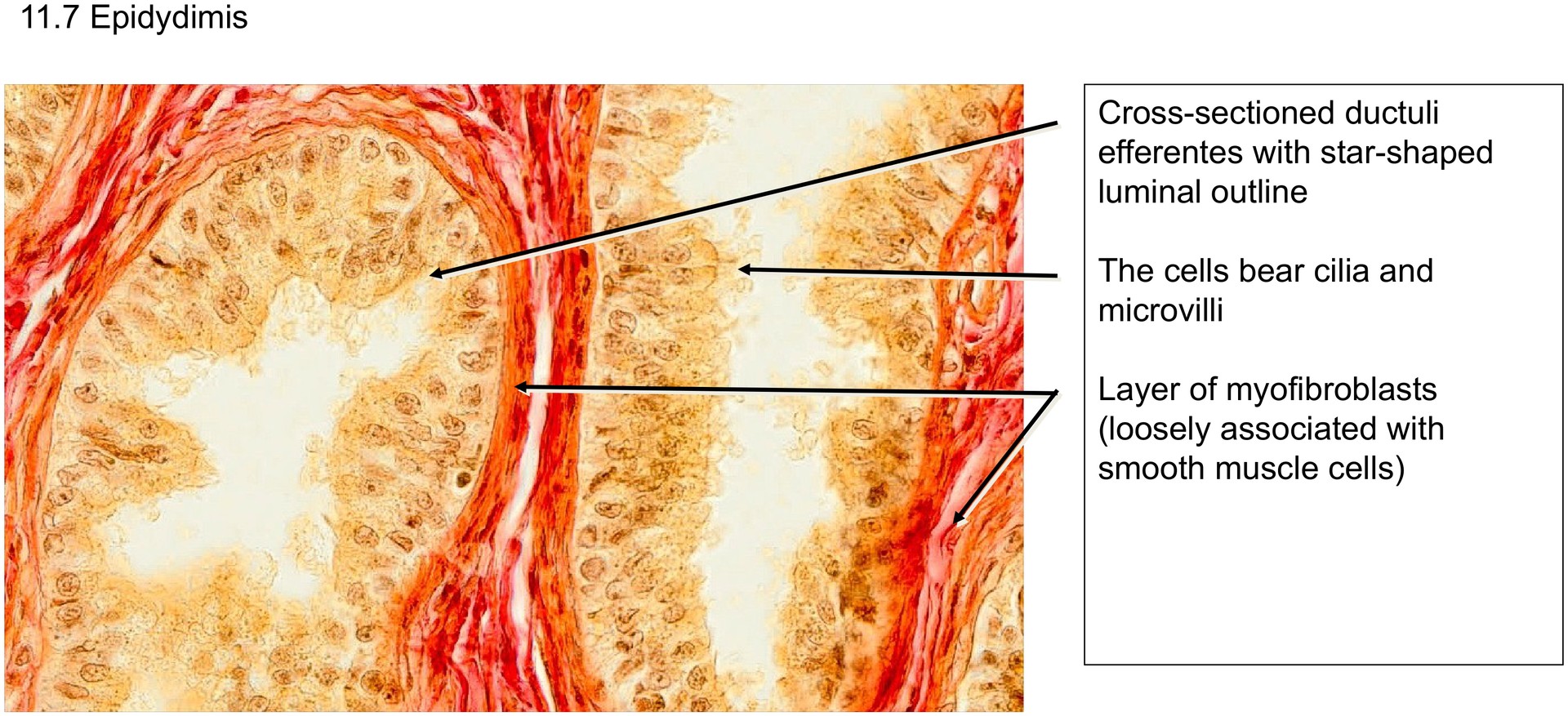
The ductuli efferentes are characterized by an uneven epithelial height, which gives their lumina a star-shaped outline in cross-section.
Their epithelium is pseudostratified ciliated columnar, containing both ciliated cells and non-ciliated cells with microvilli.
Just beneath the basal lamina lies a layer of myofibroblasts, loosely associated with smooth muscle cells.
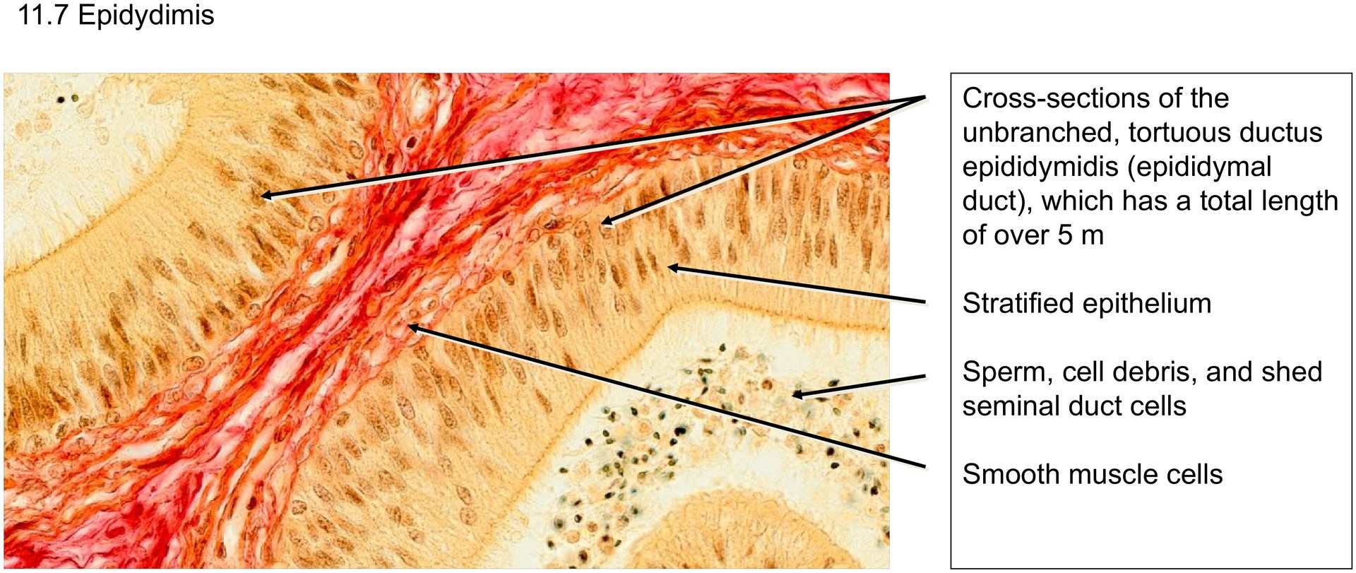
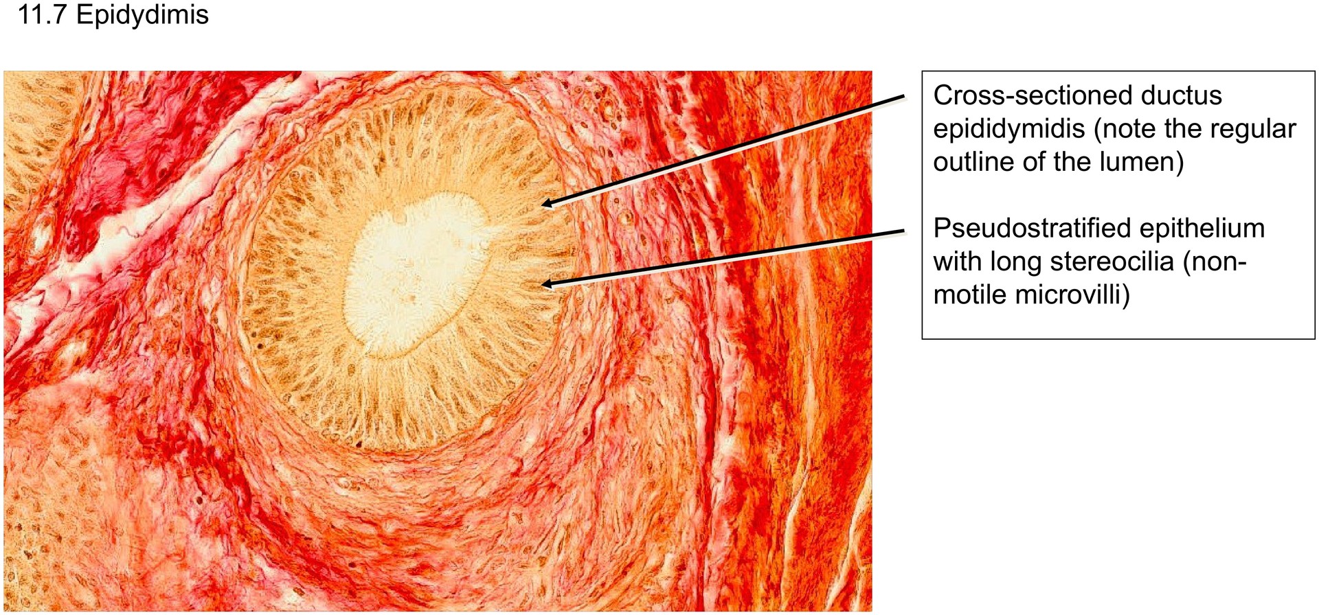
In contrast, the ductus epididymidis has a regularly defined lumen lined by a pseudostratified columnar epithelium with long stereocilia (non-motile microvilli).
All epithelial cells rest on the basement membrane, but not all reach the luminal surface.
Within the lumen, one often finds spermatozoa with condensed chromatin and shed epithelial cells from the duct lining.
Tasks:
• Identify the specimen as the epididymis by recognizing the two distinct duct systems and their epithelial differences.
• Describe the epithelial type of the ductus epididymidis. What kind of epithelium is it?
• What specialized luminal structures (differentiations) are present on the surface?
• Describe the epithelial type of the ductuli efferentes.
• How is the luminal surface of the efferent ducts constructed?
• Examine the tunica albuginea. How is it structured, and what type of connective tissue predominates?
• Search for nerve sections within the tunica albuginea or adjacent connective tissue.

License
University of Basel
Downloads